Task-specific bench model training versus basic laparoscopic skills
training for laparoscopic radical prostatectomy: a randomized
controlled study
Robert Sabbagh, BPharm, MSc, MD;
*Suman Chatterjee, MD;
†Arun Chawla, MD;
†Anil Kapoor, MD;
†Edward D. Matsumoto, MD, M.Ed
†Abstract
Background: Performing a laparoscopic urethrovesical anastomo-sis (LUA) after a radical prostatectomy is technically challenging for the novice laparoscopic surgeon. We developed a low-fidelity urethrovesical model (UVM) to allow a urologist to practise this critical step. The aim of our study was to compare the effect of task-specific bench model training (anastomotic suturing on the UVM) with that of basic laparoscopic suturing on intracorporeal urethrovesical anastomosis performance.
Methods:We recruited 28 senior surgical residents, fellows or staff surgeons for this prospective, single-blinded, randomized con-trolled study. We randomly assigned participants to an interven-tion group practising LUA on the UVM or to a control group prac-tising basic laparoscopic suturing and knot-tying on a foam pad. After practising, we videotaped participants performing 5 intra-corporeal interrupted sutures on a foam pad and a LUA on the UVM. A blinded expert scored the videotaped performance using a laparoscopic suturing checklist (CL) and a global rating scale (GRS), and timed the performance.
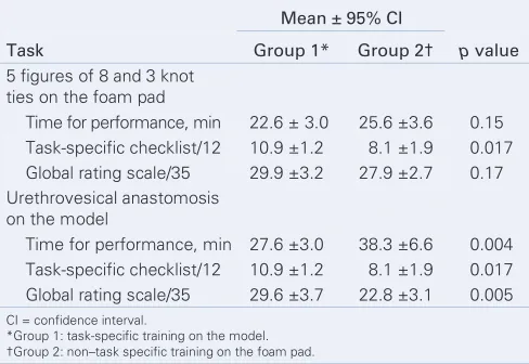
Results:On the foam pad suturing task, the group that trained on the UVM had significantly higher CL scores (10.9 v. 8.1, p = 0.017). On the LUA task, the group that trained on the UVM had significantly higher CL scores (10.9 v. 8.1, p = 0.017), GRS (29.6 v. 22.8, p = 0.005) and shorter times (27.6 v. 38.3 min, p = 0.004) than the control group.
Conclusion: Our task-specific bench model was shown to be super-ior to basic laparoscopic suturing drills on a foam pad.
Résumé
Généralités :L’anastomose urétrovésicale par voie laparoscopique (AUL) suivant une prostatectomie radicale pose certaines diffi-cultés techniques au chirurgien peu expérimenté avec la laparo-scopie. Nous avons créé un modèle urétrovésical basse-fidélité (MUB) permettant aux urologues de pratiquer cette étape cruciale de l’intervention. Le but de l’étude était de comparer l’impact d’exercices avec un modèle spécifique à la tâche (sutures
anasto-Can Urol Assoc J 2009;3(1):22-30
See related article on page 31
motiques sur le modèle urétrovésical) et d’exercices de sutures laparoscopiques sur l’aptitude à effectuer des anastomoses urétro-vésicales intracorporelles.
Méthodologie :Vingt-huit résidents séniors en chirurgie, chercheurs-boursiers et chirurgiens ont été recrutés pour cette étude prospec-tive et contrôlée, menée à simple insu avec répartition aléatoire. Les participants ont été répartis au hasard en 2 groupes, soit un groupe qui a pratiqué l’anastomose urétrovésicale sur le MUB et un groupe témoin qui a pratiqué les sutures laparoscopiques et la formation de nœuds sur un coussinet de mousse. Après la pra-tique, les participants ont été filmés pendant qu’ils effectuaient 5 suturations intracorporelles interrompues sur un coussinet de mousse et une anastomose urétrovésicale par laparoscopie à l’aide du modèle urétrovésical. Un expert ne connaissant pas le type d’exercices utilisé a ensuite évalué les chirurgiens à l’aide d’une liste de vérification des éléments clés d’une suturation laparoscopique (LV), d’un score global et du temps requis pour les suturations. Résultats :Lors de la tâche de suturation avec coussinet de mousse, le groupe qui avait pratiqué à l’aide du modèle urétrovésical a obtenu des scores LV significativement plus élevés (10,9 contre 8,1; p = 0,017). Quant à la tâche d’anastomose urétrovésicale par laparoscopie, le groupe qui avait pratiqué sur le modèle urétro-vésical a également obtenu des scores LV significativement plus élevés (10,9 contre 8,1; p = 0.017), mais aussi un score global plus élevé (29,6 contre 22,8, p = 0,005), et il a effectué la tâche en moins de temps (27,6 minutes contre 38,3 minutes, p = 0,004) par rapport au groupe témoin.
Conclusion :Nous avons élaboré un modèle spécifique à la tâche qui s’est révélé supérieur aux exercices de suturation laparo-scopique sur coussinet de mousse.
Introduction
Laparoscopic radical prostatectomy (LRP) has been intro-duced as a minimally invasive approach for the manage-ment of localized prostate cancer.1This procedure has a
improve the performance of trainees suturing a LUA, we designed a low-cost urethrovesical latex model (UVM) to teach this skill.
We hypothesized that, for complex skills such as laparoscopic urethrovesical suturing, a model incorporating task-specific surgical skills training is essential. We sought to compare the impact of task-specific bench model training (LUA suturing on UVM) to nonspecific laparoscopic practice (basic laparoscopic suturing and knot-tying) on intracor-poreal urethrovesical anastomosis performance.
Methods
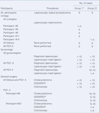
The Research Ethics Boards at St. Joseph’s Healthcare, and the Hamilton Health Sciences Corporation, Hamilton, Ont. approved our study. We recruited 28 senior surgical residents, fellows or staff surgeons in urology, general surgery and gynecology (Table 1 and Table 2) through surgi-cal rounds and advertisement. We obtained informed consent from each participant and assigned them each a study identification number. We randomly assigned participants to Group 1 (task-specific) to practise on the UVM perform-ing runnperform-ing LUA sutures (Fig. 1 and Fig. 2) or to Group 2 (control) to practise basic laparoscopic suturing and knot-tying on a foam pad. All par-ticipants completed a questionnaire on their laparoscopic experience (Appendix 1) and watched a 15-minute video of an actual
intra-operative LUA being performed. The purpose of the video was to show how the skills chosen for this study relate to the actual procedure.
Group 1 received specific instructions on angling the needle within the needle driver, approaching and holding the urethra or bladder, setting and advancing the needle into the tissue, and when to use the opposite hand or to backhand a needle. The double-armed suture consisted of two 3–0 Prolene sutures (ETHICON, Johnson & Johnson) 15 cm in length, tied tail to tail with a SH needle at each end. For orientation and communication purposes, while looking into the pelvis from the head in a supine position, the 12 o’clock position was the anterior urethra or bladder and the 6 o’clock position was the posterior urethra or bladder. We instructed par-ticipants to pass both needles out-to-in at the 6 o’clock bladder position using each arm of the suture to perform a running anastomosis up each side. They then passed 3 running anastomosis stitches connecting the bladder to the urethra from 6 to 9 o’clock on the left side and from 6 to 3 o’clock on the right side. They inserted an 18 Fr. Foley catheter from the urethral end of the model tube beyond the completed posterior half of the tomosis. Once the catheter passed across the anas-tomosis, continuing the same suturing completed the anterior part of the anastomosis. When the sutures met at 12 o’clock, both were tied together with 3 knots to complete the task.
Group 2 practised suturing on foam pads
Table 1. Distribution of surgical specialty and level of training among participants in the task-specific and control groups
No. of participants
Surgical specialty; level of training Group 1* Group 2†
Urology 8 3
Urologists 4 1
Fellows 2 0
PGY-4 2 2
Gynecology 2 5
Gynecologists 1 2
PGY-5 1 2
PGY-4 0 1
General surgery 4 6
Fellows 2 2
PGY-5 2 2
PGY-4 0 2
All participants 14 14
PGY = postgraduate year.
acquired from Limbs and Things (Bristol, UK). We instructed participants to place single, simple-interrupted sutures across the foam and tie 3 knots. Group 2 also practised running the suture across opposing straight edges of foam to practise needle-and instrument-hneedle-andling, but participants were not required to tie these sutures. They also received basic suture-handling and instrument-handling instructions similar to Group 1, but they did not perform any circular running sutures.
All participants received feedback from an experi-enced laparoscopic urologist during the practice session.
After 2 hours of training, both groups performed a post-training laparoscopic suturing test consist-ing of 2 tasks. The first task was to complete 5 fig-ures of 8 and tie 3 knots on the foam pad. The second task was to perform a running LUA on the UVM (Fig. 3). For both tasks, we videotaped and timed performances. A blinded laparoscop-ic expert evaluated the performances using a task-specific checklist (CL) and global rating scale (GRS) (Appendix 2 and Appendix 3).
We performed our statistical analysis using SigmaStat Version 3.10.0 (Systat Software Inc.). From an earlier study, looking at bench model
Table 2. Distribution of previous laparoscopic/endoscopic experience among participants in the task-specific and control groups
No. of cases
Participants Procedures Group 1* Group 2†
All participants Laparoscopic radical prostatectomy 0 0
Urology All urologists
Laparoscopic nephrectomy
Participant #2 1–4
Participant #3 0
Participant #4 0
Participant #11 0
Participant #14 1–4
All fellows None performed 0 0
All PGY-4 None performed 0 0
Gynecology All gynecologists
Diagnostic laparoscopy > 15 > 15
Laparoscopic tubal ligation > 15 > 15
All PGY- 5 Diagnostic laparoscopy > 15 > 15
Laparoscopic tubal ligation > 15 > 15
All PGY-4 Diagnostic laparoscopy 10–15
Laparoscopic tubal ligation 1–4
General surgery
All fellows and PGY- 5 Cholecystectomy > 15 > 15
OGD/ERCP > 15 > 15
Colonoscopy > 15 > 15
PGY- 4
Participant #8 Cholecystectomy 10–15
OGD/ERCP 5–10
Colonoscopy 5–10
Participant #22 Cholecystectomy 5–10
OGD/ERCP 1–4
Colonoscopy 1–4
ERCP = endoscopic retrograde cholangiopancreatography; OGD = esophagogastroduodenoscopy; PGY = postgraduate year. *Group 1: task-specific training on the model.
fidelity in ureteroscopic training, we found that the mean difference in GRS between the group that trained on the low-fidelity model and the group that underwent didactic teaching was 10.166. We calculated the pooled standard deviation as 5.0. Using t test sample size calculation, to achieve a power of 80% at an αof 0.05, our study required a minimum of 6 participants per group. To account for the possibility of dropouts and the differences in the complexity of the procedure and training intervention, compared with the original ure-teroscopy study, we recruited an additional 8 par-ticipants per group. This allowed us to detect a difference of 15% between the 2 groups. We ran-domly assigned participants using sealed envelopes prepared by our statistician. We performed com-parisons between Group 1 and Group 2 using a
Mann–Whitney test. We considered p < 0.05 to be statistically significant.
Results
At baseline, we detected no differences in previous laparoscopic experience, distribution of surgical specialty and level of training between groups (Table 1 and Table 2). None of the participants had performed an LRP. On the foam pad sutur-ing task, Group 1 had significantly higher CL scores (10.9 v. 8.1, p = 0.017), but we noted no significant
Fig. 1.The urethrovesical model sutured together with a Foley catheter passing through.
Fig. 2.Urethrovesical model positioned in the pelvic trainer simu-lated orientation within the male pelvis.
24 participants
15-minute video on laparoscopic urethrovesical anastomosis
12 participants • Task-specific laparoscopic training
• Laparoscopic circular running suture anastomosis training on the urethra bench model
• 2 hours training
12 participants • Nontask-specific laparoscopic training
• Basic laparoscopic suturing and knot tying on a foam pad
• 2 hours training
Post-training laparoscopic suturing test videotaped
1. Anastomosis on bench model 2. Foam pad
1 hour
Post-training laparoscopic suturing test videotaped
1. Anastomosis on bench model 2. Foam pad
1 hour
differences in GRS or time (Table 3). On the LUA task, Group 1 had a significantly higher task-specific CL score (10.9 v. 8.1, p = 0.017), GRS (29.6 v. 22.8, p = 0.005) and shorter times (27.6 min v. 38.3 min, p = 0.004) than the control group (Table 3).
Discussion
Reconstructive laparoscopy is technically more difficult than ablative surgery, demanding more skills and experience. With laparoscopic recon-struction, the ability to suture is a necessity. Often, suturing is performed with extreme angles and positions owing to the natural orientation of organs. In addition, suturing with standard laparoscopic needle drivers means loss of degrees of freedom. Unlike the Da Vinci robotic arm (Intuitive Surgical), which has 7 degrees of freedom because of a “wrist” within the abdominal/pelvic cavity, the pure laparoscopic surgeon has to compensate for the lack of a “wrist” by positioning and advancing a needle with a corrective angle in relation to the bladder or urethra. A urologist must learn these subtle techniques to perform a urethrovesical anas-tomosis successfully. Furthermore, working with instruments in the pelvis requires substantial mod-ification of instrument-handling, suturing and tying owing to the relatively narrow confines compared with the wide-open space for transperitoneal kid-ney surgery.
In this study, Group 1 performed the final run-ning urethrovesical anastomosis quicker and had
better CL scores and GRS compared with Group 2. This suggests that task-specific training on UVM is an effective way of learning how to perform LUA. Interestingly, Group 1 also performed the figure of 8 task better than Group 2. Training on the UVM not only led to better running urethrovesical per-formance, but also to better performance on sim-ple suturing tasks.
Understanding the critical steps of a surgical procedure is important when designing bench models. The model does not have to look realis-tic (high fidelity) to be effective as a teaching tool. In 2 separate studies, low-cost, low-fidelity mod-els have been shown to be effective teaching tools.4,5However, it is important to ensure the
model allows trainees to practise the essential steps required in the real procedure. In our study, the model reproduces almost all of the surgical steps of a LUA. This latex UVM has been designed to simulate anatomical and tissue characteristics (i.e., thickness) of the human bladder and urethra (Fig. 1). Operating in a man’s pelvis is reproduced when the UVM is inserted into a special pelvic trainer (Limbs and Things) (Fig. 2). Not only does a trainee have the opportunity to learn proper nee-dle position and handling, they also learn how to operate within the strict confines of the pelvic structure. Standing at the sides and working down the middle of the patient can be ergonomically taxing. With proper instructions and practice, trainees can anticipate the ergonomic challenges of laparoscopic pelvic surgery.
Several authors have proposed different models allowing trainees to acquire the skills necessary to perform an LUA. Teber and colleagues6
de-veloped a standardized step-by-step program to improve skills and enable trainees not experienced in laparoscopy to increase reproducible perform-ance in reconstructive laparoscopy. Katz and col-leagues7proposed a simplified 5-step model for
training LUA. By using this model and dividing complicated surgical steps into simplified tasks (e.g., passage of ligature, intracorporal knotting, intra-corporeal suturing, linear anastomosis, circular run-ning suture anastomosis with chicken skin), they were able to substantially improve trainee per-formance. Nadu and colleagues8developed a
sim-ple model using chicken skin in a pelvic trainer. After performing 20 anastomoses on this model, 2 urology fellows reduced the time required for performing the anastomosis from 75 to 20 minutes Table 3. Post-training laparoscopic suturing test results
Mean ± 95% CI
CI = confidence interval.
and were able to create a watertight running LUA in patients in a mean time of 40 minutes (range 30–55 min).8 In our study, participants who
trained on the UVM performed the urethroves-ical anastomosis in a mean time of 27.6 minutes, approaching performance in the study by Nadu and colleagues.8
Other training models studied include human cadavers, live animals and virtual reality (VR) simu-lators to teach surgical skills. Human cadavers most closely resemble live patients but they are expen-sive, have limited availability, have stiffer tissues and may transmit infections. Animals remain an alternative and have the benefit of providing living simulations that are generally faithful to opera-tive reality. However, they are expensive, neces-sitate specialized facilities and personnel, and are not always ethically acceptable.
Virtual reality simulators have been introduced as training tools and could improve the perform-ance in the operating room for laparoscopic sur-gery.9–12For example, a VR simulator named URO
Mentor (Simbionix) is commercially available for training in endourology. Several studies have shown that training on the simulator improves perform-ance.13–15However, studies validating VR
simula-tors in the operating room are limited. Simulasimula-tors also lack haptics (tactile feedback), which is essen-tial in procedures such as laparoscopy.
There were limitations with our study. The ure-throvesical anastomosis is an essential step in per-forming an LRP, yet there are preceding steps that have to be learned and consolidated before finish-ing with the anastomosis. This study focused only on a single step, and it is important to keep in mind that a successful procedure requires multiple steps to be completed successfully. From an education-al perspective, the ability to deconstruct a pro-cedure into steps and create an inventory is attract-ive, as this facilitates teaching and evaluation of complex surgeries. The challenge would be to find a teaching model that incorporates all of the crit-ical steps to teach a new procedure from begin-ning to end. With advances in graphics and vir-tual haptics, VR may provide the solution for the acquisition of complex skills.
Other limitations include the lack of intra-operative factors such as blood and urine, which can hamper surgical performance. We included only participants at an advanced level in a special-ty where laparoscopy is currently used; however,
our population is homogeneous in the sense that none of the participants had ever performed an LRP. Owing to the limited number of urologists and urology residents in our area and program, we could not rely on them exclusively to reach our sample size. Another limitation of this study is model validation. The ideal validation method would have been to perform the post-test on a patient, but for ethical reasons such as the partici-pants’ lack of experience, the transfer to this “high-fidelity model” would be unacceptable. A more suit-able high-fidelity model would be a live anesthetized pig, as the pig urethrovesical junction and the pelvic dimensions have anatomical and tissue characteris-tics similar to humans. The second phase of our study will research whether skills learned on the UVM transfer to a high-fidelity live pig model.
Conclusion
Laparoscopic urethrovesical anastomosis is one of the most challenging steps during an LRP. We have developed a task-specific bench model that allows a trainee to practise LUA. Training on this model is superior to basic laparoscopic suturing practice. The second phase of our study will assess whether skills learned on the UVM leads to a better ure-throvesical anastomosis on a live anesthetized pig.
RReeffeerreenncceess
1. Zorn KC, Lee, BR. Laparoscopic radical prostatectomy: an established minimally inva-sive procedure with proven oncologic track record. J Endourol 2008;22:2053-5. 2. Stolzenburg JU, Katsakiori PF, Liatsikos EN. Role of laparoscopy for reconstructive
urology. Curr Opin Urol 2006;16:413-8.
3. Poulakis V, Witzsch U, De Vries R, et al. Intensive laparoscopic training: the impact of a simplified pelvic-trainer model for the urethrovesical anastomosis on the learning curve. World J Urol 2006;24:331-7.
4. Anastakis D., Regehr G, Reznick R, et al. Assessment of technical skills transfer from the bench training model to the human model. Am J Surg 1999;177:167-70. 5. Matsumoto ED, Hamstra SJ, Radomski SB, et al. The effect of bench model fidelity
on endourological skills: a randomized controlled study. J Urol 2002;167:1243-7. 6. Teber D, Dekel Y, Frede T, et al. The Heilbronn laparoscopic training program for
laparo-scopic suturing: concept and validation. J Endourol 2005;19:230-8.
7. Katz R, Nadu A, Olsson LE, et al. A simplified 5-step model for training LUA. J Urol 2003;169:2041-4.
This article has been peer reviewed.
From *Sherbrooke University, Department of Surgery, Division of Urology, Sherbrooke, Que., and †McMaster University, Department of Surgery, Division of Urology, Hamilton, Ont.
8. Nadu A, Olsson L., Abbou CC. Simple model for training in the laparoscopic vesicourethral running anastomosis. J Endourol 2003;17:481-4.
9. Korndorffer JR Jr, Dunne JB, Sierra R, et al. Simulator training for laparoscopic sutur-ing ussutur-ing performance goals translates to the operatsutur-ing room. J Am Coll Surg 2005; 201:23-9.
10. Seymour NE, Gallagher AG, Roman SA, et al. Virtual reality training improves operat-ing room performance: results of a randomized, double-blinded study. Ann Surg 2002; 236:458-63.
11. Shah J, Darzi A. Virtual reality flexible cystoscopy: a validation study. BJU Int 2002; 90:828-32.
12. Ogan K, Jacomides L, Shulman MJ, et al. Virtual ureteroscopy predicts ureteroscopic proficiency of medical students on a cadaver. J Urol 2004;172:667-71. 13. Watterson JD, Beiko DT, Kuan JK, et al. Randomized prospective blinded study
vali-dating acquistion of ureteroscopy skills using computer based virtual reality endouro-logical simulator. J Urol 2002;168:1928-32.
14. Jacomides L, Ogan K, Cadeddu JA, et al. Use of a virtual reality simulator for ureteroscopy training. J Urol 2004;171:320-3.
15. Wilhelm DM, Ogan K, Roehrborn CG, et al. Assessment of basic endoscopic perform-ance using a virtual reality simulator. J Am Coll Surg 2002;195:675-81.
Correspondence:Dr. Edward D. Matsumoto, McMaster University, Department of Surgery, Division of Urology, McMaster Institute of Urology at St. Joseph’s Hospital, 50 Charlton Ave. E., Hamilton ON L8N 4A6; fax 905 308-7205; [email protected]
Appendix 1. Participant questionnaire
Candidate no.
How many of the following procedures have you performed by yourself? (i.e. > 80% of the procedure)
1. laparoscopic prostatectomy 0 1 to 4 5 to 10 10 to 15 >15
2. laparoscopic nephrectomy 0 1 to 4 5 to 10 10 to 15 >15
3. laparoscopic cholecystectomy 0 1 to 4 5 to 10 10 to 15 >15
4. OGD/ERCP 0 1 to 4 5 to 10 10 to 15 >15
5. colonoscopy 0 1 to 4 5 to 10 10 to 15 >15
6. other laparoscopic
procedure_________________
0 1 to 4 5 to 10 10 to 15 >15
How many of the following procedures have you assisted at? (i.e. < 80% of the procedure)
1. laparoscopic prostatectomy 0 1 to 4 5 to 10 10 to 15 >15
2. laparoscopic nephrectomy 0 1 to 4 5 to 10 10 to 15 >15
3. laparoscopic cholecystectomy 0 1 to 4 5 to 10 10 to 15 >15
4. OGD/ERCP 0 1 to 4 5 to 10 10 to 15 >15
5. colonoscopy 0 1 to 4 5 to 10 10 to 15 >15
6. other laparoscopic
procedure_________________
0 1 to 4 5 to 10 10 to 15 >15
How many of the following procedures have you seen?
1. laparoscopic prostatectomy 0 1 to 4 5 to 10 10 to 15 >15
2. laparoscopic nephrectomy 0 1 to 4 5 to 10 10 to 15 >15
3. laparoscopic cholecystectomy 0 1 to 4 5 to 10 10 to 15 >15
4. OGD/ERCP 0 1 to 4 5 to 10 10 to 15 >15
5. colonoscopy 0 1 to 4 5 to 10 10 to 15 >15
6. other laparoscopic
procedure_________________
0 1 to 4 5 to 10 10 to 15 >15
Appendix 2. Participant questionnaire*
Task-specific checklist
Item Not done or incorrect Done correctly
1 Needle loaded one-half to two-thirds from tip 0 1
2 Uses laparoscopic needle holder and graspers to handle needle 0 1
3 Needle enters tissues at right angles (80% of bites) 0 1
4 Single attempt at needle passage through tissues (90% of bites) 0 1
5 Follow through on curve of needle on entrance (80% of bites) 0 1
6 Follow through on curve of needle on exit (80% of bites) 0 1
7 Minimal damage with graspers 0 1
8 Equal spacing 0 1
9 Equal bites on each side (80% of bites) 0 1
10 Square knots 0 1
11 Apposition of tissues without excessive tension on suture 0 1
12 Appropriate alignment of tissues (no torsion) 0 1
Maximum total score
Total score
Appendix 3. Global rating scale Candidate identification no.
Please rate the candidate's performance on the following scale:
1 2 3 4 5
Respect for tissue
Frequently used unnecessary force on tissue or cased damage by inappropriate use of instruments
Careful handling of tissue but occasionally caused inadvertent damage
Consistently handled tissues appropriately with minimal damage
1 2 3 4 5
Time and motion
Many unnecessary moves Efficient time/motion but some
unnecessary moves
Clear economy of movement and maximum efficiency
1 2 3 4 5
Instrument
handling Repeatedly makes tentative or awkward
moves with instruments by inappropriate use of instruments
Competent use of instruments but occasionally appeared stiff or awkward
Fluid moves with instruments and no awkwardness
1 2 3 4 5
Knowledge of
instruments Frequently asked for wrong instrument or used inappropriate instrument
Knew names of most instruments and used appropriate instruments
Obviously familiar with the
instruments and their names
1 2 3 4 5
Flow of operation
Frequently stopped operating and seemed unsure of the next move
Demonstrated some forward planning with reasonable progression of procedure
Obviously planned course of operation with effortless flow from one move to the next
1 2 3 4 5
Use of assistants
Consistently placed assistants poorly or failed to use assistants
Appropriate use of assistants most of the time
Strategically used assistants to the best advantage at all times
1 2 3 4 5
Knowledge of
specific procedure Deficient knowledge. Needed specific instruction at most steps
Knew all important steps of operation Demonstrated familiarity with all aspects of operation Would you feel confident in allowing this trainee to perform this procedure in the operating room?