THE EFFECT OF CHITOSAN ON OSTEOCLAST VIABILITY, BONE RESORPTION AND RADICAL
OXYGEN PRODUCTION OF PRIMARY OSTEOCLAST CULTURE OF MOUSE BONE
DEWI FATMA SUNIARTI
1, SRI ANGKY SOEKANTO
1*, NURTAMI SOEDARSONO
1, BASRIL ABBAS
21Department of Oral Biology, Faculty of Dentistry Universitas Indonesia, Jalan Salemba Raya No 4 Jakarta Pusat 10430, Indonesia. 2Center
for Application of Isotope and Radiation Technology, National Nuclear Energy Agency, Jakarta, Indonesia. Email: sriangky@yahoo.com Received: 12 June 2015, Revised and Accepted: 27 July 2015
ABSTRACT
Objective: The purpose of this investigation was to determine the effect of chitosan on osteoclast cells by observing cell viability, bone resorption, and radical oxygen species (ROS) production.
Methods: Osteoclast cells were obtained from the primary culture of bone marrow mouse. The osteoclast cells were identified by tartrate-resistant acid phosphatase (TRAP) marker both on the cells and the culture medium. The osteoclast cell viability was observed with (3-(4,5-dimethylthiazol-2-yl)-2,5-diphenyltetrazolium bromide) tetrazolium assay and Bradford assay for total protein medium culture, while ROS production was measured with malondialdehyde (MDA) assay. Slices of cow cortical bone were used as a substrate for osteoclastic resorption and concentrated hydrochloric acid were used to activate resorption and pit formation by any osteoclasts.
Results: Osteoclast cells were identified by TRAP marker and chitosan treated group cells showed lower optical density value compared to control (p<0.05) on TRAP assay medium culture. Cell viability indicated lower on chitosan group than control (p<0.05). There was a qualitative difference of the pits formed on the bone surface between the control and the chitosan group. There was a significant difference in MDA (mmol/ml) between the control and the chitosan group with (p<0.05).
Conclusion: Based on this research, we conclude that chitosan inhibits the viability of osteoclast cells, decreases ROS production and bone resorption. Keywords: Chitosan, Osteoclast proliferation, Radical oxygen species, Bone resorption.
INTRODUCTION
Chitosan is a biodegradable and non-toxic chitin derivative with a low molecular weight of 34.8 kDa and a high molecular weight of 800-1500 kDa. Its chemical structure is similar to hyaluronate [1-3]. This biomaterial can take various forms and has various functions. It is available in a variety of useful forms as gel, membrane, fiber, beads, powder, flakes and solution. Previous research has reported that chitosan is capable of increasing hemostasis, decreasing fibroplasia, facilitating osteogenesis and increasing tissue regeneration [3-7]. In the field of dentistry, Xu et al. reported the usage of chitosan as barrier membrane material in periodontal regeneration [8]. Chitosan gel alone or its combination with demineralize bone matrix membrane is promising for periodontal regeneration [9]. Arnaud et al. reported chitosan effect on dentinal enamel demineralization: An in vitro evaluation [10]. A self-setting composite consisting of chitosan/tricalcium phosphate microparticles showed a high degree of cytocompatibility and seemed to be a “user-friendly” material for oral surgeons [11].
The healing process of bone fractures or bone defects is a complex process. The healing requires integration and coordination among specific cells. There are three specific cells, i.e. osteoblast, osteocyte and osteoclast, which have a function in the formation and remodeling of bones. The effect of chitosan on osteoblast cells has been extensively reported. Chitosan can induce the osteoblast differentiation and bone formation (in vitro) [1,3,12-14]. However, the effect of chitosan on osteoclast cells has not been much documented. Osteoclast is a giant bone cell with multiple nuclei that plays a role in bone resorption and bone remodeling. Like macrophage cells, osteoclast can produce radical oxygen that can cause damage to the surrounding tissues.
In vitro research, osteoclast can be obtained from the culture of the bone marrow of test animals [15,16]. The aim of this investigation
was to determine the effect of chitosan, an Indonesian material with special specifications produced by Indonesia National Nuclear Energy Agency (BATAN). Therefore, this study was to evaluate the interaction of chitosan with cells participating in tissue engineering, especially osteoclast. The osteoclast cells were observed by studying their cell viability, bone resorption, and radical oxygen species (ROS) production. For this research, osteoclast is obtained from the culture of the bone marrow of mice, which has already been extensively used in research on the formation of osteoclast cells with multiple nuclei from its progenitors.
METHODS
This was an in vitro study using osteoclast cell culture. The osteoclast cells were identified by osteoclast tartrate-resistant acid phosphatase (TRAP) marker (Primary Cell Co. Ltd., Sapporo, Japan) both on the cells and the culture medium. Then the osteoclast cell viability was observed with 3-(4,5-dimethylthiazol-2-yl)-2,5-diphenyltetrazolium bromide (MTT) assay (Invitrogen, Washington) and Bradford assay (Thermo Scientific, Rockford, USA) for total protein medium culture, while ROS production was measured with malondialdehyde (MDA) assay (Sigma). Concentrated hydrochloric acid (HCl 37%) 82 µl/100 ml was added to the last medium change of the osteoclast cell culture to activate the osteoclast cells [16].
Cell culture
suspension was cultured at 4 × 10⁵ in a 24 well plate. Cell cultures were
performed using Dulbecco modified eagles medium (Invitrogen) with 10% fetal bovine serum, 100 U/ml penicillin, 100 µg/ml streptomycin and amphotericin B (Biowest, South America) supplemented with
10 nM 1α,25(0H)2D3 (A.G Scientific, Inc. San Diego) as control in a
condition of 37°C and 5% CO2 for a duration of 6-8 days, according
to the method described by (1994) and Timothy (2003) [13,14]. The
culture medium was changed every day while adding 1α,25(OH)2D3.
The osteoclast cells resulting from the culture were identified by osteoclast TRAP markers (Primary Cell Co. Ltd., Sapporo, Japan) both on the osteoclast cells and the culture medium, following the manufacturer’s instruction.
Chitosan
For this research, we used chitosan produced by BATAN Indonesia, and the specifications were acetic acid soluble, molecular weight 34.8 kDa sterilized by 25 kGy of gamma irradiation and the degree of deacetylation was 72-82%. Chitosan 0.2% percent in 0.2% acetic acid was added to the treatment group by placing the chitosan on the basis of the culture dish or on top of the bone slices of the treatment group before cells were seeded in the culture dish, whereas no chitosan was administered to the control group.
MTT assay
Osteoclasts on 24 well tissue culture plates were divided into two groups; control and treatment groups. The osteoclast cells proliferation was observed with MTT assay, following the manufacturer’s instruction (Invitrogen, Washington). Briefly, 75 µl of the 5 mg/ml MTT solution was added per well (24 well plates) and incubated for additional 3 hrs. 750 µl acidified isopropanol was added per well and was shaken on a shaker at room temperature for 1 hr. The optical density (OD) of each sample was measured at 490 nm.
Bradford assay
Total protein in medium culture was observed with Bradford assay. To make a standard curve protein, we used 512 µg/ml, 256 µg/ml, 128 µg/ml, 64 µg/ml bovine serum albumin (BSA) (Albumin Standard Thermo Scientific, Rockford, USA). Then, 160 µl of culture medium or BSA was transferred to a 96 well plate, added with 40 µl Bradford reagent (Thermo Scientific, Rockford, USA), and was incubated at room temperature for 5 minutes. The total protein of the culture medium was measured with a microplate reader (Biorad, Benchmark) at 655 nm [17].
MDA assay
ROS production was measured with MDA assay. The MDA used was from Aldrich SIGMA. Briefly, 2 ml sample cell or standard MDA was added with 1 ml trichloroacetic acid 20% and 2 ml thiobarbituric acid 0.67%, and then heated in a boiling water bath for 10 minutes. Absorbance was read at 530 nm [18].
Measurement of bone resorption
Slices of cow cortical bone were used as a substrate for osteoclast resorption. Bone slices (1 cm diameter and 1 mm thick) were cut from a cleaned adult cow femur using a water-cooled diamond saw (Struers Accutom-2 and Struers LaboPol-21). The slices were washed extensively in distilled water and further sterilized by ultraviolet light radiation before use. The bone slices were placed at the bottom of a 24 well plate. The cells were allowed to settle onto the bone slices. The culture medium was replaced every 2-3 days; the final replacement was with medium acidified by the addition of 82 µl/100 ml concentrated hydrochloric acid (37%) to activate resorption and pit formation by any osteoclasts formed, according to the method described by Arnett
et al. [16]. Bone resorption was observed and the pits formed on the bone slices were observed. Bone resorption pits were qualitatively observed by phase contrast Microscope Olympus Tokyo, ×20 and Olympus F.8 Camedia C 4040 Z00m Digital Camera. Bone resorption was indicated as score 1-3: Pit bone resorption (1), lacuna bone resorption (2), and diffuse bone resorption (3).
Statistical analysis
The differences between treatment, and control using MTT, Bradford assays, MDA assay and TRAP marker identification on culture medium were tested using t-test, with p<0.05.
Ethical approval
All experiments using mouse were approved by the Faculty of Dentistry University of Indonesia Experimental Ethic Committee.
RESULTS
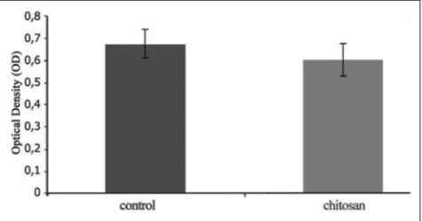
Identification of osteoclast using cells and culture medium Osteoclast cells were identified by TRAP, both on control and chitosan treatment groups. Fig. 1 shows phase contrast photomicrographs of 5 days osteoclasts culture, identified by TRAP, showed (Fig. 1a) control and (Fig. 1b) osteoclasts culture treated with chitosan. Both control and treatment groups showed positive TRAP staining, with higher cell quantity and stronger intensity in the control group. Fig. 2 shows OD value of TRAP assay from osteoclasts culture medium in the control was 0.67 ± 0.07 and chitosan treated group was 0.59 ± 0.08. T-test independent analysis showed that there was asignificant difference between the control group and chitosan-treated group (p<0.05).
Cell viability
Based t-test statistical analysis, there was a significant difference of mean OD in MTT assay between control group 1.51 ± 0.17 and chitosan treated group 0.88 ± 0.18 (p<0.05) (Fig. 3). Based on t-test statistical analysis, there was a significant difference of mean total protein in culture medium (Bradford assay) between control group 2237.62 ± 439.92 µg/ml, chitosan treated group 1963.67 ± 331.71 µg/ml (p<0.05).
Fig. 2: Optical density value of tartrate-resistant acid phosphatase assay from osteoclasts culture medium in the control and chitosan treated group (error bars show standard errors) n=3
Fig. 1: Phase contrast photomicrographs of 5 days osteoclasts culture, identified by tartrate-resistant acid phosphatase, showed (a) control and (b) osteoclasts culture treated with chitosan, (×200)
Bone resorption
Bone resorption pits were qualitatively observed by giving a microscopic score (mean score from five fields per sample). Fig. 4 showed the resorption on the bone slice. The Arnett et al. method was used for the observation of bone resorption. A phase contrast photomicrograph demonstrated pits on the surface of bone slices. Fig. 4a and b shows that osteoclasts were activated by HCl addition in the culture medium on the last medium change. From the observation, score from 1 to 3 was determined. Score 3 was determined on the pits of control group (A), and score 1 and 2 on the pit of chitosan treated group (B).
ROS product
An MDA assay was done to evaluate ROS product. This osteoclast
product was measured by a spectrophotometer on λl 530 nm. MDA
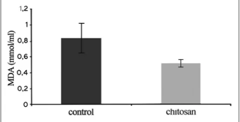
assay result in the control group was 0.83 ± 0.21 mmol/ml and chitosan treated group was 0.51 ± 0.06 mmol/ml. The independent
t-test analysis showed that there was a significant difference between the control group and chitosan treated group (p<0.05) as depicted on Fig. 5.
DISCUSSION
Chitosan is a biomaterial derived from chitin with a molecular weight of 800-1500 kDa and a chemical structure similar to hyaluronate. For this research we used chitosan (produced by BATAN, Indonesia) with special specification i.e. a molecular weight of 34.8 kDa. Previous study reported the potential stimulating ability of chitosan (BATAN) in dental pulp stromal/stem cells proliferation and early osteogenic differentiation comparable with that of dexamethasone, but no significant stimulation on mineral deposition [3]. The role of chitosan in bone regeneration and remodeling, and the healing process has already been reported, both with animals and humans [2,3,9,19-21]. Bone is a dynamic tissue that always renews and can remodel itself. The effect of chitosan on osteoblast cells has been extensively reported, but the healing process of fractured or defect bones are a complex process. The healing requires integration and coordination between specific cells. One of them is osteoclast, which in principle, plays a role in bone resorption and remodeling. Through this research, we showed that chitosan could influence osteoclast cells. Osteoclast cells were obtained through the culture of bone marrow of mouse for duration of 5-8 days by using a modified culture method previously used by Arnett et al. [16] and Soekanto et al. (1998) [15]. In order to change pre-osteoclast into immature osteoclast and subsequently into mature osteoclast that plays a function in bone resorption, a receptor activator of nuclear factor kB ligand, which is produced by osteoblast cells, is needed [15,16].
In our preliminary study for the osteoclast cell culture, two types of
controls were used, namely 1α,25(OH)2D3 only (first control) and
1α,25(OH)2D3 with a culture medium of osteoblast (second control).
This was done because in a number of previous researches, various growth factors were used. The effect of chitosan on osteoclast cell proliferation was tested with MTT assay.The results of preliminary study showed that there was no signification difference in the OD value of MTT assay between first control and second control (unpublished). This means that an increase in the growth factor does not increase osteoclast proliferation. Therefore, during the following tests, we only
used one growth factor, 1α, 25(OH)2D3.
In order to identify osteoclast cells that were formed in mouse bone marrow culture for a duration of 5-8 days, we used TRAP kit as osteoclast cell marker. TRAP is widely used as a marker of osteoclast in bone. Protein is released by osteoclast during bone resorption and therefore, TRAP can also be used as bone resorption marker in culture media [22,23]. Osteoclasts are bone cells that originate from hematopoietic stem cells that are able to resorb bone by secreting acid and proteinase [24]. The results of this research showed that TRAP positive cells, were present in the control and treatment group with a different intensity, as seen in Fig. 1. Moreover, Fig. 1a and b shows the osteoclast cell culture with the addition of HCl at the change of the last
medium (low pH). In the low pH condition, TRAP positive cells in the control group had clear color intensity and changed to their size, while in the treatment group the color intensity was unclear. From this result, it is assumed that chitosan, apart from obstructing osteoclast cell viability, also obstructs the change from fused polykaryon (immature osteoclast) to mature osteoclast. Both immature osteoclast and mature osteoclast express TRAP. The TRAP-secreted into the culture medium - was measured on a wavelength of 490 nm. A significant difference occurred in TRAP culture medium between control and
Fig. 3: Optical density value of 3-(4,5-dimethylthiazol-2-yl)-2,5-diphenyltetrazolium bromide assay, between the control and chitosan treated group (error bars show standard errors) n=3
Fig. 5: Value (mmol/ml) of malondialdehyde assay in control group and chitosan treated group (error bars show standard errors) n=4
Fig. 4: A phase contrast photomicrograph showed the pit on the surface of bone slices. Score 3 was determined in the control group (a) and score 1 and 2 in the chitosan treated group (b) (×200)
treatment group (p<0.05), as seen in Fig. 2. This result was support by Li et al., low molecular weight chitosan inhibits the formation of
TRAP positive osteoclast induced by 1α,25(OH)2D3 [25]. Rochet et al., reported that chitosan inhibits formation of TRACP positive cells. Inhibition of TRACP activity on 2% chitosan in combination with calcium phosphate cement did not result from inhibitory effect at transcription level, but might result from a inhibition of the enzymatic activity of the TRACP [21]. Chitosan influences osteoclast proliferation, as shown by the result of MTT assay (Fig. 3). The fact that the OD value of the treatment group smaller than that of the control group with significant statistical difference (p<0.05) was supported by the result of total protein in culture medium. The total protein medium of the treatment group was lower than that of the control group. This result indicated that protein production by cells in treatment groups decreased, perhaps due to inhibited osteoclast proliferation. Rochet et al. reported that 2% chitosan in combination with calcium phosphate cement does not affect the proliferation and adhesion of pre-osteoclast but inhibits the formation of TRACP positive cells and prevents the osteoclastic resorption on the composite biomaterial compared to calcium phosphate cement alone [21].
Halleen et al., reported that the ROS generating activity of TRACP may have important role both in bone resorption and in the immune defense system [26-28]. The effect of chitosan on bone resorption has been observed in bone slices 1 cm in diameter and 1 mm thick. This research showed that chitosan inhibits bone resorption. The difference between control and treatment group was observed qualitatively, namely by giving a score of 1-3 to the bone resorption picture that appeared on the bone slice. This was done because the tools for quantitative observation were limited. The patterns and amount of pit that was formed on the bone slices differed between control and treatment group. The score for the control group was higher (score 3) than the score for the treatment group (score 1 and 2) shown in Fig. 5. Bone resorption score in this research could be interpreted as bone resorption present in bone slices, but this score was not sensitive and needs more clarification on the depth and the surface area of resorption. These findings are supported by previous research, which reported resorption lacunae on 2% chitosan in combination with calcium phosphate cement were never detected. Possibly there is a negligible inhibiting effect of chitosan on osteoclastogenesis was reported by Rochet et al. [21]. In addition, chitosan can obstruct the occurrence of bone loss in a mouse model that has received ovariectomy [2]. During bone resorption, osteoclast releases various components into the resorption environment. The components include a number of acids and proteinase. Apart from this, it was reported that at the same moment osteoclast produces ROS. It was reported that superoxide anion occurred both inside and outside osteoclast in the areas of bone resorption. Therefore, it is assumed that ROS plays a role in bone resorption [29,30]. For this research, in order to understand the effect of chitosan on ROS production of osteoclast cells, we have measured MDA, which is a lipid membrane peroxydation product [18]. A significant difference in MDA value was obtained between control and treatment group, (p<0.05) (Fig. 4). Konga et al. (2009) reported that the water-soluble derivatives of chitosan and chitin were potent antioxidant and matrix metalloproteinase 2 and 9 in HT 1080 human fibrosarcoma [30]. The radical scavenging activities of chitosan depend on their degree of deacetylation and concentration. The optimal concentration of chitosan was 0.2 mg/ml [29,31].
CONCLUSION
Based on this research we can conclude that chitosan from National Nuclear Energy Agency (BATAN, Indonesia) inhibits the viability of osteoclast, decreases ROS production and bone resorption.
ACKNOWLEDGMENTS
We thank Andi Sofyan, D.D.S., M.Sc. of the Department of Dental Material, Faculty of Dentistry, Universitas Indonesia, for his cooperation in preparing bone slices and Professor Frans D. Suyatna, M.D., Ph.D., Sp.FK
from the Department of Pharmacology, Faculty of Medicine, Universitas Indonesia, for his cooperation in measuring the ROS for this research. This work was fully supported by Universitas Indonesia Research Grant (RUUI 212J/DRPM-UI/Ni.4/) for 2007 fiscal year.
REFERENCES
1. Klokkevold PR. The effect of poly-N- acetyl glucosaminoglycan (chitosan) on osteogenesis in vitro. Master Thesis of Science in Oral Biology, University of California, Los Angeles; 1995. p. 1-29. 2. Chae HJ, Lee GY, Yang SK, Kim DS, Yun KJ, Kim EC, et al. Effect of
high molecular weight water-soluble chitosan on the trabecular bone and thickness in ovariectomized rats. Immunopharmacol Immunotoxicol 2007;29(3-4):439-49.
3. Amir LR, Suniarti DF, Utami S, Abbas B. Chitosan as a potential osteogenic factor compared with dexamethasone in cultured macaque dental pulp stromal cells. Cell Tissue Res 2014;358(2):407-15. 4. Wang X, Yan Y, Zhang RA. Comparison of chitosan and collagen sponges
as hemostatic dressing. J Bioact Compat Polym 2013;21(1):39-54. 5. Azad AK, Sermsintham N, Chandrkrachang S, Stevens WF. Chitosan
membrane as a wound-healing dressing: Characterization and clinical application. J Biomed Mater Res B Appl Biomater 2004;69(2):216-22. 6. Madihally SV. Processing chitosan for tissue regeneration. J Polym Sci
2011;15:84-8.
7. Paul W, Sharma CP. Chitosan and alginate wound dressings: A short review. Trends Biomater Artif Organs 2004;18(1):18-23.
8. Xu C, Lei C, Meng L, Wang C, Song Y. Chitosan as a barrier membrane material in periodontal tissue regeneration. J Biomed Mater Res B Appl Biomater 2012;100(5):1435-43.
9. Boynuegri D, Ozcan G, Senel S, Uç D, Uraz A, Ogüs E, et al. Clinical and radiographic evaluations of chitosan gel in periodontal intraosseous defects: A pilot study. J Biomed Mater Res B Appl Biomater 2009;90(1):461-6.
10. Arnaud TM, de Barros Neto B, Diniz FB. Chitosan effect on dental enamel de-remineralization: An in vitro evaluation. J Dent 2010;38(11):848-52.
11. Bojar W, Kucharska M, Bubak G, Ciach T, Koperski L, Jastrzebski Z,
et al. Formation and preclinical evaluation of a new alloplastic injectable bone substitute material. Acta Bioeng Biomech 2012;14(1):39-44. 12. Zhang Y, Zhang M. Cell growth and function on calcium phosphate
reinforced chitosan scaffolds. J Mater Sci Mater Med 2004;15(3):255-60. 13. Guzmán-Morales J, El-Gabalawy H, Pham MH, Tran-Khanh N,
McKee MD, Wu W, et al. Effect of chitosan particles and dexamethasone on human bone marrow stromal cell osteogenesis and angiogenic factor secretion. Bone 2009;45(4):617-26.
14. Lee YM, Park YJ, Lee SJ, Ku Y, Han SB, Choi SM, et al. Tissue engineered bone formation using chitosan/tricalcium phosphate sponges. J Periodontol 2000;71(3):410-7.
15. Soekanto A, Ohya K, Ogura H. The effect of sodium salicylate on the osteoclast-like cell formation and bone resorption in a mouse bone marrow culture. Calcif Tissue Int 1994;54(4):290-5.
16. Arnett TR, Gibbons DC, Utting JC, Orriss IR, Hoebertz A, Rosendaal M,
et al. Hypoxia is a major stimulator of osteoclast formation and bone resorption. J Cell Physiol 2003;196(1):2-8.
17. Bradford MM. A rapid and sensitive method for the quantitation of microgram quantities of protein utilizing the principle of protein-dye binding. Anal Biochem 1976;72:248-54.
18. Wills ED. Evaluation of lipid peroxidation in lipid and biological membranes. Biochemical Toxicology: A Practical Approach Series. Oxford, London: IRL Press; 1987. p. 1132-4.
19. Bi L, Cheng W, Fan H, Pei G. Reconstruction of goat tibial defects using an injectable tricalcium phosphate/chitosan in combination with autologous platelet-rich plasma. Biomaterials 2010;31(12):3201-11. 20. Zhao L, Chang J. Preparation and characterization of macroporous
chitosan/wollastonite composite scaffolds for tissue engineering. J Mater Sci Mater Med 2004;15(5):625-9.
21. Rochet N, Balaguer T, Boukhechba F, Laugier JP, Quincey D, Goncalves S, et al. Differentiation and activity of human preosteoclasts on chitosan enriched calcium phosphate cement. Biomaterials 2009;30(26):4260-7.
22. Kirstein B, Chambers TJ, Fuller K. Secretion of tartrate-resistant acid phosphatase by osteoclasts correlates with resorptive behavior. J Cell Biochem 2006;98(5):1085-94.
the culture medium. Clin Chem 2000;46(11):1751-4.
24. Rissanen JP, Suominen MI, Peng Z, Halleen JM. Secreted tartrate-resistant acid phosphatase 5b is a Marker of osteoclast number in human osteoclast cultures and the rat ovariectomy model. Calcif Tissue Int 2008;82(2):108-15.
25. Li H, Miyahara T, Tezuka Y, Watanabe M, Nemoto N, Seto H, et al. The effect of low molecular weight chitosan on bone resorption in vitro and
in vivo. Phytomedicine 1999;6(5):305-10.
26. Robling AG, Castillo AB, Turner CH. Biomechanical and molecular regulation of bone remodeling. Annu Rev Biomed Eng 2006;8:455-98. 27. Halleen JM, Räisänen SR, Alatalo SL, Väänänen HK. Potential
function for the ROS-generating activity of TRACP. J Bone Miner Res
2003;18(10):1908-11.
28. Yam LT, Janckila AJ. Tartrate-resistant acid phosphatase (TRACP): A personal perspective. J Bone Miner Res 2003;18(10):1894-6.
29. Li H, Xu Q, Chen Y, Wan A. Effect of concentration and molecular weight of chitosan and its derivative on the free radical scavenging ability. J Biomed Mater Res A 2014;102(3):911-6.
30. Konga CS, Kim JA, Ahn B. Carboxymethylations of chitosan and chitin inhibit MMP expression and ROS scavenging in human fibrosarcoma cells. Process Biochem 2010;45:179-86.