Scholarship@Western
Scholarship@Western
Electronic Thesis and Dissertation Repository
8-16-2019 10:00 AM
Pattern Separation in the Ventral Visual Stream
Pattern Separation in the Ventral Visual Stream
Kayla Ferko
The University of Western Ontario Supervisor
Kohler, Stefan
The University of Western Ontario Co-Supervisor Khan, Ali
The University of Western Ontario Graduate Program in Neuroscience
A thesis submitted in partial fulfillment of the requirements for the degree in Master of Science © Kayla Ferko 2019
Follow this and additional works at: https://ir.lib.uwo.ca/etd
Part of the Neuroscience and Neurobiology Commons, and the Psychology Commons
Recommended Citation Recommended Citation
Ferko, Kayla, "Pattern Separation in the Ventral Visual Stream" (2019). Electronic Thesis and Dissertation Repository. 6485.
https://ir.lib.uwo.ca/etd/6485
This Dissertation/Thesis is brought to you for free and open access by Scholarship@Western. It has been accepted for inclusion in Electronic Thesis and Dissertation Repository by an authorized administrator of
ii
Pattern separation is a neural computation thought to underlie our ability to form distinct memories of similar events. It involves transforming overlapping inputs into less overlapping outputs. In the ventral visual stream (VVS) there is considerable evidence for hierarchical transformation from feature-based visual representations to conjunctive whole-object representations, with the latter allowing for distinct coding even when objects have
significant feature overlap. In the current study, we asked whether this transformation can be understood as pattern separation, and whether pattern separation can be observed even outside the context of classic recognition-memory tasks. To investigate pattern separation in the VVS, we combined fMRI in humans (N=23) with multivariate pattern analyses
techniques and compared representations of visual objects in a mid-level visual region, Lateral Occipital (LO) region, with those in the region proposed to be at the top of the VVS object processing hierarchy, Perirhinal Cortex (PRC). During scanning we presented images of objects from multiple categories, with differing degrees of visual similarity among
exemplars during performance of an N-Back task. Imaging results obtained using
classification revealed patterns in LO could be distinguished successfully for all categories and at the lowest level of visual similarity within category exemplars. In contrast, patterns in PRC could be distinguished at all levels of similarity within a category, but no successful category differentiations were found. Because patterns at higher levels of visual similarity are overlapping in LO, but can be differentiated in PRC, these results provide evidence for pattern separation in the VVS. More broadly, this suggests that the engagement of pattern separation may not be restricted to the hippocampus during declarative-memory tasks.
Keywords
Object Recognition, fMRI, multivariate, multi-voxel pattern analysis, perirhinal cortex,
iii
Summary for Lay Audience
The ability to distinguish two similar ‘things’ is important in everyday life. These ‘things’
can be memories, for example, finding your car in the parking garage every day. Although
the environment is very similar we are able to differentiate one day from the next. The neural
process involved here is pattern separation. Pattern separation functions by transferring
similar neural signals in one region to completely distinct neural signals in another region.
Therefore, researchers can investigate this phenomenon by measuring how similar brain
patterns are in different regions when participants complete a memory task. Previous animal
and human research has provided evidence that the hippocampus plays an important role in
separating similar signals from its input region, entorhinal cortex. But does pattern separation
occur before the hippocampus and not solely during memory tasks?
The goal of this study was to investigate whether or not pattern separation exists upstream
from the hippocampus in the ventral visual stream during object perception. To address this
goal 23 human participants were scanned in a functional MR scanner to obtain pictures of
their brain as they viewed images of objects on a screen. We presented images of objects
from multiple categories, with differing degrees of visual similarity within a category.
Imaging results obtained by analyzing the neural patterns elicited by the stimuli revealed
differences in mid-level visual region, lateral occipital region (LOC) and the region thought
to be at the top of the visual processing hierarchy, perirhinal cortex (PRC). In LO, patterns
could be distinguished successfully when they represented different categories or
within-category objects at the lowest level of visual similarity. In contrast, while all levels of visual
similarity within a category could be distinguished successfully in PRC; no categories could
non-iv
distinguishable in LO, but can be differentiated in PRC, these results provide evidence for
v
Co-Authorship Statement (where applicable)
Dr. Köhler oversaw the project and co-ordinated the efforts from the all co-authors. He
contributed significantly to the background research, motivation for the current project. Dr.
Köhler also helped in planning analyses and interpretation of results.
Dr. Khan helped in overseeing the project and contributed to the scanning protocol and
imaging processing pipelines used to analyze the functional and structural images.
Dr. Anna Blumenthal contributed significantly to the project development including the
scanning paradigm, behavioural task and the novel functional imaging task to probe pattern
separation in the ventral visual stream.
Dr. Daria Proklova helped with functional data analysis using COSMO MVPA Matlab
package.
Dr. Chris Martin also helped with the functional data analysis using COSMO MVPA Matlab
vi
Acknowledgments
Thank you to everyone in the Köhler and Khan labs for continuous support throughout this
amazing experience. Thank you to Dr. Köhler for his encouragement, constructive
discussion, kindness and time throughout my Master’s thesis. Thank you to Dr. Khan for his
innovative ideas and techniques that has allowed my coding and data analysis abilities to
grow exponentially.
Thank you to my parents, Walter and Espie Ferko, and my boyfriend, Nick Holdbrook, for
your constant love and support. Thank you for being my audience for every abstract, poster,
and presentation.
Thanks to the staff at Brain and Mind Institute and Centre for Functional and Metabolic
vii
Table of Contents
Abstract ... ii
Summary for Lay Audience ... iii
Co-Authorship Statement (where applicable) ... v
Acknowledgments ... vi
Table of Contents ... vii
List of Tables ... ix
List of Figures ... x
List of Appendices ... xii
1 Introduction ... 1
1.1 Pattern Separation in the Hippocampus ... 1
1.2 Pattern Separation beyond the Hippocampus ... 10
1.3 Perirhinal Cortex as the Apex of the Ventral Visual Stream ... 13
1.4 The Missing Link: Pattern Separation in Perirhinal Cortex ... 18
1.5 The Current Study: Approach, Goals, and Hypotheses ... 21
2 Method ... 22
2.1 Participants ... 22
2.2 Stimuli ... 23
2.3 Multi-Arrangement Task: Inverse Multi-Dimensional Scaling ... 24
2.4 fMRI Task: Variation of a 1-Back Task ... 26
2.5 MRI Data Acquisition and Preprocessing ... 28
2.6 Regions of Interest ... 29
2.7 Multi-Voxel Pattern Analysis ... 30
3 Results ... 31
viii
3.2 fMRI Results ... 33
3.2.1 Between Category Classification ... 33
3.2.2 Within Category Classification ... 34
4 Discussion ... 37
4.1 Role of Perirhinal Cortex in Object Discrimination ... 37
4.2 Role of LO in Category and Object Discrimination ... 42
4.3 Limitations and Future Directions ... 46
4.3.1 Investigating Other Regions of Interest ... 46
4.3.2 Methodological Considerations ... 49
4.3.3 Additional Insights that Could be Gained from Representational Similarity Analysis ... 50
4.4 Conclusion ... 52
References or Bibliography (if any) ... 53
Appendices ... 67
ix
List of Tables
Table 1. Overall behavioural results of the variation of a 1-back task. Trial Type is indicated by the rows, participants response proportion (averaged across all participants and rounded to nearest tenth). Correct responses are located on the diagonal and bolded. Same category trial types can be split into high-middle-low visual similarity based on each participant’s sorting. Same category response options cannot be split into high-middle-low. ... 31
x
List of Figures
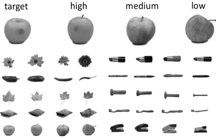
Figure 1. 40 stimuli from Migo Normative Database (Migo et al., 2016). The four object exemplars in each of the 10 categories were manipulated to be high-middle-low visual similarity compared with the an arbitrary target exemplar. ... 24
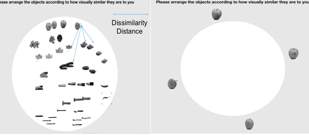
Figure 2. Inverse Multi-dimensional scaling task adapted from Kreigeskorte & Mur (2012). This task allows for the calculation of dissimilarity distances between all possible object pairs. Left side of figure depicts a sample sorting of all object stimuli. Right side of figure depicts the starting position of a category specific sorting trial. Participants completed one trial for each of the 10 categories used in the study. ... 26
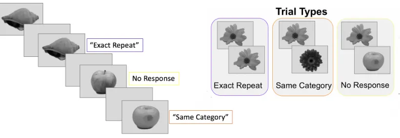
Figure 3. Variation of a 1-back task. Each image was presented for 1.2 seconds with an inter-stimulus interval of 1 second where a fixation cross was presented. Participants were asked to press one button if the object was the exact same as the one previous to it, a different button if the object was the same category as the one previous to it, and no button press if the object is a different category as the one previous to it. Each of the 40 exemplars were viewed four times--one presentation was a response trial—in each of the eight runs for a total of 24 non-response stimulus presentations of each exemplar across the entire experiment.
Corresponding neural patterns that resulted from viewing each of the 40 stimuli multiple times were averaged to obtain stable stimulus-specific estimates in fMRI analyses. ... 28
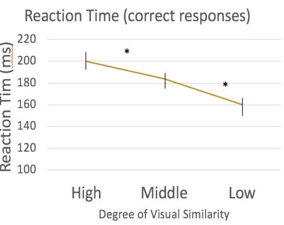
Figure 4. Reaction times for all correct “same category” button press trials. These correct trials were split by their corresponding degree of visual similarity (high-middle-low). Reaction time decreases with decreasing level of visual similarity. Behavioural results of a variation of a 1-back task indicates participants are sensitive to the visual similarity
manipulation. (* indicates p<.01) ... 32
xi
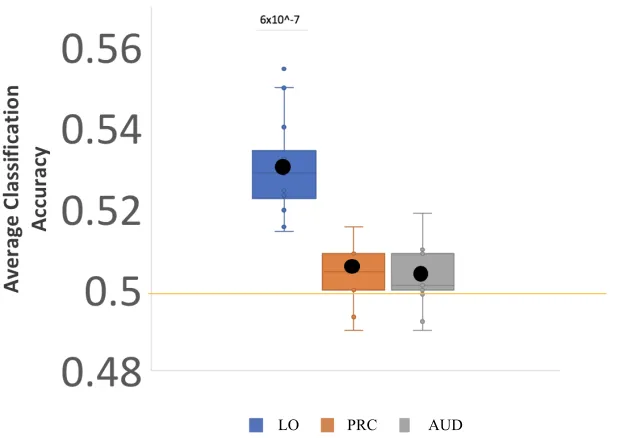
and each regions mean score (black dot) and standard deviation (shaded region of each box) plotted here. Classifier performed significantly above chance in LO only. ... 34
xii
List of Appendices
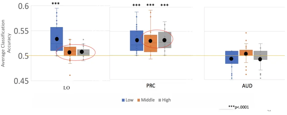
Appendix 1. Classification Accuracy of all categories in LO and PRC versus control region (Auditory Cortex) ... 67
1
Introduction
Pattern separation is a neural computation that is thought to underlie our ability to
form distinct memories of similar events. This concept was originally discussed by
Marr (1971) as a computational mechanism that functions to separate similar inputs
and reduce interference. For everyday tasks to be completed effectively, such as
remembering where you parked in your parking garage, pattern separation is required.
Although the sensory environment of the parking garage is very similar each day, you
are able to find your car by distinctly remembering each day as a new episode. Pattern
separation works here to transfer highly overlapping inputs from one brain region to
non-overlapping outputs for another region (O’Reilly and McClelland, 1994; Santoro,
2013; Neunuebel & Knierem, 2014). Extant research on pattern separation has
primarily focused on transformations of representations between entorhinal cortex
and the dentate gyrus of the hippocampus in the context of declarative memory tasks.
Pattern separation may, however, also occur in other cortical regions and may not be
limited to computations that support memory processing. This thesis will investigate
pattern separation in the ventral visual stream during object recognition using
ultra-high resolution fMRI and multi-voxel pattern analysis.
1.1 Pattern Separation in the Hippocampus
The hippocampus has been studied extensively for its role in episodic memory since the
seminal investigations in patient H.M. This individual suffered from severe anterograde
amnesia after undergoing a bilateral medial temporal lobectomy that included the
hippocampus to treat intractable epilepsy (Scoville & Milner, 1957; Penfield and Milner,
1958). Since then, countless studies have investigated the involvement of the
hippocampus in various aspects of cognition (see Moscovitch et al., 2016 for recent
reviews). More recently, researchers have discovered that this archicortical structure is
on differences in cytoarchitecture and genomic expression. The hippocampal formation
can be divided into subfields based on such differences at the cellular level. These
subfields include the dentate gyrus, subiculum, and cornu ammonis (CA) 1-4 (Ding &
Van Hoesen, 2015). They are differentially affected in diseases such as Alzheimer’s
Disease, medial temporal lobe epilepsy, and depression (e.g., Van Hoesen and Hyman,
1990; Price et al., 2001; Coras et al., 2014) and, of interest to many behavioural
cognitive neuroscientists, these subfields may perform different functional roles. One
such function is the computation of pattern separation of inputs from the neocortex via
entorhinal cortex, a major input to the hippocampus (see review by Aggleton, 2012).
Many theoretical models have pointed to the dentate gyrus subfield as a region involved
in separating similar representations from entorhinal cortex to distinguishable signals to
pass onto the CA fields (Mcnaughton & Nadel, 1989; Treves & Rolls, 1992; O’Reilly &
McClelland, 1994; Gilbert, Kesner, & Lee, 2001).
To investigate whether overlapping signals are transferred to non-overlapping signals,
scientists have used electrophysiology in non-human species to discover if
representations of similar stimuli are distinguishable in dentate gyrus. There is a
challenge, however, in recording cells in the dentate gyrus due to their sparse firing
(Jung, Weiner & McNaughton, 1994). One study by Leutgeb et al. (2007) accomplished
this difficult task when they trained rats to run in square or circular enclosures and
recorded firing activity simultaneously in CA3 putative pyramidal cells and in dentate
gyrus granule cells. The enclosure was transformed through five intermediate shapes such
that rats spent 10 minutes in a square enclosure, and then 10 minutes in each of the
enclosures with intermediate shapes until the enclosure was a circle (and vice versa).
Leutgeb et al. (2007) analyzed firing rate patterns in CA3 and dentate gyrus by stacking
the firing rate maps of all cells, such that the x and y coordinates coded a location in the
enclosure and the z coordinate coded the cell identity. Population vectors of matching
spatial locations (i.e., along the z direction) were correlated for pairs of environments in
order to examine how the representations changed as the enclosure morphed
were highly correlated representations then they were considered to be highly similar and
non-distinguishable. Population vectors for the CA3 subfield were found to be highly
overlapping for shapes 1, 2, 3, and significantly different between shape 1 versus 4, 5. In
contrast, the dentate gyrus granule cells were highly sensitive to even the smallest change
in the shape of the environment as the first intermediate shape (shape 2) enclosure was
represented differently from shape 1. Not only were the representations different—as
signaled by the lower population vector correlations—these differences were a direct
result of the cells firing at different (i.e., non-overlapping) locations. This provided strong
evidence that the dentate gyrus, and not CA3, is able to represent very similar spatial
enclosures with distinct populations of neurons. But, without sampling the
representations in the input region, entorhinal cortex, it is not possible to conclude that
the dentate gyrus is performing this transfer function, a key part of the definition of
pattern separation. In other words, researchers must provide evidence that signals are
overlapping in entorhinal cortex and are transferred to non-overlapping representations in
dentate gyrus.
In 2014, Neunuebel and Knierim did exactly that. They sampled cells in entorhinal
cortex, dentate gyrus and CA3. In this study rats ran clockwise around a track that had
four local cues on the surface of the track and six global cues around the walls of the
track. Rats were trained in a standard configuration for 16 days and then neural activity
was recorded in three standard sessions separated by two mismatch sessions. These
mismatch sessions rotated the global cues clockwise and the local cues counterclockwise,
such that the cue mismatches were 45, 90, 135, 180 degrees in size. Similar to Leutgeb et
al. (2007), the researchers then produced spatial correlation matrices by correlating the
normalized firing rate vectors between a standard session and either another standard
session or a mismatch session. If the firing rate vectors of two sessions were highly
similar, they would have high correlations and would not be differentiable. In contrast, if
the firing rate vectors of two sessions were not similar, they would have lower
correlations and would therefore be distinguishable. Results indicated that the firing rate
gyrus. This was expected and signifies that representations of spatial locations are stable
in both subfields. In comparison, firing rate patterns relating to Standard vs Mismatch
sessions were still highly correlated in CA3, but significantly less correlated in dentate
gyrus. This suggests that the representations of these spatially similar trials are not
distinguishable in CA3, but are distinguishable in dentate gyrus. In addition, a previous
article published by Neunuebel et al., (2013) used the same protocol to investigate medial
entorhinal cortex, which is a primary input source to the dentate gyrus. Medial entorhinal
cortex had highly correlated firing patterns when comparing Standard 1 vs Standard 2
and Standard vs Mismatch. Similar to CA3, these results indicate that the Standard
representations are stable across time and they do not significantly differ from the
Mismatch firing rate patterns. Because the firing patterns for similar spatial scenes in the
Standard and Mismatch conditions are not distinguishable in medial entorhinal cortex,
but can be distinguished in dentate gyrus, this provides strong, direct evidence for pattern
separation for spatial stimuli in the dentate gyrus.
While electrophysiology studies provide the gold standard in measuring this transfer of
overlapping representations to non-overlapping representations, taking such an approach
is not straightforward in humans. Taking key experimental methods from the rodent
neurophysiology methods, seminal work by Bakker et al.(2008) used high-resolution
functional magnetic resonance imaging (fMRI) in combination with an incidental
encoding task to probe pattern separation in awake adult humans. Eighteen subjects
viewed pictures of everyday objects and were asked to respond with a button press
whether an item is typically an indoor or outdoor object (e.g., light switch is typically an
indoor object). On each trial the object could either be new (not seen before), a repetition
of a previously shown object, or a lure, which is visually similar to an object seen before.
The lures differed only in visual features and not by name, which is important when
investigating how representations differ for mnemonically similar objects. They
hypothesized that if a region was involved in pattern separation the lure would be treated
like a completely new stimulus. Their prediction leveraged repetition suppression—a
mean blood-oxygen level dependent (BOLD) signal (see, for review, Grill-Spector,
Henson, & Martin, 2006; Krekelberg, Boynton, & Van Wezel, 2006; Larsson, Solomon,
& Kohn, 2015). Following their rationale, if a region is involved in pattern separation the
visually similar objects will not elicit repetition suppression as they are treated as new
objects not previously seen. To image the hippocampus and surrounding cortices,
researchers used 3 Tesla (3T) MRI to obtain 1.5 mm isotropic voxels, which allowed
them to confidently segment the hippocampus, but CA3 and dentate gyrus were
combined into one CA3/DG subregion (due to limited spatial resolution in their data).
Despite this limitation, results showed that bilateral CA3/DG did not have the decrease in
mean activity for lure trials that other regions CA1/3/DG, CA1, subiculum, or entorhinal
cortex displayed. These results indicated that only CA3/DG treated even these very
similar lures as first presentation, i.e. as new objects rather than repeat exposures. This
was the first study to provide evidence for pattern separation in the hippocampus in
humans. In their follow-up work, Lacy et al. (2011), investigated the dynamics of this
pattern separation by including more levels of similarity and comparing BOLD activity in
CA3/dentate gyrus with downstream CA1. The rationale behind these changes in protocol
followed the rodent literature where even small changes in stimuli had been shown to
elicit large differences in representations in dentate gyrus (Leutgeb et al., 2007;
Neunuebel et al., 2014). In comparison, CA1 subfield was not expected to exhibit this
transfer function; instead representations should vary continuously as similarity changes.
The authors hypothesized that only CA3/DG would represent highly similar objects
distinctly, whereas both CA3/DG and CA1 would represent low similarity objects
distinctly. Moreover, they predicted that, in CA3/DG, high and low similarity objects
would be treated as first presentations and not elicit repetition suppression. In CA1, by
contrast, high similarity objects were not expected to be discernable and therefore to elicit
repetition suppression, but low similarity objects were expected to be sufficiently
different and would not exhibit repetition suppression. Modifying the task used in Bakker
et al., (2008) by splitting the lures into high and low mnemonic similarity trials, the
high and low similarity objects displayed activation similar to completely new objects in
CA3/DG. Extending previous studies, data in CA1 showed that only low similarity
objects exhibited activation resembling first presentations, whereas high similarity lures
elicited repetition suppression similar to repeat trials. These subfields only had different
activation for one condition. Specifically, only the high similarity lures elicited
significantly different beta coefficients across regions. These results provide further
evidence that even small differences in stimuli can be transferred to distinct
representations in CA3/DG, but not CA1. Because the analyses conducted relied on
leveraging of repetition-suppression effects, however, this univariate approach does not
directly address the degree of overlap in representations within a region, and how
representations change between regions as a result of computational transformations.
Multi-voxel pattern analysis (MVPA) can provide stronger evidence for pattern
separation as it can provide quantitative estimates of how distinct patterns are across
different regions when observers view or judge similar stimuli. Innovative work that used
this multivariate approach to investigate pattern separation in the hippocampus was
published in 2016 by Berron and colleagues. This study relied on ultra-high resolution
BOLD imaging with a 7T scanner at higher spatial resolution (0.8 mm isotropic voxels)
than prior research (e.g., Bakker et al., 2008), which allowed researchers to separate CA3
and dentate gyrus as anatomically distinct structures with higher confidence. In this
experiment only two stimuli were used so that the only difference between the trial types
was that they belonged to different sequences. This was an important manipulation to
remove a confound that was present in previous studies (Bakker et al., 2008; Lacy et al.,
2011). Specifically, in those studies first presentation trials were the first presentations
within the experiment and, therefore, evidence for pattern separation from repetition
suppression may have reflected a novelty signal. Twenty young subjects viewed two
similar dining room scene stimuli, A and B, which differed in the placement of the chairs
at the table. These scenes were presented in short sequences of three to five stimulus
presentations (e.g., AABA). Participants were asked to count repetitions in the sequences
analysis, only the first two presentations in a sequence were examined such that there
were three different trial types: first presentations and then either repetitions (exact
repeats of the first presentation of stimulus A) or lures (stimulus B/different). This
allowed for similar analyses to be conducted as those reported by Bakker et al. (2008) as
a first step. The contrast of lure trials against repetitions (lures > repetitions) showed a
cluster in DG and the contrast of first presentation against repeats (first>repeats) showed
a cluster in CA1. As expected, the DG cluster did not show a decrease in neural activity
for lures, resembling the response for a first presentation. In the CA1 cluster, by contrast,
lures did elicit a decrease in neural activity similar to the repeat condition. Interestingly,
using an anatomically-defined ROI instead of these contrast clusters did not show a
decrease in repetition suppression in dentate gyrus for lures. This result provided further
motivation for the use of a multivariate approach to investigate pattern separation. Berron
et al. used a linear support vector machine with a leave-one-run-out cross-validation
classification to conduct classification of patterns of fMRI activity. The rationale behind
such an approach is that if a region is involved in pattern separation then even similar
stimuli will be represented distinctly; as a consequence the classifier may perform
significantly above chance (50%) in distinguishing corresponding patterns of activity. In
the study by Berron et al., the classifier did indeed perform significantly above chance
only in the dentate gyrus for the lure condition. In entorhinal cortex, by contrast, the
classifier did not reveal above chance performance for any condition. Therefore, the
authors interpreted these results to argue that non-distinguishable patterns of activation in
entorhinal cortex are transferred to distinguishable patterns in dentate gyrus. These
findings provide strong evidence that the dentate gyrus is involved in pattern separation
of visually similar scenes.
One limitation with human functional imaging is that the results are correlational in
nature. Thus although such research can show that dentate gyrus is capable of separating
similar inputs to distinct signals, other methods are required to show whether a given
region is necessary for that process. Recent evidence from Baker et al. (2016) provides
context of memory tasks. This study leveraged behavioural data from patient BL, a man
with selective dentate gyrus lesion, using the Mnemonic Similarity Task (Stark et al.,
2015). This recognition memory test requires participants to classify each image as “old”
if the image has been seen earlier in the experiment (trial type = target), “new” if the
image has not been seen previously (trial type = foil), or “similar” if the image is visually
similar to an object previously seen in the experiment (trial type = lures). As predicted,
BL performed similarly to the control subjects when identifying targets and foils, but was
significantly worse at identifying lures. This pattern of abnormalities is similar to
observations made in healthy older adults and those diagnosed with amnestic mild
cognitive impairment (Stark et al., 2015; Stark et al., 2013). These results further point to
the dentate gyrus as a critical region in keeping similar memories distinct. On a more
cautionary note, however, this study also highlights challenges in investigating pattern
separation in humans without any use of neuroimaging or neural recording. Arguably,
without inclusion of recording data these results cannot be classified as direct evidence
for pattern separation, given that the transfer of representations from entorhinal to the
lesioned dentate gyrus was not directly examined.
A recent study by Lohnas et al. (2018) that employed electrocorticography (ECog) aimed
to bridge the gap between neurophysiological studies of pattern separation in non-human
species with neuroimaging studies based on fMRI in humans. ECog is a type of
electrophysiological monitoring where electrodes are placed below the skull on the
cortical surface or in cortical or subcortical regions via depth electrodes. Typically, this
procedure is performed in patients who require these electrodes for clinical evaluation of
intractable epilepsy and electrodes are placed to monitor seizures and plan surgery. In the
study by Lohnas et al., participants performed two blocks of trials, and each block
contained a series of images on a computer screen. Each image was either a new object
never seen before, an exact repeat that had been seen before, or a similar image that
shared many features as a previous one. For the fine-grain task block, participants were
asked to indicate with a button press if the image was new, old, or similar. For the
be classified as old. To investigate pattern separation in awake human participants
researchers explored the temporal dynamics of high-frequency activity (HFA; 45-115Hz).
Past research has revealed strong similarities between HFA and BOLD fMRI responses
as correlates of neural activity (Fries, Reynolds, Rorie, & Desimone, 2001; Hirabayashi
et al., 2014; Manning, Jacobs, Fried, & Kahana, 2009). Results indicated that overall
HFA was significantly greater for similar items than old items in the hippocampus during
a 1.5-2 s time window. This is similar to the lack of repetition suppression for similar
items previously observed in fMRI research. Furthermore, researchers investigated
multivariate patterns to measure how HFA patterns change between regions. Results
indicated a dissimilarity in multivariate HFA activity for similar items in the
hippocampus. This was not observed in the upstream occipitotemporal cortex, therefore
providing support that similar objects elicit more distinct HFA patterns in the
hippocampus. Surprisingly, HFA in the hippocampus was found to be task-dependent.
Specifically, multivariate HFA activity was not significantly dissimilar for similar items
in the hippocampus during the coarse-grain task. The authors argued that this did not
signify an inherent difference in the task (i.e., in difficulty), because patterns in two other
regions of interest, occipitotemporal cortex and dorsal lateral prefrontal cortex, were
found to be stable across both the fine and coarse grain task. Instead, they suggested that
the structures are differentially activated for each task and that the hippocampus is
recruited to represent similar objects distinctly only when the task requires such
differentiation (i.e., fine grain task); otherwise cortical representations may be sufficient.
ECog allows for the temporal unfolding of pattern separation to be examined and the
authors cautioned that this time window of 1.5-2 seconds may be considered ‘late’ with
respect to participants’ responses. In this context, it is important to remember that the
participants examined were five patients with debilitating epilepsy, which may affect
temporal dynamics. It is also important to note that little research has been conducted
investigating the temporal dynamics of pattern separation or mnemonic reinstatement in
general, due to the limited data available and difficult electrode placement. Furthermore,
keep in mind that that electrode placements and sampling rate used in this study do not
allow for more selective localization (i.e., hippocampal subfields cannot be separated).
Despite these limitations, combining the increased temporal resolution of ECog with the
increased spatial resolution of functional imaging, allows for a more complete
understanding of pattern separation.
1.2 Pattern Separation beyond the Hippocampus
As pattern separation is defined by a transfer from overlapping to non-overlapping
representations, one may predict that this may not be a computational principle that is
relevant only to memory processing and only the dentate gyrus. This idea originated over
two decades ago (Murray & Bussey, 1999; see also Bussey and Saksida, 2002) in the
context of the Representational- Hierarchical (R-H) theory, and led to the proposal that
pattern separation may be a widespread function in many cortical regions for many
stimulus types (Kent et al., 2016). Specifically, although there is strong evidence for
pattern separation in the dentate gyrus for spatial and episodic content, other regions may
be involved for other stimulus material. To the extent that visual stimuli are concerned,
the principle is of particular relevance to the functional organization of the Ventral
Visual Stream (VVS). R-H theory proposes that during object recognition, as information
is transmitted from lower to higher regions of the VVS hierarchy, the formation of more
conjunctive features creates more distinct, non-overlapping object representations. This
transfer from similar overlapping representations in early/mid visual regions to
non-overlapping representations in late VVS can be conceptualized as pattern separation.
Perirhinal Cortex (PRC) is of particular interest in this context as it is thought to reflect
the pinnacle of object processing in the VVS that contains highly conjunctive, and in
turn, the most distinct representations of highly similar objects, as has been proposed by
Kent and colleagues (2016). In order to test specific hypotheses about pattern separation
in PRC derived from this theory, it is important to consider processing at earlier stages of
the VVS. In the following summary, emphasis will be placed on lateral occipital (LO)
probing for pattern separation The VVS has been extensively studied in both human and
animal research and extends from primary visual cortex V1 through secondary visual
areas V2, V3, V4 and then to ventral temporal cortex (Ungerleider & Mishkin, 1982; see
Grill-Spector & Weiner, 2014, for recent review). Ventral temporal cortex contains
multiple high-level visual regions, including LO, that are involved in visual perception
and recognition of objects and scenes. While low-level features such as edges or
luminance are represented in early visual cortex, increasingly more complex features
such as shape and category are represented as signals move through higher visual regions.
Research by Rust and DiCarlo (2010) using single neuron recordings in rhesus macaque
monkeys demonstrated, for example, how as information travels from V4 to inferior
temporal cortex—a likely analog to human LO (Kanwisher et al., 1996)—representations
become more selective for objects than for scrambled images. This selectivity for these
complex image features means that an image of an object on a background will produce
more activity than when those same pixels are scrambled up. Human research has
replicated the finding that mid-stream regions are more activated for objects than
scrambled images (Malach et al., 1995; Grill-Spector, 2003). Because objects are
typically defined by shape, this feature has attracted considerable attention in functional
neuroimaging research on object recognition.
In an influential early study, Kourtzi and Kanwisher (2001) used functional imaging and
repetition suppression phenomenon to understand how human LO represents perceived
object shape. Participants viewed a stream of different shapes presented either in front or
behind vertical lines (different depths). Results indicated that viewing the identical shape
at the same and different depths evoked an adaptation response. In contrast, if
participants viewed a similar shape at the same depths, adaptation response was absent.
Therefore, when the perceived shape was the same but the depths differed repetition
suppression was evoked, but no repetition suppression was evident when the shapes were
different but the depths were similar. This lack of repetition suppression for different
Objects can be discriminated based on shape, but also based on category membership.
Although primarily driven by shape, LO also elicits different responses to different
categories of objects (Grill-Spector & Weiner, 2014). To explore how regions represent
differences in shape or category membership, researchers can compare patterns evoked
by objects from different categories that are similar in shape and objects from the same
category that are different in shape. Proklova, Kaiser, and Peelen (2016) separately
modelled the degree of visual similarity and category membership (i.e., animate,
inanimate) to be compared with brain similarity patterns when viewing a set of stimuli in
the scanner. These stimuli were carefully controlled for low level features and contained
objects that had a similar shape but belonged to a different category (e.g., rope and
snake). Searchlight MVPA analysis resulted in much of the lateral occipitotemporal
cortex significantly correlated with the visual dissimilarity matrix and some regions that
are correlated with the category dissimilarity matrix. In LO, there were voxels
specifically sensitive to visual dissimilarity, but others whose response profile correlated
significantly with both visual and category dissimilarity matrices. These results provide
evidence that LO contains voxels that represent visual dissimilarity and also category
membership.
Additionally, there is evidence that LO contains both between and within category
information. A study by Eger et al (2008) leveraged fMRI classification of evoked
patterns when viewing two categories of objects, chairs & teapots, that varied in
viewpoint. Participants were asked to respond with a button press when they saw a red or
green hue to the stimuli. Interestingly, the mean signal in LO did not differ between and
within object categories. Only when patterns of activation were measured and analyzed
were differences between categories apparent. The researchers also investigated how
different numbers of voxels contained in their LO ROI affected the results. Classification
accuracy was significantly above chance for both between (average 62% at 200 voxels)
and within category (average 55% at 200 voxels). Amazingly, between category
classification was above chance with just 10 of the most discriminative voxels. These
exemplars within each category distinctly. Therefore, findings suggest that LO contains
these coarse-grained and, to a lesser extent, some fine-grained object distinctions. The
next section will focus on evidence suggesting that PRC may represent objects differently
from LO, by virtue of specifically being involved in object discrimination when objects
share many features, i.e., when there is a high degree of feature overlap.
1.3 Perirhinal Cortex as the Apex of the Ventral Visual
Stream
The Multiple Memory Systems paradigm proposes perirhinal cortex as the border region
between “memory” and “perceptual” systems (e.g., Schacter and Tulving, 1994;
Zola-Morgan, Squire, & Ramus, 1994; Tulving and Schacter, 1990). As such, recent studies
have investigated the perirhinal cortex as a potential new anterior apex of the ventral
visual stream (Murray et al., 2007; Barense et al., 2012). This was first examined in
animal lesion research such as a study reported by Bussey, Saksida, and Murrary in 2003.
In that study, researchers morphed greyscale picture stimuli creating pairs of stimuli that
shared many features and were visually similar or shared few features and were visually
dissimilar. The monkeys viewed two images and learned to associate one image with a
reward and performance was measured as percent of trials that were correct. The results
indicated that monkeys with PRC lesions were only impaired on learning to discriminate
pairs of stimuli that shared many features. This findings is part of a large body of
evidence suggesting that PRC of non-human primates is involved in perceptual
discrimination of visually similar objects (see Murrary, Bussey & Saksida, 2007 for
review).
Human lesion studies have also confirmed the role of PRC in object perception. Several
studies have compared a select group of patients that either had medial-temporal lobe
damage specific to the hippocampus or that included, but was not limited to, perirhinal
cortex (typically extending into anterior lateral temporal cortex, other parts of
parahippocampal gyrus, and the amygdala). Barense et al. (2007) investigated the
hippocampal damage and three participants with medial-temporal lobe damage that
included the PRC, as well as age-matched controls. Participants completed an oddity task
(Buckley, Booth, Rolls, & Gaffan, 2001; Lee, Buckley, Gaffan, Emery, Hodges, &
Graham, 2005) that required them to indicate which object was the odd-one-out.
Participants were asked to find and press a button corresponding to the object that did not
have an exact match in each trial. Trials ranged from low to middle to high visual
similarity. Results showed that while the hippocampal-specific lesion participants
performed similarly to controls, those with more widespread MTL damage, including
PRC, performed significantly worse than controls and HP-patients on trials with objects
of middle and the highest level of similarity. Further, these results were supported in
2012 by Barense and coworkers when these same participants completed a different
visual discrimination task. They were asked to indicate if two simultaneously presented
stimuli were a match or a non-match. The stimuli belonged to four conditions that were
defined by high or low shape ambiguity and by high or low size ambiguity. As predicted,
only patients with MTL damage that included the perirhinal cortex were impaired when
discriminating objects, and only in the high shape ambiguity condition. This follow-up
study provided evidence that this deficit in performance, in participants with MTL
damage including PRC, for highly similar objects does not generalize to size
discrimination. In these patients, however, the MTL damage is not limited to perirhinal
cortex so we cannot rule out the possibility of one or some of the other affected regions
being the site for these highly complex objects.
Lee et al. (2006) tackled this problem using functional imaging of healthy participants
while performing specific tasks. In each of three functional runs participants were asked
to compare two grids on each trial. They were either asked to determine if 1) the object
changed visually, 2) the position of the object changed, or 3) no change occurred. The
researchers then measured the activity specific to object change by subtracting the
activation in the no change condition and found that both right PRC and right posterior
hippocampus was significantly active during the object change condition. In contrast, the
this was not expected in the hippocampus, these null object position results may be due to
spatial task simplicity. For example, a previous study from this group required
participants to discriminate three-dimensional virtual reality rooms, whereas this study
required participants to pay attention to object position on the screen (Lee et al., 2005).
While this spatial task may not be suitable for probing spatial discrimination, the object
change condition has been used in many visual object discrimination tasks in the
literature. There is, however, another aspect to object discrimination besides attending to
the way the object looks. For example, we may also discriminate objects based on what
they are used for or where they are usually located and these characteristics of an object
are part of its conceptual representation. Consequently, if a change in BOLD activity
occurs in response to the object changing from apple to toothbrush, this may be due to a
change in visual features (e.g., from round to elongated) or a change in conceptual
features (e.g., from ‘used for nutrition’ to ‘used for hygiene’). Therefore, a clever
experimental design is needed to tease apart the differential effects of visual and
conceptual feature overlap.
Although many objects share both visual and conceptual features (e.g., apple and orange),
many everyday objects share only conceptual features or only visual features. Evidence
suggests PRC plays a role in representing objects conceptual features (Clarke & Tyler,
2014) in addition to visual features (e.g., Erez et al., 2016). A challenge arises when
investigating a dissociable effect of these two feature types because these features tend to
be related as form and function are typically intertwined (Mur, 2014). A great example of
this is illustrated in Martin et al (2018) that suggested that a concept of a ‘hairdryer’
shares many conceptual features with ‘comb’ (e.g., used to style hair) and shares many
visual features with ‘gun’. Because we are able to recognize these differences in both
visual and abstract conceptual features such that we do not use a gun to tame our hair in
the morning just because it looks similar to a hairdryer, therefore our brain must have a
way to integrate these representations. This study investigated how both conceptual and
visual features are represented in the brain by obtaining ratings for objects about both
investigated object representations of visual feature conjunctions during object
discrimination or conceptual feature conjunctions during semantic memory, Martin and
colleagues hypothesized that PRC is involved in both discriminating visual and
conceptual features. To test their hypothesis, participants viewed 40 words of object
concepts in the MR scanner and were asked to answer two questions about these object
concepts in a blocked design (two blocks in each of the eight functional runs). During one
block participants answered questions with a button press yes/no, pertaining to the
objects visual features such as, ‘Is the object angular?’ and the other block contained
questions about the objects conceptual features such as ‘Is this object a tool?’. Then,
representational similarity analysis (RSA) was performed to obtain representational
dissimilarity matrices (RDM) where brain patterns when viewing the different objects
was compared to the brain patterns when viewing all other objects. Therefore, if two
objects are represented distinctly then the multivoxel patterns would be distinct. In
separate studies, other participants rated visual and conceptual similarity between these
object concepts; thus the researchers obtained behavioural-based visual RDM and
behaviour-based conceptual RDM. This resulted in behaviour-based visual RDMs and
conceptual RDMs that could be compared to the visual task brain RDM and conceptual
task brain RDM. Confirming their hypothesis, both the behaviour-based visual RDM and
conceptual RDM were significantly correlated to both the brain-based visual task RDM
and conceptual task RDM. This indicates that PRC represents both visual and conceptual
features and not in a task-dependent way. In contrast, in LO, only the behaviour-based
visual RDM was significantly correlated to the brain-based RDM, only during the visual
task. This research provides evidence that both LO and PRC represent visual features, but
PRC also represents conceptual features. Seeing that both LO and PRC represent visual
features during object recognition, how do these regions differ? R-H theory would
propose that LO may be more active for more coarse-grain feature overlap and PRC may
be more active for more fine-grain distinctions (Bussey & Saksida, 2002; Kent et al.,
2016). Therefore, it is important to further explore how object representations change as a
Research has investigated PRC as a potential region that contains representations of
similar objects which share many features. To resolve this feature overlap, R-H proposes
that areas of the brain contain conjunctive representations and these conjunctive
representations have the property that “the whole is greater than the sum of its parts”
(Kent et al., 2016). At this point in the thesis, we have discussed much research that
indicates that PRC would contain these conjunctive representations, but no direct
evidence existed that these representations are in fact highly conjunctive object-based
representations and not separately represented features that are co-activated. In 2016,
Erez and colleagues provided direct evidence using fMRI to support the hierarchical
model and conjunctive representations in the ventral visual stream of humans. The
Hierarchical model predicts that early VVS regions contain low-level features and as
representations move through the visual stream these features are combined to create
increasingly complex object representations. This is in contrast to a non-local binding
mechanism where the features are represented independently and are bound by
co-activation. In this study, Erez investigated whether the representations of whole objects
differed from combined representations of its features. To do this participants viewed
different combinations of three features (A, B, C) added to a common base object (similar
to adding parts to a Mr. Potato Head toy) during a classic 1-back task where participants
pressed a button when they saw an exact repeat. Importantly, a “conjunctive contrast”
was performed to compare patterns of activation elicited by different conjunctions of
features across two objects: 1 one-feature object + 1 two-feature object (i.e., A + BC
versus B + AC versus C + AB). All combinations contained the same three features
therefore controlling for visual and mnemonic characteristics. This allowed the
researchers to create a model of conjunctive coding where patterns evoked by repetitions
of the same conjunctions were more similar than patterns evoked by the same features in
different conjunctions. If a region is highly correlated to this model then that would be
evidence that the region represents highly specific conjunctions rather than co-activating
separate feature representations. First a searchlight and then ROI-based MVPA
The ROI-based conjunction contrast showed the greatest effect size for PRC (0.24), LO
had 0.1 effect size and FFA/PPA did not have significant effects. This study provided the
first evidence for explicit conjunctive coding in PRC in humans.
1.4 The Missing Link: Pattern Separation in Perirhinal
Cortex
As reviewed in the previous sections, there is a theoretical framework and some initial
evidence that can be seen as support for the notion that pattern separation takes place
upstream of the hippocampus, including the transformation of representations from
mid-stream VVS regions to PRC. This theoretical framework was reviewed in 2012 by
Cowell in which seven major computational models of perirhinal cortex function
examined. Early computational models of PRC focused on a potential cognitive
algorithm that these neurons in this region might perform (Bogacz, Brown, &
Giraud-Carrier, 2001; Bogacz & Brown 2003). More recently, models have focused on content
rather than function (Bussey & Saksida, 2002; Cowell, Bussey, & Saksida, 2006; Cowell,
Bussey, & Saksida, 2010). This content-focused model depicts the visual pathway as a
hierarchy of layers that contain representations of an increasing number of preferred
feature conjunctions. For example, Bussey and Saksida (2002) proposed a model of
visual discrimination learning in PRC with two representational layers: caudal VVS layer
and PRC. In their study, four monkeys who underwent bilateral aspiration lesions of the
perirhinal cortex and four control monkeys completed a task where they were placed in
front of two stimuli and had to touch one of the stimuli to get a reward (the other one did
not produce a reward). The two stimuli could differ in visual similarity from minimum to
intermediate to maximum. Behavioural performance was measured by comparing how
many errors it took the monkeys to produce four correct discriminations in a row for each
of the stimulus similarity conditions. As the model predicted, the monkeys with PRC
lesions performed increasingly worse as the degree of feature overlap increased and
significantly worse than the controls in the intermediate and maximum feature overlap
highly visually similar objects. Further development of Bussey and Saksida’s (2002)
model proposed by Cowell et al. (2010) included 3 layers. In this model, Layer 1 prefers
the conjunction of two visual features, Layer 2 prefers the conjunction of three visual
features, and Layer 3 prefers the conjunction of four visual features. These layers are
thought to model successive processing stages from occipitotemporal cortex to the
anterior temporal lobe and are connected to an outcome node via weights that are
adjustable by a specific mechanism. This mechanism implies that each layer has a
preferred feature complexity of a stimulus, and as such later layers are maximally active
for stimuli with increasing feature overlap. Additionally, this mechanism can vary based
on the stimuli used and the task design, including both the task instructions and its
representational demands. Previous research indicates that representations in LO contain
coarse-grained distinctions (i.e., category), and some additional finer-grained distinctions
(i.e., within-category) between objects. In contrast, much evidence has supported PRC as
a region that contains the more fine-grain distinctions and less so for coarse-grain
distinctions. However, there remains a missing link to the existing extensive literature on
pattern separation in the hippocampus. Currently, methods that have been used to probe
pattern separation in the hippocampus during episodic memory tasks differ from those
that have been employed to probe object discrimination with high feature overlap during
object perception. No studies, to our knowledge, have used the methods from the
hippocampal-memory literature and tailored them to the content that may be pattern
separated in the ventral visual stream. Specifically, R-H model would propose that LO is
similar to earlier layers and preferentially activated for low-feature overlap conditions
and more downstream there is a region that prefers high-feature overlap conditions
(Bussey & Saksida, 2002; Cowell 2010; Kent et al., 2016). Previous studies that have
indicated that this downstream region is perirhinal cortex as it performs conjunctive
coding (Erez et al., 2016) of visual and conceptual features (Martin et al., 2018). Some of
these studies might not yet have revealed pattern separation directly in the ventral visual
stream because they were not tailored to probe similarities between objects outside of the
R-H theory proposes that the dentate gyrus is unlikely to maintain all levels of
representations for all stimulus materials. Therefore, pattern separation is thought to take
place in the dentate gyrus for spatial and episodic material, but high level visual object
perception may occur at the top of the VVS hierarchy (i.e., in the perirhinal cortex).
Currently, the only study that has revealed pattern separation in the dentate gyrus without
a novelty confound is by Berron et al., 2016 and that study used scene stimuli. It would
be desirable to have evidence from a similar paradigm that avoids novelty confounds
when probing object representations and pattern separation in perirhinal cortex. To
measure pattern separation in the visual stream we must use stimuli that are represented
there. One approach to show that there is pattern separation in the VVS, specifically with
respect to input for PRC, might be to use object stimuli given that PRC has been shown
to represent objects in numerous studies (Lee et al., 2006; Erez et al., 2016; Martin et al.,
2018). In contrast to the hippocampal-memory literature, in would also be critical to use
tasks other than those traditionally employed to probe declarative memory functioning.
A promising task to consider in this context is the 1-back task, which is more commonly
used in VVS functioning (e.g., Reddy & Kanwisher, 2007). A 1-back tack has no
declarative memory component, and allows for probing of object representations that
support object perception and working memory over brief delays .
Studies have used a task that was not focused on declarative memory (e.g., Martin et al.,
2018) did not manipulate similarity along a continuous dimension. The use of a varying
range of stimuli visual similarity is especially important when investigating the
differences of representations between LO and PRC because they may represent different
grains of similarity, coarse and fine, respectively. Moreover, these studies did not
examine whether their manipulation affected perceived similarity. When viewing objects
there is the objective or physical characteristics, but also a subjective or perceptual
experience of that object. A measure of perceived similarity might provide a another
means to probe for pattern separation in the ventral visual stream. There is a subjective,
individual-specific component in representational object space (Charest et al., 2014) and
1.5 The Current Study: Approach, Goals, and Hypotheses
The general goal of the current study is to test whether there is pattern separationupstream from the hippocampus and if it can be observed even when participants do not
perform a declarative memory task. The approach to address this goal is to probe pattern
separation with a focus on classification of activation patterns of objects with different
degrees of similarity in the ventral visual stream. The current study has two more specific
goals, each with their own set of hypotheses and predictions. The first goal is to develop a
behavioural paradigm to probe pattern separation in relation to fine-grained object
discrimination in the Ventral Visual Stream. To achieve this, the paradigm must
challenge participants’ object discrimination such that participants cannot use low level
features to complete the task. To foreshadow our approach, we modified the classic
1-back task so that participants were required to indicate with a button press if the object is
the exact same as the one previous and a different button press if the object is the same
category as the one previous. Because exemplars within a category share the same name
(e.g., apple, toothbrush, leaf) this probes the object level of object processing, while
controlling for mnemonic similarity. We hypothesize that behavioural performance will
track subjectively perceived similarity. We predict that as perceptual similarity increases,
the number of errors and the reaction time (for correct responses) will also increase.
The second goal of this study is to seek evidence for pattern separation using fMRI
MVPA classification. We use multi-voxel pattern analysis which allows us to investigate
differences in representations of visually similar objects between LO and PRC. In line
with the R-H model, we hypothesize that more coarse-grain differences such as
categories will be represented in LO and more fine-grain differences such as
within-category exemplars will be represented in PRC. Specifically, we hypothesize that object
representation patterns of visually similar objects are more distinct in PRC than in LOC.
Therefore, we predict the support vector machine classifier will perform significantly
above chance discriminating categories and within-category exemplars with low visual
similarity in LOC. But the classifier will perform at chance when discriminating
will then be resolved in PRC, with the classifier performing significantly above chance in
PRC and distinguishing representations of highly similar within-category exemplars.
2
Method
2.1 Participants
23 participants (12 females) were recruited from Western University, London, Canada.
All participants were 18 to 35 years old, right-handed, native English speakers with no
known history of psychiatric or neurological disorders. Participants had normal or
corrected-to-normal vision. This study was conducted with Western’s Human Research
experiment and participants were debriefed and given monetary compensation upon
completion of the experiment.
2.2 Stimuli
Similar to studies that investigate pattern separation in the dentate gyrus (e.g., Bakker et
al., 2008; Lacy et al., 2011), the current study used stimuli that have varying levels of
visual similarity. This allowed for the identification of the “cut-off” level of visual
similarity that is distinguishable in each of the regions of interest. Stimuli were carefully
chosen from the Migo Normative Database (Migo et al., 2016). 40 greyscale images of
objects from 10 categories were selected after careful piloting of 40 participants. Stimuli
were selected to range from high-middle-low similarity compared with a target object
(see Figure 1). Low similarity objects ranged from 910 to 1300, middle similarity objects
ranged from 3260 to 3840 and high similarity objects ranged from 5420 to 6030 on the
Migo normative database. Pilot work was conducted with the purpose of choosing stimuli
that correlated closely with the Migo database normative ratings. At first, pair-wise
ratings of visual similarity were used. We reasoned that using a measurement of the
entire visual similarity space would also be beneficial in order to consider similarities
within and between categories. For this purpose, we employed an Inverse
Multi-Dimensional Scaling task that was modified from Kriegeskorte & Mur (2012). The
stimuli selected after piloting were in high agreement with the Migo normative database
although some inter-individual differences were present. Our study design allowed us to
capture these inter-individual differences using inverse multi-dimensional scaling with
individual-specific measurements, which outputs a representational dissimilarity matrix
that shows the relative distances between any and all object pairs, in all participants (see
section 2.3; Kriegeskorte & Mur, 2012). In the scanner images were projected onto the
center of a screen onto a white background and participants watched them through a
mirror mounted on the head coil, at a visual angle of 25 degrees. Stimulus presentation
Figure 1. 40 stimuli from Migo Normative Database (Migo et al., 2016). The four object exemplars in each of the 10 categories were manipulated to be high-middle-low visual similarity compared with the an arbitrary target exemplar.
2.3 Multi-Arrangement Task: Inverse Multi-Dimensional
Scaling
To obtain a metric for subjective similarity space, participants completed a behavioural
task on a computer outside of the scanner. After obtaining informed consent, participants
were seated in front of a monitor to complete a variation of a multi-arrangement sorting
task, where participants were asked to click-drag-drop objects within the white circle
based on how visually similar they think the objects are (Kriegeskorte & Mur, 2012; see
Figure 2). The closer two objects were placed, the more visually similar the participant
thought the objects were. After carefully completing the sorting of all 40 exemplars,
participants completed 10 category-specific trials where they were asked to sort the four
calculated distances from all trials and we obtained participant-specific perceptual visual
similarity ratings to be used in future analyses to compare to brain dissimilarity matrices
when viewing these same objects. Specifically, the sorting of four exemplars results in
six unique pairwise dissimilarity distances (1&2; 1&3; 1&4; 2&3; 2&4; 3&4). For our
pattern separation classification analysis (see section 3.2.2 Fine-Grained Representations:
Exemplar Classification), we split these six unique distances into the two shortest
distances, the two middle distances, and the two longest distances to create high, middle,
and low visual similarity, respectively. Moreover, we ensured the greatest separation
between the similarity levels by excluding any values that was not at least .06
dissimilarity (10% of the highest dissimilarity which ranged from 0 – 0.6) between each
of the successive levels (i.e., high-middle, middle-low). Out of the 690 data points (23
participants * 3 levels of similarity * 10 categories), 8 data points were removed and were
2.4 fMRI Task: Variation of a 1-Back Task
To address the first goal of this thesis, we aimed to develop a task to probe pattern
separation using techniques similar to those used in the hippocampal-memory literature.
Therefore, a 1-back task was modified to suit the demands of investigating fine-grained
object discrimination. After completing the inverse multi-dimensional scaling task,
participants completed a 1 minute training session on the variation of a 1-back task using
2-D images of the object stimuli (see section 2.2 Stimuli) on a computer before entering
the MR scanner. Like in a classic 1-back task, participants pressed a button when they
saw an exact repeat of the object previous to it and no response when consecutive objects
were from different categories (Figure 3). Our novel twist was the addition of a second
but a different exemplar. Participants used their right index and middle finger with
counter-balancing of response assignments across participants. Of the three trial types—
exact repeat, same category, no response—only the no response trials were used in the
fMRI analysis to avoid motor confounds. The two other trial types served as catch trials
to keep participants’ attention focused on differences between objects across consecutive
trials and to assess behavioural performance (see 3.1 Behavioural Results section).
Importantly, these modifications of the classic 1-back task were introduced to ensure that
participants attended closely to each object and engaged in object processing at the
exemplar and category level. That is to say, this task cannot be completed using only
low-level features such as local changes in luminance or local changes in shape across
consecutive trials.
Additional pilot work went in to making this task as robust as possible. At first, pilot
participants were asked to complete 4 functional runs that lasted eight minutes each. This
seemed to fatigue the participants because it resulted in poor performance, especially
towards the end of the run. Therefore, we shortened the run duration to four minutes and
split the functional runs such that participants would complete 4-four minute runs, rest
(structural scan), and complete the remaining 4-four minute functional runs. In this way,
we were able to increase behavioural performance and not sacrifice individual
presentations of objects. We ensured that each object was viewed a total of 32 times
across the entire experiment: three no response trials and one response trial, either exact
repeat or same category trial, per exemplar per run. Therefore, 24 non-response
presentations of each object was used in the MVPA ensuring adequate signal for each
stimulus. Additionally, same category response trials were manipulated to be of equal