Copyright © 2002, American Society for Microbiology. All Rights Reserved.
Determination of the Nucleotide Sequences of Heat Shock Operon
groESL
and the Citrate Synthase Gene (
gltA
) of
Anaplasma
(
Ehrlichia
)
platys
for Phylogenetic and Diagnostic Studies
Hisashi Inokuma,
1* Kaori Fujii,
1Masaru Okuda,
1Takafumi Onishi,
1Jean-Pierre Beaufils,
2Didier Raoult,
3and Philippe Brouqui
3Laboratory of Veterinary Internal Medicine, Faculty of Agriculture, Yamaguchi University, 753-8515 Yamaguchi, Japan,1and Clinique Ve´te´rinaire, route de Salinelles, 30250 Somieres,2and Unite´ des Rickettsies,
Faculte´ de Me´decine, Universite´ de la Me´diterrane´e, Marseille Ce´dex 5,3France
Received 29 January 2002/Returned for modification 8 May 2002/Accepted 3 June 2002
The 1,670-bp nucleotide sequence of the heat shock operongroESLand the 1,236-bp sequence of the citrate synthase gene (gltA) ofAnaplasma(Ehrlichia)platyswere determined. The topology of thegroEL- andgltA-based phylogenetic tree was similar to that derived from 16S rRNA gene analyses with distances. BothgroESL- and
gltA-based PCRs specific toA. platyswere also developed based upon the alignment data.
Anaplasma(Ehrlichia) platys is a bacterial parasite of dog
platelets that causes infectious cyclic thrombocytopenia (8).A.
platyshas been shown to be closely related toAnaplasma mar-ginale,Anaplasma centrale, andAnaplasma phagocytophila, in-cluding the former human granulocytic ehrlichia (HGE)
agentsEhrlichia equi and Ehrlichia phagocytophila, based on
16S rRNA gene sequences (7); however, little information is available regarding the natural history of the pathogen. The
16S rRNA gene had been the only known gene sequence ofA.
platys before the heat shock protein gene (groEL) was
se-quenced recently (21). AgroELsequence analysis supported
the phylogenetic relationship between A. platys and related
species. ThegroESLoperon contains a spacer region between
groESandgroELwhich is thought to be more divergent than the coding regions (18, 19). However, the nucleotide sequence
of the spacer region ofA. platyshas not been studied yet. Thus,
the nucleotide sequences ofgroESand the spacer region
be-tweengroESandgroELwere analyzed for additional
phyloge-netic characterization of A. platys. More recently, we
se-quenced the citrate synthase gene (gltA) of 13 species,
includingEhrlichia,Anaplasma, andNeorickettsia, for
phyloge-netic analyses and found higher variation than for the 16S
rRNA gene (10). The topology of thegltA-based phylogenetic
tree confirmed the reorganization of genera in the families
RickettsiaceaeandAnaplsmataceaereported recently (7).
How-ever, thegltAsequence ofA. platyshas yet to be determined.
Thus, the nucleotide sequences of A. platys gltA were also
analyzed to support the phylogenetic relationship ofA. platys
among related species. We also propose to use sequence data fromgroESLandgltAwith greater differences among species
to develop anA. platys-specific PCR method. New PCR
prim-ers to specifically detect A. platysfragments were developed
based on the alignment data of these two genes for a diagnostic assay.
TheA. platysDNA analyzed in this study was supplied from a dog infected in Somieres, France (2). The dog had a history of a clotting disorder. At the time of bleeding, the platelet
count was 256,000/l, which was in the normal range, andA.
platys was observed within 58% of platelets on a Giemsa-stained peripheral blood smear. To evaluate the
species-spe-cific PCR designed in this study, DNA from other strains ofA.
platyswere used. DNA from a dog infected withA. platys in Venezuela was kindly provided by E. B. Breitschwerdt, North Carolina State University (17). DNA from a dog infected with
A. platysin Okinawa, Japan, was also used (9).
For amplification of the groESL operon of A. platys,
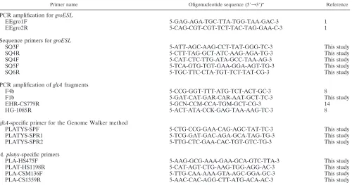
EEgro1F and EEgro2R were used to amplify an approximately 1,700-bp fragment with an annealing temperature of 55°C (3) (Table 1). The amplification products were purified using the QIAquick PCR purification kit (Qiagen GmbH, Hilden, Ger-many) and sequenced. Five primers shown in Table 1 were
used to complete the sequence of groESL.
Fluorescence-la-beled dideoxynucleotide technology was used for DNA se-quencing reactions (Perkin-Elmer, Applied Biosystems Divi-sion, Foster City, Calif.). Samples were then sequenced using a Perkin-Elmer ABI Prism 377 automated DNA sequencer at the DNA Core Facility of the Center for Gene Research, Yamaguchi University.
The strategy for determining thegltAsequence was similar
to that used in our previous report (10). A partial sequence of
A. platys gltAwas first determined using two sets of degenerate primers, F1b and EHR-778R, and F1b and HG1085R (10, 15) (Table 1). These primers were designed based upon the
se-quence ofgltAofA. phagocytophila,A. marginale, andA.
cen-trale. The amplification conditions were the same as in the previous study (10), with an annealing temperature of 53°C. The amplification products were purified and sequenced as described above. After a partial determination of the
se-quence, the unknown areas of the 3⬘and 5⬘ends of the gene
were determined using the Universal Genome Walker kit (Clontech Laboratories, Palo Alto, Calif.). Briefly, genomic
DNA was digested withEcoRV,DraI,PvuII,StuI, andScaI.
DNA fragments were ligated with a Genome Walker adaptor,
* Corresponding author. Mailing address: Laboratory of Veterinary Internal Medicine, Faculty of Agriculture, Yamaguchi University, 753-8515 Yamaguchi, Japan. Phone and fax: (81) 83-933-5895. E-mail: [email protected].
1132
on August 17, 2020 by guest
http://cvi.asm.org/
which had one blunt end and one end with a 5⬘ overhang. A ligation mixture of the adaptor and ehrlichial genomic DNA fragments was used as a template for PCR. This PCR was performed using an adaptor primer supplied by the
manufac-turer andA. platys gltA-specific primers to walk downstream on
the DNA sequence (Table 1). For the amplification, the con-ditions were as in our previous report (10).
The sequences ofA. platysand the registered sequences of
other related species deposited in GenBank were analyzed for phylogenetic relationships. Multiple alignment analysis, the calculation of distance matrices, and the construction of phy-logenetic trees were performed with the ClustalW program (20) version 1.8 in the DNA Data Bank of Japan (Mishima, Japan; http://www.ddbj.nig.ac.jp/htmls/E-mail/clustalw-e.html). The distance matrices for the aligned sequences with all gaps ignored were calculated using the Kimura two-parameter method (11), and the neighbor-joining method was used for constructing a phylogenetic tree (16). The stability of the tree obtained was estimated by bootstrap analysis for 100 replica-tions using the same program. Tree figures were generated using the TreeView program, version 1.61 (14).
A primer set, forward primer PLA-HS475F and reverse primer PLA-HS1198R, was designed based upon the
align-ment data to specifically amplify anA. platys groESLfragment.
Another set of primers, PLA-CSM136F and PLA-CS1359R,
was also designed based upon the alignment data ofgltA. PCR
conditions were the same as described above but with an an-nealing temperature of 58°C and the use of 40 cycles. The specificity of the reaction was tested with DNA extracted from
the three strains ofA. platysand related species, includingA.
phagocytophila(formerly the HGE strain Webster) (J. S.
Dum-ler),E. equi strain California (J. E. Madigan),E.
phagocyto-philastrain 1602 (A. Garcia-Perez),A. marginalestrain Florida
(G. H. Palmer),E. canisstrain Oklahoma (J. Dawson),
Wol-bachia pipientis(M. Taylor), andNeorickettsia helminthoeca(Y. Rikihisa). The sensitivities of both PCR systems were also
examined using DNA fromA. platysstrain France. The DNA
was diluted 10-fold from 1:1 to 1:10,000 with distilled water.
A 1,670-bpgroESLfragment ofA. platyswas determined to
contain 41 bp of the partialgroES, 51 bp of the spacer region,
and 1,577 bp of thegroELcoding region. ThegroESLoperon
was compared with that of other Anaplasma and Ehrlichia
bacteria reported previously and was found to be closely
re-lated to the operon ofA. phagocytophilaandA. marginale, with
81.4 and 78.8% identity, respectively. The level of similarity
amonggroESLsequences was much lower than that for the 16S
rRNA gene sequence in the same species (98.6% with A.
phagocytophila and 96.1% with A. marginale). The spacer length of 51 nucleotides was similar to that for related
Anaplasmaspecies: 52 bp forA. phagocytophilaand 47 bp for
A. marginale. The percent identities of the nucleotide sequence
in the spacer region ofA. platyscompared to that ofA.
phago-cytophila and to that of A. marginale were 74.5 and 72.3%, respectively, revealing a greater degree of divergence than for
the entiregroELcoding region.
After the initial identification of the 955-bp partial sequence ofA. platys gltAusing the degenerate PCR strategy, the full-length open reading frame extending from the ATG start codon to the TAA stop codon was determined using the
Ge-nome Walker PCR method. The length of thegltAopen
read-ing frame was 1,236 bp and encoded a protein of 411 amino
acids. The completegltAsequence was compared with that of
TABLE 1. Oligonucleotide sequences of primers used in this study
Primer name Oligonucleotide sequence (5⬘33⬘)a Reference
PCR amplification forgroESL
EEgro1F 5-GAG-AGA-TGC-TTA-TGG-TAA-GAC-3 1
EEgro2R 5-CAG-CGT-CGT-TCT-TAC-TAG-GAA-C-3 1
Sequence primers forgroESL
SQ3F 5-ATT-AGC-AAG-CCT-TAT-GGG-TC-3 This study
SQ4R 5-CTT-TAG-GCT-ATC-AAG-AGA-TG-3 This study
SQ4F 5-CAT-CTC-TTG-ATA-GCC-TAA-AG-3 This study
SQ5F 5-TCA-GTG-TGT-GAA-GGA-AGT-TG-3 This study
SQ6R 5-TGC-TTC-CTA-TGT-TCT-TAT-CG-3 This study
PCR amplification ofgltAfragments
F4b 5-CCG-GGT-TTT-ATG-TCT-ACT-GC-3 8
F1b 5-GAT-CAT-GAR-CAR-AAT-GCT-TC-3 This study
EHR-CS779R 5-GCN-CCM-CCA-TGM-GCT-CG-3 14
HG-1085R 5-ACT-ATA-CCK-GAG-TAA-AAG-TC-3 8
gltA-specific primer for the Genome Walker method
PLATYS-SPF 5-CTG-CCG-GAA-CAG-AGC-TAT-TC-3 This study
PLATYS-SPR1 5-TCG-GAT-GAC-AGA-GCA-TAG-TG-3 This study
PLATYS-SPR2 5-TTG-CTC-GAA-CAC-TGT-GTC-TG-3 This study
A. platys-specific primers
PLA-HS475F 5-AAG-GCG-AAA-GAA-GCA-GTC-TTA-3 This study
PLAT-HS1198R 5-CAT-AGT-CTG-AAG-TGG-AGG-AC-3 This study
PLA-CSM136F 5-TTG-CAA-AAA-GTA-AGC-GGA-GC-3 This study
PLA-CS1359R 5-AAC-CAC-AGG-CTT-ATG-ACA-AC-3 This study
aK, G or T; R, G or A; M, C or A; N, G, C, A, or T.
on August 17, 2020 by guest
http://cvi.asm.org/
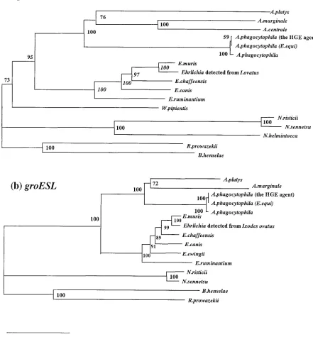
FIG. 1. Phylogenetic relationship of variousAnaplasma,Ehrlichia, andNeorickettsiaspp. based on the nucleotide sequences ofgltA(a) and
groESL(b) genes. The neighbor-joining method was used to construct the phylogenetic tree with the ClustalW program. The scale bar represents 10% divergence. The numbers at nodes are the proportions of 100 bootstrap resamplings that support the topology shown. The GenBank accession numbers of thegroESLsequences used to construct the phylogenetic tree and aligned data are as follows:A. phagocytophila(strain HGE agent), AF165812;E. equi, AF172162;E. phagocytophila, U96729;A. marginale, AF165812;E. chaffeensis, L1 0917;E. canis, U96731;E. muris, AF210459;
Ehrlichiasp. detected fromI. ovatus, AB032711;Ehrlichia(Cowdria)ruminantium, U13638;Neorickettsia(Ehrlichia)risticii, U96732;Neorickettsia
(Ehrlichia)sennetsu, U88092;Rickettsia prowazekii, Y15783;Bartonella henselae, U96734. The GenBank accession numbers of thegltAsequences used for comparative analysis are as follows:A. phagocytophila(strain HGE agent), AF304136;E. equi, AF304137;E. phagocytophila, AF304138;
A. marginale, AF304139;A. centrale, AF304141;E. chaffeensis, AF304142;E. canis, AF304143;E. muris, AF304144;Ehrlichiasp. detected fromI. ovatus, AF304145;E. (C.)ruminantium, AF304146;N. (E.) risticii, AF304147; N. (E.) sennetsu, AF304148:N. helminthoeca, AF304149; R. prowazekii, M17149;B. henselae, U78514.
on August 17, 2020 by guest
http://cvi.asm.org/
other Anaplasma and Ehrlichia bacteria reported previously
(10) and found to be closely related to those ofA.
phagocyto-philaandA. marginale, with 62.7 and 63.2% identity,
respec-tively. The level of similarity among ehrlichialgltAwas much
lower than that for the 16S rRNA gene sequence in the same
species. Thus, the sequence of gltA is much more variable
among these species than isgroESL. The length of thegltA
sequence of A. platys (1,236 bp) is the same as that of A.
phagocytophila but slightly shorter than that ofA. marginale
andA. centrale(1,254 bp).
In topology, the gltA-based phylogenetic tree (Fig. 1a) was
very similar to the tree derived from analysis of the 16S rRNA
gene analyses (3) and thegroEL-based tree reported previously
(21). However, the trees constructed from gltA and groESL
nucleotide sequences showed more distance than the 16S
rRNA-based trees. These findings also support the use ofgltA
-based andgroESL-based comparisons in determining the
phy-logeny ofAnaplasma, Ehrlichia, and Neorickettsiaagents and
strengthen the 16S rRNA- and groESL-based phylogeny
re-ported recently (7). There have been reports ofA. platys-like
organisms identified based on 16S rRNA analysis, including bacteria from white-tailed deer in North America (6) and from a cow in South Africa (7). It would be interest to determine the
phylogenetic position of these agents by analyzing thegltAand
groESLgene sequences.
PCR is a powerful tool for epidemiological or diagnostic purposes because of its high sensitivity and specificity;
how-ever, there are few molecular tools available forA. platys. All
PCR assays reported previously were developed based upon 16S rRNA gene sequences (4, 5, 9, 12, 13). In the present
study, new PCR primers were designed to amplify A. platys
DNA specifically and were based upon the alignment data of
groESLandgltAofA. platysand closely relatedAnaplasmaand
Ehrlichiaspecies. The PCR produced a fragment of 724 bp fromgroESLand 1,459 bp fromgltA with the DNA fromA. platysstrain France (Fig. 2). The sensitivities of the PCR
sys-tems were examined by using diluted DNA. BothgroESL- and
gltA-based PCRs detected DNA diluted 1:100 (Fig. 2), similar
to the 16S rRNA-based method (9). As 5l of the original
DNA solution contained genomic DNA from approximately
375 platelets infected with A. platys, both PCR systems can
detect DNA from 3.75 infected platelets in a reaction mixture. The specificity of both systems was also examined using DNA
of threeA. platysstrains from different geographic locations,
France, Japan, and Venezuela, and using DNA from related
species, includingA. phagocytophilaandA. marginale. Figure 3
shows that both PCR systems were specific forA. platys.
Fur-thermore, the gltA-based PCR amplifies the whole gltA
se-quence ofA. platys, which contains both start and stop codons.
As the sequences ofgltA and groESLhave greater variation
than the sequence of 16S rRNA, the sequence analysis of PCR products may supply useful information for phylogenic studies
of the agents. Our findings suggest that both groESL- and
gltA-based PCRs are useful for the specific detection of A.
platysDNA, and they would be additional molecular tools for both phylogenetic study and diagnosis in veterinary medicine.
Nucleotide sequence accession numbers.The nucleotide
se-quences ofgroESLand gltA ofA. platysstrain France
deter-mined herein have been deposited in the GenBank database under the accession numbers AY044161 and AB058782, re-spectively.
We acknowledge the technical expertise of the DNA Core Facility of the Center for Gene Research, Yamaguchi University, which is sup-ported by a grant-in-aid from the Ministry of Education, Science, Sports and Culture of Japan. This work was supported in part by a grant for an international joint research project from Institut National de la Sante et de la Recherche Medicale in France and the Japan Society for the Promotion of Science and a grant-in-aid for scientific research from the Japan Society for the Promotion of Science (grant no. 14360190).
We also thank E. B. Breitschwerdt for theA. platysDNA from a dog in Venezuela and J. S. Dumler for scientific suggestions and English correction.
REFERENCES
1.Allsopp, M. T. E. P., E. S. Visser, J. L. du Plessis, S. W. Vogel, and B. A. Allsopp.1997. Different organisms associated with heartwater as shown by analysis of 16S ribosomal RNA gene sequences. Vet. Parasitol.71:283–300. 2.Beaufils, J.-P., H. Inokuma, J. Martin-Granel, P. Jumelle, M.
Barbault-FIG. 2. The sensitivities of theA. platys-specific PCR based upon thegroESL(lanes 1 to 6) andgltA(lanes 7 to 12) genes were evaluated. DNA equivalent to that from 375, 37.5, 3.75, 0.375, and 0.0375 infected platelets was used as a template for the amplicons demonstrated in lanes 1 and 7, 2 and 8, 3 and 9, 4 and 10, and 5 and 11, respectively. Distilled water was used in lanes 6 and 12 as a negative control. Lane M, 100-bp DNA ladder.
FIG. 3.A. platys-specific PCRs based upon thegroESL(A) andgltA
(B) genes were evaluated for their specificity with DNA fromA. platys
strains from Somieres, France (lane 1), Okinawa, Japan (lane 2), and Venezuela (lane 3), and fromA. marginale(lane 4),A. centrale(lane 5),
A. phagocytophila strain HGE agent (lane 6), E. equi (lane 7), E. phagocytophila(lane 8),W. pipientis(lane 9),E. canis(lane 10), andN. helminthoeca(lane 11), with distilled water as a negative control (lane N). Positive bands (arrows) were observed only with DNA of the three strains ofA. platysin bothgroESL- andgltA-based PCRs.
on August 17, 2020 by guest
http://cvi.asm.org/
Jumelle, and P. Brouqui.2002.Anaplasma platys(Ehrlichia platys) infection in a dog in France: description of the case, and characterization of the agent. Rev. Med. Vet.153:85–90.
3.Chae, J.-S., J. E. Foley, J. S. Dumler, and J. E. Madigan.2000. Comparison of the nucleotide sequence of 16S rRNA, 444Ep-ank, andgroESLheat shock operon genes in naturally occurringEhrlichia equiand human granulocytic ehrlichiosis agent isolates from northern California. J. Clin. Microbiol.38:
1364–1369.
4.Chang, A. C. H., W. L. Chang, C. T. Lin, M. J. Pan, and S. C. Lee.1996. Canine infectious cyclic thrombocytopenia found in Taiwan. J. Vet. Med. Sci.
58:473–476.
5.Chang, W. L., and M. J. Pan.1996. Specific amplification ofEhrlichia platys DNA from blood specimen by two-step PCR. J. Clin. Microbiol.34:3142– 3146.
6.Dowson, J. E., C. K. Warner, V. Baker, S. A. Ewing, D. E. Stallknecht, W. R. Davidson, A. A. Kocan, J. M. Lockhart, and J. G. Olsen.1996. Ehrlichia-like 16S rDNA sequence from wild white-tailed deer (Odoceileus virginianus). J. Parasitol.82:52–58.
7.Dumler, J. S., A. F. Barbet, C. P. J. Bekker, G. A. Dasch, G. H. Pulmer, S. C. Ray, Y. Rikihisa, and F. R. Rurangirwa.2001. Reorganization of genera in the familiesRickettsiaceaeandAnaplasmataceaein the orderRickettsiales: unification of some species ofEhrlichiawithAnaplasma,Cowdriawith Ehr-lichia, andEhrlichiawithNeorickettsia, descriptions of six new species com-bination and designation ofEhrlichia equiand⬘HGE agent’ as subjective synonyms ofEhrlichia phagocytophila. Int. J. Syst. Evol. Microbiol.51:2145– 2165.
8.Harvey, J. W., C. F. Simpson, and J. M. Gaskin.1978. Cyclic thrombocyto-penia induced by a rickettsia-like agent in dogs. J. Infect. Dis.137:182–188. 9.Inokuma, H., K. Ohno, T. Onishi, D. Raoult, and P. Brouqui.2001. Detec-tion of ehrlichial infecDetec-tion by PCR in dogs from Yamaguchi and Okinawa Prefecture. Jpn. J. Vet. Med. Sci.63:815–817.
10.Inokuma, H., P. Brouqui, M. Drancourt, and D. Raoult.2001. Citrate syn-thase gene sequences: a new tool for phylogenetic and identification of Ehrlichia. J. Clin. Microbiol.39:3031–3039.
11.Kimura, M.1980. A simple method for estimating evolutional rates of base
substitutions through comparative studies of nucleotide sequence. J. Mol. Evol.16:111–120.
12.Kordick, S. K., E. B. Breitschwerdt, B. C. Hegarty, K. L. Southwick, C. M. Colitz, S. I. Hancock, J. M. Bradley, R. Rumbough, J. T. McPherson, and J. N. MacCormack.1999. Coinfection with multiple tick-borne pathogens in a Walker Hound kennel in North Carolina. J. Clin. Microbiol.37:2631–2638. 13.Mathew, J. S., S. A. Ewing, G. L. Murphy, K. M. Kocan, R. E. Corstvet, and J. C. Fox.1997. Characterization of a new isolate of Ehrlichia platys (Order Rickettsiales) using electron microscopy and polymerase chain reaction. Vet. Parasitol.68:1–10.
14.Page, R. D. M.1996. TREEVIEW: an application to display phylogenetic trees on personal computers. Comput. Appl. Biosci.12:357–358. 15.Parola, P., H. Inokuma, J.-L. Camicas, P. Brouqui, and D. Raoult.2001.
Detection of spotted fever groupRickettsiaeandEhrlichiaein African ticks. Emerg. Infect. Dis.7:1014–1017.
16.Saitou, N., and M. Nei.1987. The neighbor-joining method: a new method for reconstructing phylogenetic trees. Med. Biol. Evol.4:406–425. 17.Suksawat, J., C. Pitulle, C. Arraga-Alvarado, K. Madridal, S. I. Hancock,
and E. B. Breitschwerdt.2001. Coinfection with threeEhrlichiaspecies in dogs from Thailand and Venezuela with emphasis on consideration of 16S ribosomal DNA secondary structure. J. Clin. Microbiol.39:90–93. 18.Sumner, J. W., W. L. Nicholson, and R. F. Massung.1997. PCR
amplifica-tion and comparison of nucleotide sequences from groESL heat shock operon ofEhrlichiaspecies. J. Clin. Microbiol.35:2087–2092.
19.Sumner, J. W., G. A. Storch, R. S. Buller, A. M. Liddell, S. L. Stockham, Y. Rikihisa, S. Messenger, and C. D. Paddock.2000. PCR amplification and phylogenetic analysis ofgroESLoperon sequences fromEhrlichia ewingiiand Ehrlichia muris. J. Clin. Microbiol.38:2746–2749.
20.Thompson, J. D., D. G. Higgins, and T. J. Gibson.1994. CLUSTAL W: improving the sensitivity of progressive multiple sequence alignment through sequence weighting, position specific gap penalties and weight matrix choice. Nucleic Acid Res.22:4673–4680.
21.Yu, X. J., X.-F. Zhang, J. W. McBride, Y. Zhang, and D. H. Walker.2001. Phylogenetic relationship ofAnaplasma marginaleand⬙Ehrlichia platys’ to otherEhrlichiaspecies determined bygroELamino acid sequences. Int. J. Syst. Evol. Microbiol.51:1143–1146.