Coexistence of
Bos taurus
and
B. indicus
Mitochondrial DNAs in Nuclear
Transfer-Derived Somatic Cattle Clones
Ralf Steinborn,*
,†,1,2Pamela Schinogl,*
,2David N. Wells,
‡Andreas Bergthaler,*
Mathias Mu
¨ller* and Gottfried Brem*
,†,§*Institute of Animal Breeding and Genetics, University of Veterinary Medicine, A-1210 Vienna, Austria,†Ludwig-Boltzmann Institute for Immuno-, Cyto- and Molecular Genetic Research, A-1210 Vienna, Austria,‡Reproductive Technologies
Group, AgResearch, PB3123, Hamilton, New Zealand and§Department of Biotechnology in Animal Production, Institute for Agrobiotechnology, A-3430 Tulln, Austria
Manuscript received May 2, 2002 Accepted for publication July 11, 2002
ABSTRACT
We investigated the mitochondrial DNA (mtDNA) composition in one of the largest adult somatic mammalian clones (n⫽20) reported so far. The healthy cloned cattle were derived from nuclear transfer of an identical nuclear genetic background (mural granulosa donor cells including surrounding cytoplasm) into enucleated oocytes with eitherBos indicusor B. taurus mtDNA. Here we report the first cases of coexisting mtDNAs of two closely related subspecies following nuclear transfer. Heteroplasmy (0.6–2.8%) was found in 4 out of 11 cross-subspecies cloned cattle. Quantitation was performed using “amplification refractory mutation system (ARMS) allele-specific real-time PCR.” We determined that the ratio of donor cell to recipient cytoplast mtDNA copy number was 0.9% before nuclear transfer. Therefore, we concluded that the percentage of donor cell mtDNA in the heteroplasmic intersubspecific cloned animals is in accordance with neutral transmission of donor mtDNA. We determined an amino acid sequence divergence of up to 1.3% for the two subspecies-specific mtDNA haplotypes. In addition, intrasubspecificB. indicus heteroplasmy ofⵑ1% (but up to 7.3 and 12.7% in muscle and follicular cells of one animal) was detected in 7 out of the 9B. indicusintrasubspecific cloned cattle.
M
ITOCHONDRIAL DNA (mtDNA) heteroplasmy et al. 1996) or embryonic pronuclei surrounded by some represents a naturally rarely occurring phenome- cytoplasm (Meirelles andSmith 1997). Interspecific non (Chinneryet al. 2000). Following intraspecific sex- heteroplasmic mice were also produced by microinjec-ual crosses mammalian sperm mitochondria generate tion of somatic mitochondria into pronucleus-stage em-initial heteroplasmy, but are selectively destroyed be- bryos (Shitaraet al. 2000). Moreover, embryonic stem tween the four- and the eight-cell stage of development cell cybrids were used to introduce mtDNA mutations (Sutovsky et al. 1999, and references therein). It is into the mouse female germline (Slighet al. 2000). Clon-proposed that the ubiquitination of sperm mitochon- ing by nuclear transfer of embryonic donor cells resulted dria that occurs during spermatogenesis tags them for in the production of some heteroplasmic cloned animals species-specific extinction by the embryo’s proteasomes (Steinbornet al. 1998c;Hiendlederet al. 1999), whereas and lysosomes (Sutovsky et al. 1999). Remarkably, in other embryonic cell-derived cloned animals mtDNA sperm mitochondria persist in mammalian interspecies arises exclusively from recipient oocytes (Steinborn et crosses as demonstrated for murine (Gyllenstenet al. al. 1998c; Takeda et al. 1999). Similar contradictory 1991;Kanedaet al. 1995) and bovine (Sutovskyet al. findings were found after nuclear transfer of fetal or 1999) hybrids. Heteroplasmy can be generated by a adult somatic donor cells (Evanset al. 1999;Steinborn number of cell manipulation techniques. Intraspecific et al. 2000). In addition to the neutral transmission of heteroplasmic mice have been generated by fusing em- donor cell mtDNA (heteroplasmy atⵑ1% according to bryonic cytoplasts with either one-cell embryos (Jenuth Steinbornet al. 2000) reported for cattle but not sheep,a complete absence of donor cell mtDNA transmission has been observed (Evanset al. 1999;Steinborn et al.
Sequence data from this article have been deposited with the
2000).
EMBL/GenBank Data Libraries under accession nos. AF361442–
AF361461, AY029263–AY029268, AY052631, AF416451, AF419237, This work describes the fate ofB. indicus donor cell
AF384025, AF419238, AF384022, and AF384026. mitochondria from a single genetic origin following 1Corresponding author:Institute of Animal Breeding and Genetics,
nuclear transfer into 20 cytoplasts of random genetic
University of Veterinary Medicine, Veterina¨rplatz 1, A-1210 Vienna,
origin, which harbored either Bos indicus or B. taurus
Austria. E-mail: [email protected]
2These authors contributed equally to this work. mtDNA haplotypes.
MATERIALS AND METHODS gories of rate heterogeneity. The phylogenetic tree was graphi-cally displayed using TREEVIEW, version 1.6.5. TheB. gaurus
Biological material:Two sets of adult somatic cloned ani- mtDNA (GenBank accession no. AF083371) was used as an
mals were produced by nuclear transfer using primary cultures outgroup. The two cattle taxa B. taurusand B. indicus are of quiescent (presumptive G0) mural granulosa cells derived represented in the tree by the GenBank accession nos. J01394 from the genetic origin “E” (denoted E-donor). The cloned and L27733.
animals of set I (E1 and E4–E12;Wellset al. 1999) and set Quantitation of mtDNA copy number ratios and hetero-II (E13–E22, D. N.Wells, unpublished data) derived from plasmy:Real-time PCR quantitation was performed using flu-donor cells at passage three (E1 at passage five) and eight of oresence measurement by the ABI PRISM 7700 sequence de-culture, respectively. The embryos that resulted in the cloned tection system (Applied Biosystems) and the 5⬘-3⬘exonuclease animals of set II were either culturedin vitroin an improved activity of theTaqpolymerase. The 25-l amplification reac-media formulation termed AgResearch Synthetic Oviduct tions contained 67.7 mmTris-HCl, pH 8.8; 16.6 mm(NH4)2SO4; Fluid medium (AgR SOF; AgResearch, Hamilton, New 0.01% Tween 20; 0.2 mmdATP, dCTP, dGTP, and 0.4 mm Zealand; used to produce E13–E16, E21, and E22) or cultured dUTP; 1.25 units Taq polymerase; 4.5 mm MgCl2; 200 nm in vivoin the oviducts of temporary recipient sheep (E17–E20). TaqMan probe; 300 nmof each primer; and 10–50 ng total AgR SOF is a modified formulation to that described byGard- cellular DNA. In general, each sample was amplified in
tripli-ner et al. (1994), containing 8 mg/ml BSA. On day 4–5 of cate with standard deviations of threshold cycle (Ct) values in vitroculture, embryos were transferred to fresh AgR SOF not exceeding 0.5 (for Ct values⬍33). Quantitation was per-containing 10m2,4-dinitrophenol, acting as an uncoupler formed at an annealing temperature of 62⬚or 63⬚(or lower of oxidative phosphorylation (Thompsonet al. 2000). for primers with a reduced GC content).
In addition, we analyzed the mtDNA composition in the The ratio of donor cell to recipient cytoplast mtDNA copy blood of five other adult somatic cloned cattle [cloned animal numbers before nuclear transfer was determined by the equa-L2 and ongoing pregnancies (Wellset al. 1998; here referred tion Ratio⫽ 100%/(1⫹ E)⌬Ct, where E⫽ 10⫺1/m ⫺1 and to as cloned animals L5, L6, L8, and L9)] derived from the ⌬Ct is the difference between the Ct values of the fusion genetic origin “L” (denoted L-donor). Quiescent (presump- partners detected in the quantitative real-time PCR assay. The tive G0) granulosa donor cells between passages four and slope (m) being a measure for the efficiency (E) of the real-eight of culture were used for their production. time PCR is determined by the standard curvey ⫽mx⫹b. The standard curve is generated by plotting the Ct values for The lack of a priori heteroplasmy in recipient cytoplasts
was determined in 12 oocyte aliquots (nine randomly taken serial template dilutions in relation to the logarithm of the dilution factor.
genetic origins of the “Austrian Simmental” breed), each
con-sisting of 10–15in vitromatured and denuded oocytes. For quantitation of heteroplasmy two sets of experiments were run: (i) The total amount of mtDNA was determined by Straws containing bovine sperm were thawed, and cells were
spun down and resuspended in 200 l phosphate-buffered real-time quantitative PCR (Steinbornet al. 1998c) and (ii) the less frequent mtDNA type was quantitated by amplification saline (PBS) buffer. Four 50-l drops were blotted onto
What-man paper, dried, and individually wrapped for convenient refractory mutation system (ARMS;Newtonet al. 1989;Wu
et al. 1989) allele-specific real-time PCR (Steinborn et al. transport.
DNA isolation:The cloned bovine embryos produced pre- 1998c, 2000). The limitation of the dynamic range in the
ARMS allele-specific real-time PCR assay is caused by the late-viously (Steinbornet al. 1998b) and the sample containing
exactly 15 cells of the E-donor were lysed in 50l K-buffer cycle amplification from the nontarget allele. This phenome-non is an inherent feature of ARMS-PCR. The discrimination (20 mmTris-HCl, pH 8.3, 1.5 mmMgCl2, 25 mmKCl, 0.5%
Tween 20). Subsequently, total cellular DNA was isolated as of the allele-specific quantitation assay,i.e., the limitation of the dynamic range, was controlled by the use of two homoplas-described (Steinbornet al. 1998b). DNA isolation from blood,
tissues, oocyte aliquots, and sperm spots was performed using mic nontarget allele controls (bovine total cellular DNA), both carrying the alternative base for the single-nucleotide the respective DNA isolation kit provided by Macherey & Nagel
(Dueren, Germany). For DNA isolation from sperm cells we polymorphism (SNP) under study. We proved that the abso-lute value of the slope in the equation obtained by plotting the additionally applied 27 mm dithiothreitol to support their
lysis. log inputvs.⌬Ct calculated from total mtDNA copy number
quantitation and the specific allele-specific quantitation assay
Microsatellite analysis: Nine microsatellite loci were
ana-lyzed, using the second version of the StockMarks kit for cattle was⬍0.1. This allowed the use of the “comparative Ct method” (ABI PRISM 7700 sequence detection system, Applied Biosys-paternity PCR typing (Applied Biosystems, Foster City, CA),
which is based on four-dye fluorescent labeling. The fluores- tems, 1997) for quantitation of heteroplasmy without running standard curves on the same plate. Briefly, the percentage of cence data collected by the ABI PRISM 310 genetic analyzer
were subsequently analyzed using the ABI PRISM Genotyper donor cell mtDNA in a cloned animal in relation to the total mtDNA content in the donor cell sample, i.e., the level software for automated genotyping (Applied Biosystems).
Analysis of the mtDNA control region:We amplified the com- of heteroplasmy, was determined by the equation Hetero-plasmy⫽100%/(1⫹E)⫺⌬⌬Ct.
plete bovine mtDNA control region as described (Steinbornet
al. 1998a) and sequenced the PCR products using the ABI Primers and probes:Nucleotide positions in the names of the oligonucleotides given below represent their localization PRISM BigDye terminator cycle sequencing kit and the ABI
PRISM 377 sequencer (Applied Biosystems). Note that a po- on the bovine mtDNA genome (numbering according to the GenBank accession no. J01394). For amplification and se-tential heteroplasmy of 5–10% in a clone would not impair
the determination of the recipient cytoplast-derived mtDNA quencing of the mtDNA control region we used the primers F15,747-15,766, R383-364, and F16,168-16,185. The fluoro-sequence by sequencing of PCR products.
Demonstration of parental mtDNA diversity using phyloge- genic probes (MWG-Biotech AG, Germany) for real-time PCR
consisted of an oligonucleotide with the 5⬘-reporter dye
netic analysis: The phylogeny of mtDNA control region
se-quences of the E-donor and its cloned offspring was recon- 6-carboxyfluorescein (FAM) and the quencher dye 6-carboxy-tetramethylrhodamine (TAMRA) attached to the 3⬘end. For structed with the software package TREE-PUZZLE, version 5.0
(Strimmerandvon Haeseler1996), using the HKY model total mtDNA quantitation we used the probe P99-74 and the
cate-Figure 1.—Localization of the fluorogenic probes and ARMS primers (designated ARMS) used for ARMS allele-specific real-time PCR quan-titation. Nucleotide positions correspond to the numbering in the GenBank accession no. J01394.
used in combination with the probe P99-74 and with one out of cellular distribution of the oocyte mitochondria before three ARMS primers (ARMS16,211-16,229, ARMS16,283-16,301, enucleation and the amount of the missing part of the and ARMS16,056-16,074) in three separate ARMS allele-specific
ooplasm that is removed from the recipient oocyte
dur-real-time quantitative PCR assays. The probe P15,900-15,929 was
ing the enucleation procedure. We determined the
fol-used for ARMS allele-specific real-time PCR in combination with
F15,873-15,898 and ARMS16,071-16,050. We used the following lowing mean Ct values⫾standard deviation for the 15
primer pairs to amplify and sequence the genes coding for E-donor cells and from the three embryo samples: 27.71⫾ NADH dehydrogenase 3 (MTND3), NADH dehydrogenase 6 0.01 and 24.59⫾0.44 (all four samples were amplified (MTND6), cytochrome b (MTCYB), cytochrome c oxidase II
in duplicates in the same real-time PCR experiment).
(MTCO2), cytochrome c oxidase III (MTCO3), and ATP
syn-Considering the slope of m ⫽ ⫺3.56 we calculated a
thase 6 (MTATP6): F9,785-9,804/R10,218-10,194 and
F13,860-13,881/R14,468-14,449, F14,383-14,402/R15,757-15,740, F7, ratio of donor cell to recipient cytoplast mtDNA copy
266-7,286/R8,132-8,115, F8,944-8,963/R9,885-9,868, and F8, number before nuclear transfer of 0.9%.
132-8,152/R9,314-9,297, respectively. In addition we applied Absence ofa prioriheteroplasmy in the recipient cyto-the primers F14,871-14,890 and R14,955-14,934 for internal
plasts: Human oocytes have been shown to contain
sequencing ofMTCYB.
mtDNA molecules with homopolymeric tract
hetero-Determination of the phylogenetic divergence between
pairs of species:The estimation of the pairwise relatedness of plasmy and a small proportion of oocyte mtDNA
mole-Ovis aries(GenBank accession no. AF010406)/O. aries musimon cules harbor the 4977-bp deletion (Nagley and Wei
(D84203), B. taurus (V00654)/B. indicus (AF419237), Mus 1998 and references therein). Although we have used musculus ( J01420)/M. spretus (AB033700), Pan troglodytes
neither homopolymeric tract polymorphisms nor
inser-(NC_001643)/Homo sapiens (AF347015), and B. gaurus
tion/deletion substitution to differentiate our target
al-(AF348596)/B. taurus(V00654) was performed by
determin-ing their phylogenetic divergence (percentage) on the basis leles we wanted to exclude the possibility that the
hetero-of cytochrome b amino acid sequences. plasmy was already present in the recipient cytoplasts.
Accession numbers:Sequences of the mtDNA control re- Therefore, we analyzed 12 samples, each of which
con-gion of the recipient cytoplasts and the two donor cell origins
tained 10–15in vitromatured and denuded oocytes from
used for nuclear transfer were submitted to GenBank under
nine randomly chosen slaughtered cows (from three
the following accession numbers: AF361442–AF361461 (cloned
cows two aliquots were obtained). The target allele
animals E1–E22; note that E2 and E3 were not investigated) and
AY029263–AY029268 (L-donor cell and cloned animals produced AC16,300/1GTwas used for ARMS allele-specific
real-to preserve the L-donor). We submitted the following mtDNA- time PCR (assay sensitivity 0.1%). As expected, we could encoded genes of theB. indicusdonor to the GenBank:MTND3
show the absence of heteroplasmy in these oocytes (Ct
(GenBank accession no. AY052631), MTND6 (AF416451),
values for the oocyte aliquots between 22.5 and 24.6;
MTCYB(AF419237),MTCO2(AF384025),MTCO3(AF419238),
standard deviations obtained for duplicate
amplifica-andMTATP6(AF384022). AMTCO2variant found in theB.
taurus mtDNA haplotype of the cloned animal E12 was also tionⱕ0.5).
submitted (AF384026). Analysis of mtDNA composition in the cloned
ani-mals:First, using microsatellite analysis we confirmed that nuclear DNA in all cloned animals matched the
RESULTS
nuclear DNA isolated from the donor cell line (data
mtDNA copy number ratios of the fusion partners be- not shown). To identify E-donor-specific SNPs necessary
fore nuclear transfer:Real-time PCR quantitation (Fig- for the quantitation of heteroplasmy in the cloned ani-ure 1) was used to determine the ratio of donor cell to mals we performed sequencing analysis of the mtDNA recipient cytoplast mtDNA copy number before nuclear control region. Regardless of a clearB. taurus morphol-transfer. Therefore we compared the Ct values obtained ogy it revealed the unexpected occurrence ofB. indicus
intra-TABLE 1
Intersubspecific heteroplasmy:B. indicusdonor mtDNA in B. taurusrecipient oocytes
Tissues analyzed (%B. indicus
Cloned animals donor mtDNA)
E6, E9, E10, E12 Blood (0)a, muscle (0)a, skin (0)a, follicular cells (0)a
E14, E19, E21 Blood (0)a
E15 Blood (0.6)
E18 Blood (1.1)
E20 Blood (1.8)
E22 Blood (2.8)
aAssay sensitivity, 0.1%.
sensitivity levels of 0.1% were routinely achieved for ARMS allele-specific quantitative real-time PCR (assays in Steinborn et al. 2000; assay for the target allele
AC16,300/1GTof this work). The reduced sensitivity/al-lelic discrimination of theB. indicus-allele-specific quan-titation assay designed for the target allele C16,050T
(0.5%) is most probably due to the low GC content (reduced annealing temperature of 43⬚) of the genomic
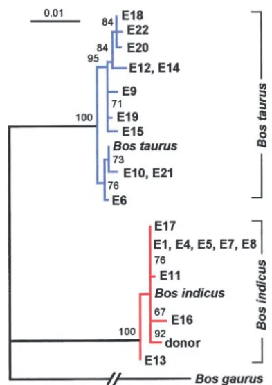
Figure2.—Maximum-likelihood tree of complete mtDNA
environment surrounding this SNP. Additional
se-control region sequences from the cloned cattle E1–E22 and
their donor cell. AB. gaurussequence was used as an outgroup. quencing of 1683 bases (MTCO2,MTATP6, and
adja-For each of the two cattle taxa B. taurus and B. indicus, a cent 318 bp of MTCO3) did not reveal another SNP representative sequence obtained from GenBank was included between the E-donor and the cloned animals E1, E4, in the tree. Measures of support are quartet puzzling values.
E5, E8, and E11.
Recently, the use of adult somatic nuclear transfer in animal conservation was demonstrated by cloning the last surviving cow (denoted L-donor in this work) of TheB. taurusmtDNA haplotypes exhibited pronounced
variation with only two pairs (one individual from set I the Enderby Island cattle breed to preserve the female genetics of this endangered breed (Wellset al. 1998). and set II within each pair) of identical sequence. The
B. indicussequences were less variable since one haplo- Enderby Island Shorthorns had adapted to harsh sub-antarctic conditions. Here we analyzed the mtDNAs of type was shared by 5 cloned animals. These 5 cattle
produced in set I of the nuclear transfer experiments five cloned cattle derived from the L-donor. Nuclear genetic identity between the L-donor cell and the five potentially originated from a single pair of ovaries or
from cows belonging to one maternal lineage. cloned animals derived from this donor was demon-strated by microsatellite analyses (data not shown). Se-Using quantitative ARMS allele-specific PCR we
ana-lyzed the mtDNA composition of adult cloned cattle quencing of the mitochondrial control region of the five cloned animals yielded twoB. taurushaplotypes (cloned produced by nuclear transfer of the E-donor into
enu-cleated oocytes of random genetic origin. The allele- animals L2 and L9) and three B. indicus haplotypes, whereas the L-donor harbored a B. indicushaplotype. specific assays were designed for donor-specific SNPs
(Figure 1) to quantitate the contribution of the donor Only L9 was heteroplasmic, harboring 0.9% donor mtDNA, whereas the L2 sample was homoplasmic, ex-mtDNA. We found the first four cases of intersubspecific
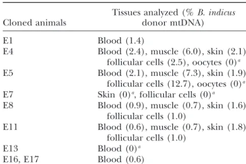
B. indicus/B. taurusheteroplasmy (n⫽11, Table 1). We hibiting only the B. taurus recipient cytoplast-derived mtDNA. The quantitation of the L-donor mtDNA in the also detected intrasubspecific B. indicus heteroplasmy
in 7 animals (n⫽9, Table 2). In contrast, we detected three cloned individuals with cytoplast-derivedB. indicus
mtDNA was not possible due to the absence of polymor-a complete polymor-absence of heteroplpolymor-asmy following either
inter- or intrasubspecific nuclear transfer in 9 (7 and 2 phisms. Since the control region represents the most variable part of the mtDNA, we did not continue to cloned animals, respectively) of the 20 cloned animals
derived from the E-donor (Tables 1 and 2). No traces search for SNPs in the coding region.
In addition to the last surviving Enderby Island cow, of donor mtDNA were detectable in the female germline
of the intrasubspecific cloned animals E4 and E5, al- analysis of samples stored from other now deceased Enderby Island animals revealed 12B. indicusmtDNAs though their investigated tissues showed respective
TABLE 3 TABLE 2
IntrasubspecificB. indicusheteroplasmy Type of amino acid differences in mtDNA-encoded subunits
betweenB. taurusandB. indicus
Tissues analyzed (%B. indicus
Cloned animals donor mtDNA) Amino acid Amino acid
Complex position substitution
E1 Blood (1.4)
E4 Blood (2.4), muscle (6.0), skin (2.1), I MTND3
follicular cells (2.5), oocytes (0)a 82 A→T
E5 Blood (2.1), muscle (7.3), skin (1.9), I MTND6 No
follicular cells (12.7), oocytes (0)a III MTCYB
E7 Skin (0)a, follicular cells (0)a 356 V→I
E8 Blood (0.9), muscle (0.7), skin (1.6), 372 I→V
follicular cells (1.0) IV MTFCO2
E11 Blood (0.6), muscle (0.7), skin (1.8), 57 D→Na
follicular cells (1.0) IV MTCO3
E13 Blood (0)a 171 V→I
E16, E17 Blood (0.6) 238 G→A
V MTATP6
aAssay sensitivity, 0.5%.
7 T→A
69 T→A
154 M→V
Phylogenetic divergence betweenB. taurusandB.
indi-The amino acid substitutions (single-letter code) occurred
cus based on mtDNA-encoded peptides: To estimate in the complexes I–IV of the electron transport system or in the extent of the possible mtDNA sequence divergence the ATP synthase complex (complex V). Numbers indicate between two (sub)species, which may allow the genera- the amino acid position inside each subunit.
aSubstitution found only in the cloned animal E12 (B. taurus
tion of inter(sub)specific heteroplasmy, sequencing of
haplotype).
parts of theB. indicusmtDNA was performed. On the basis of the haplotype classification presented in Figure 2 we selected the E-donor and randomly two
representa-similar to those reported by us earlier for heteroplasmic tiveB. indicusmtDNAs (cloned animals E1 and E11) for
cloned B. taurus cattle (Steinborn et al. 2000). We sequencing and subsequent comparison with theB. taurus
showed thatB. indicusmtDNA was not presenta priori
reference. We analyzed six randomly selected
mtDNA-in the recipient oocytes used for nuclear transfer. More-encoded proteins from the electron transport system
over, each heteroplasmic cloned individual was quanti-complexes I, III, and IV (there is no mtDNA-encoded
tated at two sites (donor cell-specific ARMS primers subunit of complex II) and the ATP synthase complex
ARMS16,211-16,229 and ARMS16,283-16,301 for detec-(complex V). Among them are three highly conserved
tion of intersubspecific heteroplasmy and primers proteins (encoded by MTCO2, MTCO3, and MTCYB;
ARMS16,056-16,074 and ARMS16,071-16,050 for quan-classification reviewed inBlieret al. 2001) and proteins
titation of intrasubspecificB. indicusheteroplasmy). At that are regarded as more variable [encoded byMTND6
each of these four sites the donor mtDNA exhibited a andMTATP6(Blieret al. 2001) andMTND3]. We found
rare polymorphism compared to the cloned animals intersubspecific differences in four of the six protein
(Steinbornet al. 1998a and data not shown). Thus, it is sequences investigated (Table 3), yielding divergence
very unlikely that these rare mutations were, by chance, rates between 0% (MTND6) and 1.3% (MTATP6). While
presenta priori in the recipient cytoplasts used for nu-nonconservative changes were not detected, out of a
clear transfer. The fact that the percentage of donor total of eight differences, three were conservative
(be-mtDNA in our heteroplasmic cloned cattle was at a level tween amino acids of the same family) and five were
expected for neutral transmission of parental mtDNAs semiconservatives (hydrophobic or charged to neutral
makes the theory ofa prioriheteroplasmy of the recipi-amino acids). By chance, we additionally detected aB.
ent cytoplasts even more unlikely. Together with the
taurus MTCO2variant (Table 3, semiconservative, 0.4%
fact that homo- and heteroplasmy occurred within one divergence).
clonal origin (i.e., the same nuclear DNA), the utiliza-tion of a nontarget allele control (bovine total cellular DNA carrying the alternative base for the SNP under
DISCUSSION
study) excludes a hypothetical attribution of nuclear We report intrasubspecificB. indicusand intersubspe- mitochondrial pseudogenes (for review seeBensasson
cific heteroplasmy in adult somatic cattle cloned by nu- et al. 2001) to the heteroplasmic signal.
clear transfer. The ratios quantitated for the coexisting The heteroplasmic intersubspecific adult somatic
the Enderby Island cattle, separated under harsh sub- crosses show reduced fitness, indicating a misbalanced nuclear-mitochondrial interaction (Nagaoet al. 1998). antarctic conditions for 150 years (Bunn 1998), we
found haplotypes from both subspecies. The unex- Recently, two wild endangered species, a gaur (B. gaur-us) and a European mouflon (O. aries musimon: http:// pected occurrence of B. indicusmtDNA haplotypes in
the New Zealand population with European cattle (B. www.ncbi.nlm.nih.gov/htbin-post/taxonomy/), were suc-cessfully cloned by either interspecific or
intersubspe-taurus) morphology was confirmed by another group
(S. H.Phua, K. G.Dodds, R.Spelmanand A. M.Craw- cific nuclear transfer using recipient oocytes collected from B. taurus and European O. aries, respectively
ford, personal communication) reporting a similarB.
indicus/B. taurushaplotype ratio for New Zealand Jersey (Lanzaet al. 2000;Loiet al. 2001). The gaur clone died soon after birth. Comparing cytochrome b amino acid cattle. The B. taurus/B. indicus nuclear-mitochondrial
compatibility is also in accordance with reports on the sequences a divergence of 3.2% (partial sequences), 0.3%, and 0.5% was revealed for O. aries musimon/O.
descendants of hybridization betweenB. indicusandB.
taurus (Troyet al. 2001) and can be concluded from aries and B. indicus/B. taurus, respectively. For the B. gaurus/B. taurusinterspecies nuclear transfer clone an the successful intersubspecific embryonic cell cloning
between B. indicus andB. taurus, which for unknown impairment of nuclear-mitochondrial interaction can-not be excluded, whereas the divergence between B.
reason(s) led to a shift to homoplasmy during
em-bryogenesis (Meirelleset al. 2001). indicus/B. taurusandO. aries musimon/O. ariesseems to be tolerated.
In general, the reason(s) leading to heteroplasmy or
homoplasmy in the described inter- or intrasubspecific Due to the limited supply of oocytes and surrogate animals, the cloning of highly endangered or extinct nuclear transfers remains unknown. The mtDNA
trans-mission pattern in the cloned animals did not correlate species will require inter(sub)specific nuclear transfer. However, the nuclear-cytoplasmic composition should with donor cell passage similarly as reported previously
(Steinbornet al. 2000) or with mtDNA haplotype. The be considered if repopulation under specific (extreme) environmental conditions is attempted (see above). In comparison of the set I (conventionalin vitroculturing
of cloned embryos) and the set II (improved media light of these limitations, future research may reveal whether inter(sub)specific cloning can participate in formulation or in vivo culturing) animals that were
cloned from the E-donor showed an increased occur- the efforts to conserve endangered species.
rence of intersubspecific heteroplasmy (heteroplasmic We thank C. Schlo¨tterer for discussion of phylogenetic data; G. animals/total number of animals, 0/4 vs.4/7, respec- Muir for comments on the manuscript; G. Mo¨sslacher, E. Dworak, N. Katic, J. Forsyth, K. Cockrem, M. Berg, J. Oliver, T. Day, and M. Ashby
tively). It remains unclear whether the occurrence of
for technical assistance; and the Austrian Science Fund (FWF) for
heteroplasmy in intersubspecific cattle reflects a
renor-funding (project P14840 to R.S.).
malization in the expression of genes involved in mito-chondrial biogenesis due to the improvement of culture conditions. Genetic changes and perturbations in
devel-LITERATURE CITED
opment that result from manipulating mammalian
em-bryos are well documented (references in Wilmut Bensasson, D., D. Zhang, D. L. HartlandG. M. Hewitt, 2001 Mitochondrial pseudogenes: evolution’s misplaced witnesses.
2002).
Trends Ecol. Evol.16:314–321.
Our finding of intersubspecific heteroplasmy in Blier, P. U., F. DufresneandR. S. Burton, 2001 Natural selection
and the evolution of mtDNA-encoded peptides: evidence for
healthy cloned mammals has important implications in
intergenomic co-adaptation. Trends Genet.17:400–406.
view of the ongoing discussion concerning the use of
Bunn, T., 1998 Lady saves the day. N. Z. Geogr.39:8–13.
animal cloning for preserving genetic variation, recreat- Chinnery, P. F., D. R. Thorburn, D. C. Samuels, S. L. White,
H. M. Dahlet al., 2000 The inheritance of mitochondrial DNA
ing species and populations already extinct or on the
heteroplasmy: Random drift, selection or both? Trends Genet.
verge of extinction (Lanzaet al. 2000;Lee2001). It is
16:500–505.
a first demonstration that the mtDNA of the endangered Evans, M. J., C. Gurer, J. D. Loike, I. Wilmut, A. E. Schnieke
et al., 1999 Mitochondrial DNA genotypes in nuclear
transfer-species can also be rescued by nuclear transfer (in
addi-derived cloned sheep. Nat. Genet.23:90–93.
tion to the nuclear genome of the donor cell) to derive
Gardner, D. K., M. Lane, A. SpitzerandP. A. Batt, 1994 Enhanced
offspring with the original mitochondria. The necessity rates of cleavage and development for sheep zygotes cultured to
the blastocyst stage in vitro in the absence of serum and somatic
to ascertain that the mtDNA of the endangered species
cells: amino acids, vitamins, and culturing embryos in groups
is also rescued was discussed recently (Meirelleset al.
stimulate development. Biol. Reprod.50:390–400.
2001). Segregation toward donor mtDNA might be pos- Gyllensten, U., D. Wharton, A. JosefssonandA. C. Wilson, 1991
Paternal inheritance of mitochondrial DNA in mice. Nature352:
sible in subsequent generations due to random genetic
255–257.
drift as shown for intraspecific mice (Jenuthet al. 1996).
Hasegawa, M., H. KishinoandT. Yano, 1985 Dating of the
human-However, tissue-specific and age-related selection for ape splitting by a molecular clock of mitochondrial DNA. J. Mol.
Evol.22:160–174.
different mtDNA genotypes in those mice (Jenuth et
Hiendleder, S., S. M. Schmutz, G. Erhardt, R. D. Green and
al. 1997) or even lack of mtDNA maintenance (Moraes
Y. Plante, 1999 Transmitochondrial differences and varying
et al. 1999) in interspecific hybrids of closely related levels of heteroplasmy in nuclear transfer cloned cattle. Mol.
Reprod. Dev.54:24–31.
Jenuth, J. P., A. C. Peterson, K. FuandE. A. Shoubridge, 1996 Sligh, J. E., S. E. Levy, K. G. Waymire, P. Allard, D. L. Dillehay
et al., 2000 Maternal germ-line transmission of mutant mtDNAs Random genetic drift in the female germline explains the rapid
from embryonic stem cell-derived chimeric mice. Proc. Natl. segregation of mammalian mitochondrial DNA. Nat. Genet.14:
Acad. Sci. USA97:14461–14466. 146–151.
Steinborn, R., M. Mu¨ llerandG. Brem, 1998a Genetic variation Jenuth, J. P., A. C. PetersonandE. A. Shoubridge, 1997
Tissue-in functionally important domaTissue-ins of the bovTissue-ine mtDNA control specific selection for different mtDNA genotypes in
heteroplas-region. Biochim. Biophys. Acta1397:295–304. mic mice. Nat. Genet.16:93–95.
Steinborn, R., V. Zakhartchenko, J. Jelyazkov, D. Klein, E. Wolf Kaneda, H., J.-I. Hayashi, S. Takahama, C. Taya, K. Fischer-
Lin-et al., 1998b Composition of parental mitochondrial DNA in dahlet al., 1995 Elimination of paternal mitochondrial DNA
cloned bovine embryos. FEBS Lett.426:352–356. in intraspecific crosses during early mouse embryogenesis. Proc.
Steinborn, R., V. Zakhartchenko, E. Wolf, M. Mu¨ llerand G. Natl. Acad. Sci. USA92:4542–4546.
Brem, 1998c Non-balanced mix of mitochondrial DNA in Lanza, R. P., J. B. Cibelli, F. Diaz, C. T. Moraes, P. W. Farinet al.,
cloned cattle produced by cytoplast-blastomere fusion. FEBS Lett. 2000 Cloning of endangered species (Bos gaurus) using
interspe-426:357–361. cies nuclear transfer. Cloning2:79–90.
Steinborn, R., P. Schinogl, V. Zakhartchenko, R. Achmann, W. Lee, K., 2001 Can cloning save endangered species? Curr. Biol.11:
Schernthaneret al., 2000 Mitochondrial DNA heteroplasmy R245.
in cloned cattle produced by fetal and adult cell cloning. Nat. Loi, P., G. Ptak, B. Barboni, J. J. Fulka, P. Cappaiet al., 2001 Ge- Genet.25:255–257.
netic rescue of an endangered mammal by cross-species nuclear Strimmer, K., andA. von Haeseler, 1996 Quartet puzzling: a quar-transfer using post-mortem somatic cells. Nat. Biotechnol. 19: tet maximum likelihood method for reconstructing tree
topolo-962–964. gies. Mol. Biol. Evol.13:964–969.
Meirelles, F. V., andL. C. Smith, 1997 Mitochondrial genotype Sutovsky, P., R. D. Moreno, J. Ramalho-Santos, T. Dominko, C. segregation in a mouse heteroplasmic lineage produced by em- Simerlyet al., 1999 Ubiquitin tag for sperm mitochondria. Na-bryonic karyoplast transplantation. Genetics145:445–451. ture402:371–372.
Meirelles, F. V., V. Bordignon, Y. Watanabe, M. Watanabe, A. Takeda, K., S. Takahashi, A. Onishi, Y. Goto, A. Miyazawaet Dayanet al., 2001 Complete replacement of the mitochondrial al., 1999 Dominant distribution of mitochondrial DNA from genotype in aBos indicuscalf reconstructed by nuclear transfer recipient oocytes in bovine embryos and offspring after nuclear to aBos taurusoocyte. Genetics158:351–356. transfer. Reprod. Fertil.116:253–259.
Moraes, C. T., L. KenyonandH. Hao, 1999 Mechanisms of human Thompson, J. G., C. McNaughton, B. Gasparrini, L. T. McGowan mitochondrial DNA maintenance: the determining role of pri- andH. R. Tervit, 2000 Effect of inhibitors and uncouplers of mary sequence and length over function. Mol. Biol. Cell 10: oxidative phosphorylation during compaction and blastulation of bovine embryos cultured in vitro. J. Reprod. Fertil.118:47–55. 3345–3356.
Troy, C., D. MacHugh, J. Bailey, D. Magee, R. Loftuset al., 2001 Nagao, Y., Y. Totsuka, Y. Atomi, H. Kaneda, K. F. Lindahlet al.,
Genetic evidence for Near-Eastern origins of European cattle. 1998 Decreased physical performance of congenic mice with
Nature410:1088–1091. mismatch between the nuclear and the mitochondrial genome.
Wells, D. N., P. M. Misica, H. R. TervitandW. H. Vivanco, 1998 Genes Genet. Syst.73:21–27.
Adult somatic cell nuclear transfer is used to preserve the last Nagley, P., andY. H. Wei, 1998 Ageing and mammalian
mitochon-surviving cow of the Enderby Island cattle breed. Reprod. Fertil. drial genetics. Trends Genet.14:513–517.
Dev.10:369–378. Newton, C. R., A. Graham, L. E. Heptinstall, S. J. Powell, C.
Wells, D. N., P. M. MisicaandH. R. Tervit, 1999 Production of Summerset al., 1989 Analysis of any point mutation in DNA.
cloned calves following nuclear transfer with cultured adult mural The amplification refractory mutation system (ARMS). Nucleic
granulosa cells. Biol. Reprod.60:996–1005. Acids Res.17:2503–2516.
Wilmut, I., 2002 Are there any normal cloned mammals? Nat. Med. Page, R. D. M., 1996 TREEVIEW: an application to display
phyloge-8:215–216.
netic trees on personal computers. Comput. Appl. Biosci. 12: Wu, D. Y., L. Ugozzoli, B. K. PalandR. B. Wallace, 1989
Allele-357–358. specific enzymatic amplification of beta-globin genomic DNA for
Shitara, H., H. Kaneda, A. Sato, K. Inoue, A. Oguraet al., 2000 diagnosis of sickle cell anemia. Proc. Natl. Acad. Sci. USA86: Selective and continuous elimination of mitochondria microin- 2757–2760.
jected into mouse eggs from spermatids, but not from liver cells,