Transposon Mutagenesis of the Mouse Germline
Corey M. Carlson,*
,†Adam J. Dupuy,*
,†Sabine Fritz,*
,†Kevin J. Roberg-Perez,
†Colin F. Fletcher
‡and David A. Largaespada*
,†,1*The Arnold and Mabel Beckman Center for Transposon Research, Institute of Human Genetics, Department of Genetics, Cell Biology, and Development,†University of Minnesota Cancer Center, University of Minnesota, Minneapolis, Minnesota 55455 and
‡Genomics Institute of the Novartis Research Foundation, San Diego, California 92121
Manuscript received October 30, 2002 Accepted for publication April 30, 2003
ABSTRACT
Sleeping Beautyis a synthetic “cut-and-paste” transposon of the Tc1/mariner class. TheSleeping Beauty transposase (SB) was constructed on the basis of a consensus sequence obtained from an alignment of 12 remnant elements cloned from the genomes of eight different fish species. Transposition ofSleeping Beautyelements has been observed in cultured cells, hepatocytes of adult mice, one-cell mouse embryos, and the germline of mice. SB has potential as a random germline insertional mutagen useful forin vivo gene trapping in mice. Previous work in our lab has demonstrated transposition in the male germline of mice and transmission of novel inserted transposons in offspring. To determine sequence preferences and mutagenicity of SB-mediated transposition, we cloned and analyzed 44 gene-trap transposon insertion sites from a panel of 30 mice. The distribution and sequence content flanking these cloned insertion sites was compared to 44 mock insertion sites randomly selected from the genome. We find that germline SB transposon insertion sites are AT-rich and the sequence ANNTANNT is favored compared to other TA dinucleotides. Local transposition occurs with insertions closely linked to the donor site roughly one-third of the time. We find that ⵑ27% of the transposon insertions are in transcription units. Finally, we characterize an embryonic lethal mutation caused by endogenous splicing disruption in mice carrying a particular intron-inserted gene-trap transposon.
M
EMBERS of the Tc1/mariner family of transpos- mouse germline offers the potential for in vivoinser-tional mutagenesis and gene trapping. TheP element
able elements have been found in a diverse group
of species (Plasterk et al.1999). Several active Tc1/ has been mobilized in the fruit flyDrosophila melanogaster and played an important role in Drosophila functional mariner family transposons have been cloned and
stud-ied for potential use as insertional mutagens (Eideand genetics as a random insertional mutagen. Methods us-ing gene-trap technology with ES cells have proven use-Anderson1985;Medhora et al.1991;van Luenenet
al.1993;Pelicic et al. 2000; Hartl2001). There has ful for generating novel mutants and for assigning
func-tions to mouse genes (Skarnes et al. 1992; Townley
been much interest in the Tc1/mariner transposons as
insertional mutagens because they have a small target et al. 1997). However, these methods are technically
demanding and time consuming and do not provide the site for integration and seem to require few host cell
factors for their activity. TheSleeping Beauty(SB) trans- potential for forward genetic screens. ES cell libraries generated thus far also show evidence of bias for certain posase is one member of the Tc1/mariner family that
was recently engineered on the basis of a consensus loci.N-ethyl-N-nitrosourea (ENU) is an effective random
chemical mutagen of the male germline of the mouse sequence obtained from 12 remnant Tc1/mariner
ele-ments from eight different fish species (Ivics et al. for generating novel phenotypes (Hrabe de Angelis
et al. 2000;Nolan et al. 2000). However, detection of 1997). The activity of theSleeping Beautytransposase has
been demonstrated in cultured mammalian cells (Ivics ENU-induced single-base-pair changes responsible for
a given mutant phenotype is difficult and necessitates et al.1997;Izsvak et al.2000), mouse embryonic stem
(ES) cells (Luoet al.1998), mouse hepatocytes (Yant laborious techniques such as positional cloning.Sleeping Beautyprovides us with a useful system to mimic random et al. 2000), the one-cell mouse embryo (Dupuyet al.
2002), and the mouse germline (Dupuy et al. 2001; P-element mutagenesis of Drosophila in the mouse
with-out these drawbacks. Multiple gene disruptions can be Fischeret al.2001;Horieet al.2001).
Transposition of Sleeping Beauty transposons in the generated from a single mouse linein vivo, and determi-nation of loci affected by a given insertion is facilitated by sequences within the transposon vector that provide a molecular tag. In addition, the loci vulnerable to
dif-1Corresponding author:Room 6-160, Jackson Hall, 321 Church St.,
S.E. Minneapolis, MN 55455. E-mail: larga002@umn.edu ferent methods of functional inactivation may vary, and
transgenic mouse was isolated and sent to SeeDNA Biotech for of theSBtransposase must be determined. The insertion
fluorescentin situ hybridization analysis. Lymphocytes were site preferences of the DrosophilaPelement have been
isolated from the spleen of a transgenic mouse and cultured most thoroughly studied (for review see Spradlinget at 37⬚in RPMI 1640 medium supplemented with 15% fetal calf al.1995). TheP-element transposase recognizes an 8-bp serum, 3g/ml concanavalin A, 10g/ml lipopolysaccharide and 5 ⫻ 105mmercaptoethanol. After 44 hr, the cultured
sequence in the insertion site (O’Hare and Rubin
lymphocytes were treated with 0.18 mg/ml BrdU for an addi-1983). However, the target sequences within a region
tional 14 hr. The synchronized cells were washed and recul-of DNA do not have an equal chance recul-of being disrupted tured at 37⬚for 4 hr in␣-MEM with thymidine (2.5g/ml).
because the P-element transposase preferentially tar- Chromosome slides were made by a conventional method of
gets the 5⬘ untranslated or promoter regions of genes preparation (hypotonic treatment, fixation, and air dry). A plasmid containing the T/MPT-eGFPF transposon was biotin-(Spradling et al.1995). It has also been shown thatP
ylated with dATP using the BRL BioNick labeling kit (15⬚, 1 elements insert withinⵑ100 kb of the donor site at a
hr;Henget al.1992). The procedure for fluorescencein situ
rate 46- to 67-fold higher than that of regions outside hybridization (FISH) detection was performed according to that interval (Tower et al. 1993). In addition, there Henget al.(1992) andHengandTsui(1993). Briefly, slides were baked at 55⬚for 1 hr. After Rnase A treatment, the slides may be additional sequence requirements flanking an
were denatured in 70% formamide in 2⫻ SSC for 2 min at insertion site that create favorable molecular
interac-70⬚followed by dehydration with ethanol. Probes were dena-tions between the target site and theP-element
transpo-tured at 75⬚ for 5 min in a hybridization mix consisting of sase (Liaoet al. 2000). The insertion site preferences 50% formamide and 10% dextran sulfate and mouse cot I of the Tc1 and Tc3 transposable elements have been DNA and prehybridized for 15 min at 37⬚. Probes were loaded on the denatured slides. After overnight hybridization, slides studied, although not to the extent that thePelement
were washed and detected as well as amplified as described
has (van LuenenandPlasterk 1994). The exact
na-inHeng et al.(1992); FISH signals and the 4⬘ ,6-diamidino-ture of insertion site preference of the Tc1/mariner
2-phenylindole (DAPI) banding pattern were recorded sepa-family has not been elucidated, and variations are ob- rately. Images were captured and combined by CCD camera, served within the family. Aside from insertion into TA and the assignment of the FISH mapping data with chromo-somal bands was achieved by superimposing FISH signals with dinucleotides, recent analysis of Sleeping
Beauty-medi-DAPI-banded chromosomes (HengandTsui1993). ated transposon insertion sites in HeLa cells suggests
Splinkerette PCR using blocking primers:Genomic DNAs that the sequence ANNTANNT is favored (Vigdalet al.
from tail-clips from offspring of doubly transgenic founders 2002). It also appears that SB transposons demonstrate a were each digested withNlaIII at a concentration of 50 ng/ “local transposition” phenomenon as seen with P-ele- g. The Sau3AI digestions are useful for cloning from the IR/DR(L) using the primers described previously (Dupuyet
ment transposase (Tower et al. 1993;Luo et al. 1998;
al.2001). TheNlaIII enzyme is used to clone from the IR/ Fischeret al.2001).
DR(R) using primers described below. Splinkerettes were Work in our lab has produced a panel of mice harbor- made by heating equimolar amounts of the primerette-long
ing novel gene-trap transposon insertions (Dupuyet al. (5⬘-CCTCCACTACGACTCACTGAAGGGCAAGCAGTCCTA
2001). We cloned and mapped 44 transposon insertions ACAACCATG-3⬘) with the appropriate splink to 80⬚for 5 min and allowing them to cool to room temperature (splinkNlaIII, obtained after germline transposition to better
charac-5⬘-GTTGTTAGGACTGCTTGGAGGGGAAATCAATCC
terize the SB transposase insertion site preference and
CCT-3⬘, 5⬘-phosphate; splink Sau3AI, 5⬘-GATCCATGGTT
to assess the ability of gene-trap transposons to mutate GTTAGGACTGGAGGGGAAATCAATCCCCT-3⬘, 5⬘-phosphate).
genes for functional genomics studies. The insertion Splinkerettes were then ligated to the ends of the
restriction-sites were compared to 44 randomly selected TA dinu- endonuclease-treated genomic DNA. Ligation was performed
at a splinkerette concentration of 7.5mand a DNA concen-cleotides from the mouse genome for differences in
tration of 25 ng/l using T4 DNA Ligase (New England Bio-sequence content as well as distance and position
rela-labs, Beverly, MA). Primary PCR entailed primerette-short
tive to genes. Among the cloned insertion sites, 12 were (5⬘-CCTCCACTACGACTCACTGAAGGGC-3⬘) in conjunction
within known or predicted genes. We have demon- with the long IR/DR(R) primer (5⬘-GCTTGTGGAAGGCTAC
TCGAAATGTTTGACCC-3⬘). In addition to these primers, two strated stable germline inheritance of 3 transposon
in-blocking primers were added to the reaction at a final concen-sertions by Southern blotting with probes from the
dis-tration twice that of the PCR primers, AD-003 (5⬘-ATTACG rupted loci. We also show that transposon gene traps
CCAAGCTCGAAATTAACCCTCACTAAAGGGAACAAAA are capable of efficiently disrupting the splicing of en- GCTG-3⬘, 3⬘-phosphate) and AD-004 (5⬘-TAGGGGATCCT
dogenous genes by splicing to a splice acceptor in the CTAGCTAGAGTCGACCTCGAGGGGGGGCCCGGTACC-3⬘,
3⬘-phosphate). Primary PCR involved 10 cycles of 95⬚ for 5 transposon vector. Finally, we describe an embryonic
sec and 70⬚(⫺0.5⬚/cycle) for 2 min followed by 20 cycles of lethal mutant phenotype arising from a specific
3⬘) in conjunction with IR/DR(R)KJC1(5⬘-CCACTGGGAAT TCATTATT-3⬘. PCR products were gel purified and cloned into the pCR2.1-TOPO vector (Invitrogen). Southern blotting GTGATGAAAGAAATAAAAGC-3⬘). Blocking primers AD-003
and AD-004 were also included in the nested PCR reaction was performed essentially as previously described (Jenkinset al.1982). Genomic DNA was digested withEcoRV, run out on at twice the concentration of the PCR primers. Nested PCR
involved 30 cycles of 95⬚for 5 sec, 61⬚for 30 sec, and 70⬚for a 1% agarose gel, and transferred to a membrane.
RT-PCR: Tissues (liver, lung, spleen, thymus) were ex-90 sec. Both primary and nested PCRs incorporated a hot
tracted from wild type and mice heterozygous for insertion 01-start at 95⬚for 1 min and a final extension at 70⬚for 10 min.
0032, and total RNA was extracted using Trizol (Invitrogen). The PCR was run on a 1% agarose gel, the bands were cut
Primers were designed for RT-PCR using predicted exon se-out, and gel was extracted. They were cloned into the pCR
quences from the Celera whole mouse genome assembly. To 2.1-TOPO vector using the TOPO TA (Invitrogen, San Diego)
assess upstream splicing of the poly(A) trap, primers were cloning kit. Positive clones were sequenced and analyzed.
designed specific for sequences just upstream of the poly(A) Insertion mapping and annotation pipeline: Mapping of
signal (5⬘-TTAGGAAAGGACAGTGGGAGTG-3⬘) and within insertions using public data sets was performed with an
auto-an upstream exon of the endogenous gene (5⬘-TCAAACCCG mated pipeline. Each insertion was compared to the mouse
TGAAGCACA-3⬘). Splicing of the green fluorescent protein genome (ref., MGSC_2002 April11_V3) using the BLAST
algo-(GFP) reporter into a downstream exon was also assessed rithm (Schafferet al.2001) with default settings except that
using primers within GFP (5⬘-CTGCCCGACAACCACTA the number of descriptions and alignments was limited to
CCT-3⬘) and the predicted exon (5⬘-AGACACCTGTGCCC five each. The resulting BLAST reports were subjected to the
TCTGCT-3⬘). Gapdh primers are as follows: 5⬘-TGTCTCC BioPerl blast parser accessed through the Bio::SearchIO
mod-TGCGACTTCAACAGC-3⬘ and 5⬘-TGTAGGCCATGAGGTC ule (Stajichet al.2002). Genomic position data were derived
CACCAC-3⬘. RT-PCR was performed with 0.5g of total RNA from the best BLAST hit using the contig position from the
using the QIAGEN OneStep RT-PCR kit. Amplification con-BLAST report and the assembly_contig table from the
sisted of reverse transcription (50⬚, 30 min), initial denatur-mus_musculus_core_7_3b Ensembl database (Clamp et al.
ation (95⬚, 15 min), polymerase chain reaction (94⬚, 1 min; 2003). Quality comments were assigned to each mapping on
61⬚, 1 min; 72⬚, 1 min; 35 cycles), and a final extension (72⬚, the basis of previously described criteria (Roberg-Perezet al.
10 min). RT-PCR products were gel purified using the 2003). Of the 44 insertion-flanking sequences examined, all
Q-BIOgene GENECLEAN II kit and cloned into the pCR4 had a best BLAST hit with 95% or greater identity. A total of
TOPO Vector (Invitrogen). Sequencing was performed using 5 insertion-flanking sequences (0005, 0007, 0010,
01-M13 forward (⫺20; 5⬘-GTAAAACGACGGCCAG-3⬘) and M13 0023, and 01-0043) were found to have a second-best BLAST
reverse primers (5⬘-CAGGAAACAGCTATGAC-3⬘). hit with a match lengthⱖ90% of the first, and a fraction of
Northern blotting:Fifty micrograms of total RNA was elec-identical residuesⱖ95% of the first. These were flagged as
trophoresed on a 1.3% agarose, 1⫻MOPS, 18% formaldehyde “best blast hits are very similar” and should be considered
gel with 1⫻MOPS running buffer at 4⬚. RNA was transferred with caution. The remaining sequences passed these criteria
to Amersham Pharmacia Biotech Hybond-N⫹nitrocellulose and were considered distinct. BLAST reports and quality
com-in 10⫻ SSC and hybridized with the appropriate probes. ments are available through http://mouse.ccgb.umn.edu/
Probes were generated by RT-PCR of predicted exon se-transposon.
quences from 0.5g wild-type total RNA. The upper (5⬘-CCG Nearest gene information based on the mouse Ensembl
GAAGTAGTTGCTCCA-3⬘) and lower (5⬘-CATGTGCTTCAC and Ensembl_espressed sequence tag (EST) gene annotation
GGGTTT-3⬘) primers generated a probe of ⵑ387 bp using assignments were determined using the mus_musculus_
the QIAGEN one-step RT-PCR kit as described above with an core_gene and the mus_musculus_est_gene_gene tables of
annealing temperature of 59.4⬚. This product was cloned, the ensembl_mart_7_3 database. The position of the insertion
subsequently labeled with [32P]dCTP isotope, and hybridized relative to the nearest was determined using the mus_ to the nitrocellulose containing the total RNA. The blot was musculus_core_gene_structure and the mus_musculus_est_ subsequently stripped and probed with a GAPDH probe. gene_gene_structure tables of the same database. To facilitate Quantitative real-time RT-PCR:cDNA was produced from the use of the National Center for Biotechnology Information 500 ng total RNA using the SuperScript II first strand synthesis (NCBI) gene annotation assignments, gene identifier, posi- for RT-PCR kit and treated with RNase H. A total of 10 ng tion, and structure information were extracted from the cDNA was subsequently amplified using 100 nmof primers chr_GenomeScan.gtf file (downloaded using the NCBI ftp listed above for RT-PCR specific for immediately flanking ex-serve from /genomes/M_musculus/MGSCv3_Release1/maps) ons and primers for GAPDH (also listed above) as a reference and formatted to match the Ensembl tables mentioned above. cDNA. Reactions (25l) were performed with the SYBR Green Nearest genes were identified by querying for gene termini Master mix and run/analyzed on the ABI Prism 7700 Q-PCR present within a given range from the insertion site. The machine.
search was initiated with a range of 500 kb. If no genes were Genotyping PCR:Mouse genomic DNA was used in a three-identified, the search range was progressively increased in 500- primer PCR with two primers flanking a given transposon kb increments until genes were found. If a set of genes was insertion and one within the transposon. Primers for insertion identified, the gene closest to the insertion site was selected. 01-0032 used in Figure 9 include 0032 upper (5⬘-CCAGGC Finally, positions within genes were defined as being in either ATGAGAAATCTTCTTTTG-3⬘), 0032 lower (5⬘-ATGGAGAT exons or introns by querying the appropriate gene_structure AGGAATCACACTGGTT G-3⬘), and 0032 transposon lower
table. (5⬘-CCTAACTGACCTTAAGACAGGGAATCT-3⬘). PCR
en-Generation of probes and Southern blotting:Primers were tailed 5 min at 94⬚; 30–35 cycles of 30 sec at 94⬚, 57⬚, and 68⬚; designed against sequence flanking each transposon. Stan- and a final extension of 10 min at 68⬚. The wild-type product dard PCR conditions were used to amplify probes from wild- is 476 bp and the transposon insertion yields a 374-bp product. type FVB/n strain mouse DNA. For insertion 01-0001, primers
were 5⬘-TCGACGGAGTTGGCAGAAA-3⬘and 5⬘-AAGTGTGG
GCCCTGAGTGTC-3⬘. For insertion 01-0004, primers were RESULTS
5⬘-CAAGCAACGCATCTACAAAT-3⬘ and 5⬘-ACTTGCCACAC
Generation of transgenic mouse lines and mapping
AACCTCTAA-3⬘. For insertion 01-0024, primers used were 5⬘
trans-Figure 1.—Transgene constructs and FISH mapping of transgene insertions. (A) We created a mutagenic poly(A) trap transposon (pT/MPT-GFP) and a cassette that expresses the SB transpo-sase from the ubiquitous CAGGS promoter (Dupuyet al.2001). (B) Splenocytes from a trans-genic mouse were used for fluorescentin situ hy-bridization to map the transposon transgene in-sertion to chromosome 9A2-3.
genic lines of FVB/n strain mice, one that ubiquitously genomic DNA was digested with either the NlaIII or
the Sau3AI restriction enzyme and ligated to a linker
expresses the Sleeping Beauty transposase from the
CAGGS promoter (Niwaet al.1991) and another that containing a region of nonhomology called a
splinker-ette. Ligated products were then used as a template for harbors a mutagenic gene-trap transposon (Figure 1A;
Dupuyet al.2001). Previous work from our lab demon- a PCR reaction using a transposon-specific primer along with a splinkerette-specific primer. However, unmobi-strated germline mobilization of transposons to novel
genomic sites (Dupuyet al.2001). Before cloning and lized transposons from within the concatomer would
yield a repetitive PCR product that competes with the mapping these novel sites, we first used FISH to map the
transposon concatomer to chromosome 9A2-3 and the novel transposon insertions for amplification (Figure
2). To reduce this background amplification, we in-transposase concatomer to chromosome 3 (Figure 1B).
Cloning and sequencing transposon insertion sites: cluded two 47-bp primers in the reaction that are com-plementary to the plasmid vector sequence flanking the We used splinkerette PCR to amplify transposon
junc-tions from genomic DNA of mice harboring novel transposons in the concatomer (Figure 2). These
prim-ers were phosphorylated at the 3⬘end and designed to transposon insertions (Dupuyet al.2001, 2002). Briefly,
Figure 2.—Splinkerette PCR using
TABLE 1
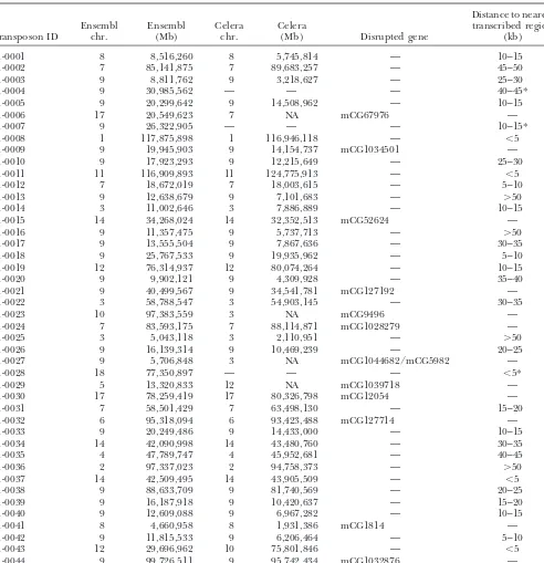
Mapping of transposon insertions using the Ensembl and Celera databases
Distance to nearest
Ensembl Ensembl Celera Celera transcribed region
Transposon ID chr. (Mb) chr. (Mb) Disrupted gene (kb)
01-0001 8 8,516,260 8 5,745,814 — 10–15
01-0002 7 85,141,875 7 89,683,257 — 45–50
01-0003 9 8,811,762 9 3,218,627 — 25–30
01-0004 9 30,985,562 — — — 40–45*
01-0005 9 20,299,642 9 14,508,962 — 10–15
01-0006 17 20,549,623 7 NA mCG67976 —
01-0007 9 26,322,905 — — — 10–15*
01-0008 1 117,875,898 1 116,946,118 — ⬍5
01-0009 9 19,945,903 9 14,154,737 mCG1034501 —
01-0010 9 17,923,293 9 12,215,649 — 25–30
01-0011 11 116,909,893 11 124,775,913 — ⬍5
01-0012 7 18,672,019 7 18,003,615 — 5–10
01-0013 9 12,638,679 9 7,101,683 — ⬎50
01-0014 3 11,002,646 3 7,886,889 — 10–15
01-0015 14 34,268,024 14 32,352,513 mCG52624 —
01-0016 9 11,357,475 9 5,737,713 — ⬎50
01-0017 9 13,555,504 9 7,867,636 — 30–35
01-0018 9 25,767,533 9 19,935,962 — 5–10
01-0019 12 76,314,937 12 80,074,264 — 10–15
01-0020 9 9,902,121 9 4,309,928 — 35–40
01-0021 9 40,499,567 9 34,541,781 mCG127192 —
01-0022 3 58,788,547 3 54,903,145 — 30–35
01-0023 10 97,383,559 3 NA mCG9496 —
01-0024 7 83,593,175 7 88,114,871 mCG1028279 —
01-0025 3 5,043,118 3 2,110,951 — ⬎50
01-0026 9 16,139,314 9 10,469,239 — 20–25
01-0027 9 5,706,848 3 NA mCG1044682/mCG5982 —
01-0028 18 77,350,897 — — — ⬍5*
01-0029 5 13,320,833 12 NA mCG1039718 —
01-0030 17 78,259,419 17 80,326,798 mCG12054 —
01-0031 7 58,501,429 7 63,498,130 — 15–20
01-0032 6 95,318,094 6 93,423,488 mCG127714 —
01-0033 9 20,249,486 9 14,433,000 — 10–15
01-0034 14 42,090,998 14 43,480,760 — 30–35
01-0035 4 47,789,747 4 45,952,681 — 40–45
01-0036 2 97,337,023 2 94,758,373 — ⬎50
01-0037 14 42,509,495 14 43,905,509 — ⬍5
01-0038 9 88,633,709 9 81,740,569 — 20–25
01-0039 9 16,187,918 9 10,420,637 — 15–20
01-0040 9 12,609,088 9 6,967,282 — 10–15
01-0041 8 4,660,958 8 1,931,386 mCG1814 —
01-0042 9 11,815,533 9 6,206,464 — 5–10
01-0043 12 29,696,962 10 75,801,846 — ⬍5
01-0044 9 99,726,511 9 95,742,434 mCG1032876 —
Chromosome and megabase position are shown for both assemblies if available. In addition, distance from the nearest transcribed region and genes disrupted are shown for the Celera assembly. An asterisk indicates a distance based on Ensembl. Updated insertion position information using the NCBI30 version of the mouse genome and recent genomic annotation data sets are available at the Mouse Transposon Insertion Database (http://mouse.ccgb.umn.edu/transposon).
have melting temperatures at least 10⬚higher than those for positive clones was obtained by high-throughput
preparation of DNA followed by sequencing. Sequences used for splinkerette PCR. During the PCR reaction,
these “blocking primers” should anneal to the DNA were then processed to remove remaining transposon
and splinkerette sequences prior to mapping. fragments from the concatomer and block extension of
the splinkerette primers through these regions. Follow- Mapping novel transposon insertion sites:Cloned
Figure 3.—Map position of transposon insertion sites. (A) We obtained 44 novel transposon in-sertions sites and mapped them using Celera’s whole mouse ge-nome assembly. The position of each insertion is shown by a red line with the transposon ID to the right. The map position of known genes is indicated by a black line with the gene name shown to the left. (B) An enlarged view of chro-mosome 9. The position of the concatomer is indicated as deter-mined by FISH analysis.
genome assembly using the BLAST search tool (Alt- each insertion as well as the distance to the nearest
transcribed region according to Celera for those inser-schulet al.1990). The average length of cloned
flank-ing sequence analyzed was ⵑ133 bp, and the average tions not in genes (Table 1). Clones were eliminated
from further analysis if the map position was ambiguous percentage of identity with specific sites in the Celera
assembly wasⵑ99%. Insertions were also later mapped due to the presence of repetitive sequence or if the
clone represented an insertion into an adjacent transpo-using the Ensembl database (http://www.ensembl.org;
Figure 4.—Distribution of transpo-son insertions relative to genes. The dis-tance to the nearest transcription unit was calculated for each insertion as well as for each randomly selected TA dinu-cleotide. These distances were put into groups of 5 Mb. The percentage of inser-tions for each category as well as the frequency of gene hits is shown.
to chromosome 9 where the transposon concatomer is tions relative to transcription units is nearly the same as randomly selected TAs, indicating the randomness of located (Figure 3B). The remaining insertions occurred
on other chromosomes without any obvious preference transposition. Previous work has demonstrated that P
elements preferentially integrate into the 5⬘region of for chromosome or region (Figure 3A).
Of the 19 insertions that mapped to chromosome 9, genes (Spradling et al. 1995). We examined more
closely those insertions that occurred within 20 kb of a 13 are within the interval containing the transposon
concatomer (Figure 1B). These transposition events can known transcribed region. Of the 16 insertions
oc-curring near genes, we determined that 8 of them were be attributed to local transposition in which an excised
transposon tends to integrate near the donor site. How- 5⬘ while the remaining 8 were 3⬘ of the nearest gene (data not shown).
ever, unlike P elements in which local transposition
occurs over a 100-kb interval,Sleeping Beautytransposons We also compared the sequence flanking the TA dinu-cleotide between the transposon insertion and control have a much larger local transposition interval (Tower
et al.1993). TheSleeping Beautylocal transposition inter- groups to detect any differences in nucleotide content. Transposable elements in the Tc1/mariner family re-val appears to be between 5 and 15 Mb, depending on
the exact location of the concatomer within band 9A2-3. quire only a TA dinucleotide for insertion (Plasterk
et al.1999). If this were the sole requirement, we would The cluster of local transposition events detected
oc-curred between theTrrp6gene (3.2 Mb, 1 cM) and the expect to find no differences in the sequence flanking
the TA dinucleotides in either group. We compared 25
Pin1gene (14.5 Mb, 4 cM).
Determination of insertion site preferences:To deter- bp flanking both sides of each transposon integration site and randomly selected TA dinucleotides. An un-mine any bias in transposon insertion sites, we used a
random number generator to select 44 TA dinucleotides pairedt-test was performed to compare the nucleotide
content of flanking sequence from each group. Analysis from the genome. Random TA dinucleotides were noted
if they occurred within a transcribed region. Otherwise, of each individual nucleotide revealed a decrease in the percentage of cytosine in the sequence flanking the the distance to the nearest transcribed region was
deter-mined. The number of hits within known and predicted transposon insertions (P⫽0.0091). We also found that
the transposon insertion sites occurred in regions with transcription units in the control group was 34% (14
of 44) compared to 27% (12 of 44) for the transposon higher AT content (P⫽ 0.015) and lower GC content
(P⫽ 0.015) when compared to the control group.
insertion group (Figure 4). Outside transcribed regions,
there does not appear to be any obvious preference for Although these differences were statistically
signifi-cant, they were slight. We aligned the junction se-SB transposons to insert near or distant from genes. As
Figure 5.—Nucleotide content in regions flanking the site of insertion. A total of 25 bp of sequence flanking each side of the insertion site was analyzed. The percentage of each nucleotide at each position was calculated and graphed for both the (A) transposon insertion group and the (B) randomly selected TA dinucleotide group. (Green) A, (red) T, (black) G, (blue) C.
content could be attributed to a consensus, other than supported by homology to mouse or human cDNA
clones (Figure 6). Eight of the predicted transcripts do the TA dinucleotide, used by the transposase (Figure
5). Although we did not find any consensus nucleotides not contain a complete open reading frame. Thus, other
transposon insertions that mapped close to transcrip-strictly required, other than the TA, we did detect strong
preferences. Most of the insertions had an adenine at tion units may in fact be within them, because unidenti-fied upstream or downstream exons exist.
position⫺3 and a thymine at position⫹3 (82 and 61%,
respectively). Therefore, the SB transposase appears to Germline transmission of transposon insertions:
Sev-eral of the mice harboring novel transposon insertions prefer an expanded consensus of ANNTANNT,
consis-tent with a recent report for SB transposon insertions were bred to demonstrate germline transmission of the
transposons. We performed PCR on wild-type mouse in zeocin-selected HeLa cells (Vigdalet al.2002).
Analysis of transposon insertions in genes:We cloned genomic DNA to amplify sequences flanking transposon insertion sites for use as probes. Southern blotting was 12 transposon insertions that are within 13 genes
ac-cording to the Celera mouse genome assembly (Figure performed on both offspring and parental tail-biopsy
DNA was digested with EcoRV restriction enzyme. We
6). Of these insertions, 6 are in the same orientation
as transcription and would be predicted to disrupt the were able to demonstrate germline transmission in
roughly Mendelian ratios (Figure 7). The rearranged gene. All 12 insertions are within introns and are spread
throughout the length of genes. Twelve of the insertions and wild-type bands corresponded to the predicted sizes on the basis of the sequence obtained from the Celera are within introns flanked by coding exons and 1 is
within the 3⬘untranslated region (mCG1814). Of the database (data not shown).
Figure 6.—Distribution and orientation of transposon insertions in genes. (Top) Twelve transposons are inserted within transcription units of known or predicted genes. The transposon ID is shown next to a diagram of each insertion. The direction of transcription is from left to right for each gene, and exons are represented with black vertical lines. The approximate location of each transposon is also indicated. The arrow represents the orientation of the mutagenic cassette within the transposon. (Bottom) Additional information for each gene is listed.
tagged mutagenesis to be successful in the mouse, gene- and real-time quantitative RT-PCR. Insertion 01-0032
lies within the mCG127714 transcription unit as anno-trapping elements within the transposon must be
capa-ble of producing a mutant transcript upon gene inser- tated by the Celera whole mouse genome assembly
(Fig-ure 6). Predicted exons of this locus were analyzed with tion. The gene trap used here is designed to truncate
endogenous transcripts via splicing and polyadenyla- the NCBI BLAST search tool for identity to
character-ized ESTs with known expression patterns to determine tion. If the vector functions as designed, then splicing
from an upstream exon of the disrupted gene will join appropriate tissues for transcript analysis. The predicted mCG127714 exons showed significant identity to ESTs it to the splice acceptor within the transposon vector.
The vector includes stop codons in all three frames and isolated from brain, intestine, liver, lung, spleen, testes, and thymus.
a polyadenylation signal (Figure 8A). The downstream
portion of the transposon is a poly(A) trap that is pre- RT-PCR was employed to assess the efficiency of both
the upstream splice acceptor and the downstream splice dicted to express GFP when it is provided with a poly(A)
signal from an endogenous gene via splicing to a down- donor within the transposon vector. Primers were
de-signed for the predicted upstream and downstream ex-stream exon. The GFP gene is driven by the ubiquitous
ROSA26 promoter (Kissenberthet al.1999), followed ons as well as for sequences predicted to be transcribed
within the gene-trap vector (Figure 8A). RT-PCR was by a splice donor from the HPRT gene. To test the
splicing efficiency of intron-inserted gene-trap transpo- performed on total RNA of heterozygous mice (Figure
this particular transcription unit actually results in re-duction of the amount of wild-type transcript produced, real-time quantitative RT-PCR was performed on liver and spleen RNA from heterozygous carrier and wild-type mice. Primers specific for exons immediately flank-ing the intron into which the transposon inserted were used to amplify a wild-type cDNA. These primers are incapable of amplifying a product from the mutant tran-script cDNA due to its premature truncation. Trantran-script levels of the gene in question were compared toGapdh as a reference and revealed that wild-type transcript levels were reduced by half within the liver of carrier mice relative to wild type (Figure 8E). Levels were also decreased in the spleen to a lesser extent. These results prove that transposon gene traps can decrease the amount of wild-type transcript produced when inserted into an intron.
Figure 7.—Southern blotting of parental and offspring
Mouse phenotype analysis: Finally, we attempted to DNA. Several mice harboring mapped insertion sites were
bred to wild-type mice, and offspring were genotyped by South- generate mice homozygous for several insertions to as-ern blotting using probes specific for the sequence flanking sess any resultant phenotypes. Heterozygous carrier mice one side of each insertion. Southern blotting results are shown were intercrossed and offspring were genotyped by for three mice. In each case, parental DNA is shown at the
three-primer PCR (Dupuy et al. 2002). Figure 9A dis-left with the offspring DNA in the adjacent lanes. The
wild-plays the genotypic results of intercrosses for several type allele is indicated by “WT” and the transposon allele is
indicated by “T.” The transposon ID corresponding to each different transposon insertions within predicted genes.
probe is also indicated. The lack of mice homozygous for insertion 01-0032 is
statistically significant (P⫽0.011), indicating an embry-onic lethal phenotype presumably caused by the loss
stream splicing of the gene-trap vector with the endoge- of the disrupted gene’s product. We performed timed
nous gene with no detectable product in the wild-type pregnancies for the intercross of mice heterozygous for
controls. Cloning and sequencing of RT-PCR products insertion 01-0032 to further define the phenotype. We
revealed the expected sequence resulting from the were able to isolate embryos at E8.5 that were
homozy-given splicing reactions (Figure 8C), including the pres- gous for the insertion and Figure 9B shows the appearance
ence of stop codons in all three reading frames from of these embryos compared to wild-type and heterozygous
within the upstream chimeric transcript. Thus, the gene- littermates. Although gastrulation and somitogenesis
ap-trap transposon is capable of splicing with endogenous pears to have initiated, mutant embryos are growth
re-genes to create chimeric transcripts. tarded, underdeveloped, and starting to recess. The
Since RT-PCR has extraordinary sensitivity, the fre- gene mutated by insertion 01-0032 is a mitochondrial
quency of transcript mutation needed to be demon- carrier protein-related gene with the predicted amino
strated by other means. Thus, a Northern blot was per- acid sequence indicated in Figure 9C. This predicted
formed on total liver and spleen RNA from wild-type gene has not been fully characterized, but the predicted
mice and heterozygous mice carrying insertion 01-0032 protein contains sequence motifs and a tripartite
struc-to determine whether the mutant transcript occurs at ture characteristic of the mitochondrial carrier protein
a significant frequency relative to the wild-type tran- family (Palmieri 1994). Similar to insertion 01-0032,
script. A probe composed of predicted exon sequences we have been unable to obtain mice homozygous for
upstream of the gene-trap insertion was produced by insertion 01-0009, suggesting that it also causes a
reces-RT-PCR and thus is predicted to hybridize with both sive embryonic lethal mutant phenotype. In contrast,
the wild-type and the predicted truncated transcript. homozygous mice for several insertions within and
out-Figure 8D shows the Northern blot, which reveals the side of genes have been generated. Notably, these
inser-presence of a novel, smaller transcript within the liver tions are in an antisense orientation with respect to the
and spleen RNA of the heterozygous carrier mouse, but disrupted gene. The insertions noted in Figure 9A that
which is absent in the wild type. This indicates that produce viable homozygotes show no significant
splic-Figure 8.—RNA analysis of gene disruption. Insertion 01-0032 was assessed for the ability of the transposon gene trap to function as designed for putative transcriptional disruption of the gene in question. (A) Primers (indicated on figure) were designed to assess splicing of the upstream exon with the splice acceptor within the transposon. Likewise, splicing of the splice donor within the transposon with the downstream endogenous exon was examined using the same assay with appropriate primers. (B) Total RNA was extracted from appropriate tissues of wild-type mice and mice heterozygous for insertion 01-0032 and subjected to RT-PCR. (C) RT-PCR products were cloned and sequenced to demonstrate the expected spliced products as shown. (D) Northern blot analysis of total RNA from wild type and mice heterozygous for insertion 01-0032 was also performed with a probe specific for exons upstream of the gene-trap insertion, which should recognize the wild-type full-length transcript and the mutant truncated transcript. (E) Real-time quantitative RT-PCR was performed with primers that amplify across the intron disrupted by the gene-trap transposon insertion on RNA from wild-type and heterozygous tissues; an average of two experiments is shown.
of these partially suppress gene expression. We can con- that gene-trap transposons are capable of mutating
dis-clude from insertion 01-0032 that SB transposon gene rupted endogenous genes upon intronic insertion,
re-traps can be very efficient insertional mutagens yielding ducing wild-type transcript levels, and producing
mu-novel phenotypes. tant phenotypes. These data suggest thatSleeping Beauty
will be useful as a random germline insertional mutagen in mice.
DISCUSSION It is apparent that the SB transposase displays a local
transposition tendency similar toP-element transposase The insertion site preferences for several commonly
(Toweret al.1993). We estimate the local transposition
used transposable elements have been examined (van
interval for SB transposase to be between 5 and 15 Mb LuenenandPlasterk 1994;Liao et al.2000). These
compared to 100 kb for theP-element transposase. Al-studies have indicated that there are sequences outside
thoughⵑ43% of our insertions mapped to the donor
the consensus target site that are preferred by the
trans-chromosome, one-third of our total mapped insertions posase. We have cloned and mapped 44Sleeping Beauty
are within our estimated “local hopping” interval. The transposon insertions from a panel of 30 mice to
deter-reported frequency of local transposition using the Sleep-mine if the SB transposase displays any insertion site
ing Beauty transposon system has varied between 50% preferences. Generally, we are unable to identify any
(Luoet al.1998;Dupuyet al.2001) and 83% (Fischer insertion site preference that would severely restrict the
et al.2001). It is not clear whether these differences are number of potential genomic integration sites for
Thus, it would appear that we were equally successful in cloning local transposon insertions in both groups and that no bias exists.
Analysis of the sequence flanking each insertion site did reveal a tendency for SB transposase to select TA dinucleotides that occurred within AT-rich regions (Fig-ure 5). Although this difference was statistically signifi-cant, the AT content flanking the transposon insertion sites was only 10% higher than that of the sequence flanking randomly selected TA dinucleotides. The SB transposase also appears to prefer the consensus AN-NTANNT. In this regard, SB transposase seems to be more similar to Tc1 than to Tc3 in its insertion site
preference (van Luenen and Plasterk 1994).
How-ever, the AT content of the sequence flanking transpo-son insertion sites is still significantly higher even if the
⫺3 and ⫹3 positions are excluded from the analysis
(P ⫽ 0.021). Therefore the preferred consensus site
does not entirely account for the increase in AT content in the sequence around transposon insertion sites. AT richness and sequence preferences at ⫺3 and ⫹3 are both preserved even when insertions mapped to chro-mosome 9 are excluded from the data set (data not shown). This indicates that our results are not biased by any altered sequence content in the 9A2-3 region of Figure9.—Genotype and phenotype analysis of insertions.
the mouse genome. Other published transposon inser-(A) Male and female mice heterozygous for given insertions
were bred in attempts to generate homozygous mice for each tion junction sequences tend to support our observa-gene insertion; genotype results from such breedings are indi- tions of the preferred consensus site (data not shown; cated. (B) Since no viable homozygous mice could be
pro-Ivicset al.1997;Luoet al.1998;Yantet al.2000;Fischer duced for insertion 01-0032, timed pregnancies were
per-et al.2001;Horieet al.2001;Dupuyet al.2002). Recent formed to isolate homozygous embryos at E8.5. Pictures of
data from cloned sequences flanking SB-mediated trans-the embryos (not to scale) and three-primer genotyping PCR
results are represented (T, transposon allele;⫹, wild-type al- position events in HeLa cells selected in zeocin show a lele). (C) The predicted amino acid sequence of the predicted strong tendency of transposons to insert within an AT-gene disrupted by insertion 01-0032 is shown; amino acid
repeat: ATATATAT with the center TA as the site of residues that are underlined and in boldface type indicate
insertion (Vigdal et al. 2002). The data presented
sequence motifs characteristic of the mitochondrial carrier
herein are consistent with this trend, but the TA appears protein family.
to be the only sequence absolutely necessary for transpo-son insertion in both data sets. Additional work will be required to determine if this insertion tendency will locus) may affect this rate. However, in this work the
amplification of novel insertion sites in mice or cells with significantly reduce the mutagenicity of the Sleeping Beautytransposon system. However, our data reveal that a donor locus consisting of a multicopy concatomer of
transposons, rather than of single-copy elements as inLuo SB transposons can insert into a variety of genes in regions all over the genome.
et al.(1998) andFischeret al. (2001), proved to be difficult
and necessitated the use of blocking primers to inhibit The transposon insertion sites do not appear to differ notably from randomly selected TA dinucleotides in amplification of transgene vector sequences. The
effi-ciency of these blocking primers is unknown and thus their position relative to transcribed sequence. We did
not expect to see a significant difference in the number amplification of novel insertions linked to the donor
locus could have been compromised in these mice by of SB insertions within genesvs.random TAs when we
compared the two groups, and this was verified by our the presence of the concatomer, biasing toward a lower
observed incidence of local transposition. Despite this data. It is important to note that transcribed regions are most likely underrepresented in the Celera mouse potential bias, 45% (13/29) of the insertion sites cloned
from mice that retained the transposon conatomer were genome assembly. Many predicted gene transcripts lack
trans-the transposon insertions that occurred near genes may mutateⵑ30 genes per gamete in treated males (Justice et al.2000), we will need to produce between 135 and have actually occurred within the transcription unit. In
fact, two of the insertions not mapped within genes (01- 270 insertions per gamete. This number accounts for
roughly one-third of the insertions in transcription units 0001 and 01-0020) appear to be within introns of specific
EST clones (data not shown). It is thus evident that (30⫻ 3⫽ 90), subtracts all local transposition events
(90⫻1.5⫽135), and can be calculated for a gene trap informatics issues confuse the number of gene
inser-tions detected in our analysis. The gene inserinser-tions noted that functions in both orientations (135) or in only one orientation (135⫻2⫽270). Of course, rapid mutant here are an estimate of the total number of transcription
units actually disrupted by transposons in this screen. gene identification in the downstream part of any screen compensates for the reduced mutagenicity of SB trans-All 44 germline transposon insertions were reanalyzed
using a variety of gene-calling algorithms. As mentioned, poson mobilization. Nevertheless, we are attempting to improve transposition frequency 10-fold using improved we have mapped the transposon insertions using the
public version of the mouse genome assembly (MGSC transposase and transposon transgenes. Like ENU, our
preliminary data suggest that SB transposon insertions V3) using our insertion mapping and annotation
pipe-line (IMAP), which automatically maps insertion sites will be found to cause hypomorphic alleles in some
cases. Unlike ENU, SB transposon vectors could be engi-(Roberg-Perezet al.2003). Using IMAP we have been
able to map all 44 transposons to specific chromosomal neered to express useful reporter molecules such as
GFP, -galactosidase, or the Cre recombinase in the and nucleotide positions on the public version of the
mouse genome (Table 1). Of the 37 insertions that were temporal and spatial pattern of the disrupted
endoge-nous gene. successfully mapped to a chromosome and nucleotide
position with the Celera system, all but one is mapped It should be possible to utilize the local transposition phenomenon that we observed to focus transposon mu-to the same chromosome using IMAP. Furthermore,
the nucleotide positions assigned for these insertions tagenesis into defined regions of the genome of high
biological interest. Used in this way, saturation mutagen-never differed⬎8 Mb between IMAP and Celera
assign-ments with an average difference of 4 Mb (K.Roberg- esis could be performed in a 5- to 15-Mb region
sur-rounding a transposon concatomer array. In the mouse Perez, unpublished data). Consistency between the
assemblies is further indicated by the colinearity of as- this corresponds toⵑ2–7 cM. In these experiments, we observed two new insertions per gamete with approxi-signed insertion positions, with the exception of two
adjacent insertions in the chromosome 9 cluster. These mately one-third of those attributed to local transposi-tion. Thus, it will be feasible to achieve a 1⫻coverage results confirm and extend our initial mapping work
using the Celera mouse genome assembly. of a 10-Mb region, with transposon insertions every 20
kb, in as few as 750 mice. In addition, mobilization of Furthermore, it is clear that the gene trap and poly(A)
trap of the transposon function as predicted to effec- transposons within the germline of mice could be
uti-lized for chromosome engineering, mobilizing border tively disrupt endogenous wild-type gene expression.
Stop codons within all three frames of the gene-trap elements, and to further our understanding of gene
clusters. transposon spliced into the upstream exons of an
endog-enous gene are predicted to cause a truncated protein We thank the University of Minnesota Mouse Genetics Laboratory product to be generated upon translation. The resultant for their assistance and Steve Buganski for his mouse husbandry. We also thank Dr. William Shawlot for his input and assistance with the
mutant transcript should demonstrate expression
pat-E8.5 embryos and Craig Eckfeldt for his guidance with quantitative
terns identical to the full-length endogenous transcript,
real-time PCR. This work was supported by the Arnold and Mabel
but when translated may lack the function of the wild- Beckman Foundation and the National Institutes of Health (NIDA
type protein product of the gene. We have demon- R01DA14764).
strated the ability of our transposon gene trap to effi-ciently mutate a gene upon intronic insertion, eliciting
a mutant phenotype, and are currently assessing the LITERATURE CITED
remaining insertions for their effects at the sequence
Altschul, S. F., W. Gish, W. Miller, E. W. MyersandD. J. Lipman,
and phenotype level. 1990 Basic local alignment search tool. J. Mol. Biol.215:403–
410.
Taken together, these results suggest that randomin
Clamp, M., D. Andrews, D. Barker, P. Bevan, G. Cameronet al.,
vivogermline transposon-tagged mutagenesis is a
feasi-2003 Ensembl 2002: accommodating comparative genomics.
ble approach to functional genomics in the mouse. Nucleic Acids Res.31:38–42.
Dupuy, A. J., S. FritzandD. A. Largaespada, 2001 Transposition
Given the transposition frequency we have obtained in
and gene disruption in the male germline of the mouse. Genesis
the mouse male germline (Dupuy et al. 2001), along
30:82–88.
with the frequency of gene insertion predicted here, Dupuy, A. J., K. Clark, C. M. Carlson, S. Fritz, A. E. Davidsonet al., 2002 Mammalian germ-line transgenesis by transposition.
theSleeping Beautysystem does not appear to be efficient
Proc. Natl. Acad. Sci. USA99:4495–4499.
enough to perform a genome-wide mutagenesis screen
Eide, D., andP. Anderson, 1985 Transposition of Tc1 in the
nema-without a substantial increase in transposition fre- tode Caenorhabditis elegans. Proc. Natl. Acad. Sci. USA82:1756–
1760.
743–750.
48–54.
Hartl, D., 2001 Discovery of the transposable element mariner.
Pelicic, V., S. Morelle, D. LampeandX. Nassif, 2000 Mutagenesis
Genetics157:471–476.
of Neisseria meningitidis by in vitro transposition of Himar1
Heng, H. H., andL. C. Tsui, 1993 Modes of DAPI banding and
mariner. J. Bacteriol.182:5391–5398. simultaneous in situ hybridization. Chromosoma102:325–332.
Plasterk, R. H., Z. IzsvakandZ. Ivics, 1999 Resident aliens: the
Heng, H. H., J. SquireandL. C. Tsui, 1992 High-resolution
map-Tc1/mariner superfamily of transposable elements. Trends ping of mammalian genes by in situ hybridization to free
chroma-Genet.15:326–332. tin. Proc. Natl. Acad. Sci. USA89:9509–9513.
Roberg-Perez, K., C. M. CarlsonandD. A. Largaespada, 2003
Horie, K., A. Kuroiwa, M. Ikawa, M. Okabe, G. Kondoh et al.,
MTID: a database ofSleeping Beautytransposon insertions in mice. 2001 Efficient chromosomal transposition of a
Tc1/mariner-Nucleic Acids Res.31:78–81. like transposon Sleeping Beauty in mice. Proc. Natl. Acad. Sci.
Schaffer, A. A., L. Aravind, T. L. Madden, S. Shavirin, J. L. Spouge
USA98:9191–9196. et al., 2001 Improving PSI-BLAST protein database search
sensi-Hrabe de Angelis, M., H. Flaswinkel, H. Fuchs, B. Rathkolb, D. tivity with composition-based statistics and other refinements.
Soewartoet al., 2000 Genome-wide, large-scale production of Nucleic Acids Res.29:2994–3005.
mutant mice by ENU mutagenesis. Nat. Genet.25:444–447. Skarnes, W. C., B. A. AuerbachandA. L. Joyner, 1992 A gene
Hubbard, T., D. Barker, E. Birney, G. Cameron, Y. Chenet al., trap approach in mouse embryonic stem cells: the lacZ reporter
2002 The Ensembl genome database project. Nucleic Acids Res. is activated by splicing, reflects endogenous gene expression, and
30:38–41. is mutagenic in mice. Genes Dev.6:903–918.
Ivics, Z., P. B. Hackett, R. H. PlasterkandZ. Izsvak, 1997 Molec- Spradling, A. C., D. M. Stern, I. Kiss, J. Roote, T. Laverty et
ular reconstruction of Sleeping Beauty, a Tc1-like transposon al., 1995 Gene disruptions using P transposable elements: an from fish, and its transposition in human cells. Cell91:501–510. integral component of the Drosophila genome project. Proc.
Izsvak, Z., Z. IvicsandR. H. Plasterk, 2000 Sleeping Beauty, a Natl. Acad. Sci. USA92:10824–10830.
wide host-range transposon vector for genetic transformation in Stajich, J. E., D. Block, K. Boulez, S. E. Brenner, S. A. Chervitzet
vertebrates. J. Mol. Biol.302:93–102. al., 2002 The Bioperl toolkit: Perl modules for the life sciences.
Jenkins, N. A., N. G. Copeland, B. A. Taylor, H. G. Bedigianand Genome Res.12:1611–1618.
Tower, J., G. H. Karpen, N. CraigandA. C. Spradling, 1993
Pref-B. K. Lee, 1982 Ecotropic murine leukemia virus DNA content
erential transposition of DrosophilaPelements to nearby chro-of normal and lymphomatous tissues chro-of BXH-2 recombinant
in-mosomal sites. Genetics133:347–359. bred mice. J. Virol.42:379–388.
Townley, D. J., B. J. Avery, B. Rosen and W. C. Skarnes,
Justice, M. J., D. A. Carpenter, J. Favor, A. Neuhauser-Klaus, M.
1997 Rapid sequence analysis of gene trap integrations to
gener-Hrabe de Angeliset al., 2000 Effects of ENU dosage on mouse
ate a resource of insertional mutations in mice. Genome Res.7:
strains. Mamm. Genome11:484–488.
293–298.
Kissenberth, W. C., N. T. Brettingen, J. K. LohseandE. P.
Sand-van Luenen, H. G., andR. H. Plasterk, 1994 Target site choice of
gren, 1999 Ubiquitous expression of marker transgenes in mice
the related transposable elements Tc1 and Tc3 of Caenorhabditis and rats. Dev. Biol.214:128–138.
elegans. Nucleic Acids Res.22:262–269.
Liao, G. C., E. J. RehmandG. M. Rubin, 2000 Insertion site
prefer-van Luenen, H. G., S. D. CollomsandR. H. Plasterk, 1993
Mobili-ences of the P transposable element in Drosophila melanogaster.
zation of quiet, endogenous Tc3 transposons of Caenorhabditis Proc. Natl. Acad. Sci. USA97:3347–3351.
elegans by forced expression of Tc3 transposase. EMBO J.12:
Luo, G., Z. Ivics, Z. IzsvakandA. Bradley, 1998 Chromosomal
2513–2520. transposition of a Tc1/mariner-like element in mouse embryonic
Vigdal, T. J., C. D. Kaufman, Z. Izsva´k, D. F. VoytasandZ. Ivics,
stem cells. Proc. Natl. Acad. Sci. USA95:10769–10773.
2002 Common physical properties of DNA affecting target site
Medhora, M., K. Maruyama and D. L. Hartl, 1991 Molecular selection ofSleeping Beautyand other Tc1/marinertransposable
and functional analysis of the mariner mutator element Mos1 in elements. J. Mol. Biol.323:441–452.
Drosophila. Genetics128:311–318. Yant, S. R., L. Meuse, W. Chiu, Z. Ivics, Z. Izsvaket al., 2000
So-Niwa, H., K. YamamuraandJ. Miyazaki, 1991 Efficient selection matic integration and long-term transgene expression in normal
for high-expression transfectants with a novel eukaryotic vector. and haemophilic mice using a DNA transposon system. Nat. Gene108:193–200. Genet.25:35–41.
Nolan, P. M., J. Peters, M. Strivens, D. Rogers, J. Haganet al.,