Electronic Thesis and Dissertation Repository
4-17-2012 12:00 AM
Structural Characterization of Protein Folding Intermediates by
Structural Characterization of Protein Folding Intermediates by
Oxidative Labeling and Mass Spectrometry
Oxidative Labeling and Mass Spectrometry
Bradley B. Stocks
The University of Western Ontario
Supervisor Lars Konermann
The University of Western Ontario Graduate Program in Biochemistry
A thesis submitted in partial fulfillment of the requirements for the degree in Doctor of Philosophy
© Bradley B. Stocks 2012
Follow this and additional works at: https://ir.lib.uwo.ca/etd
Part of the Biochemistry, Biophysics, and Structural Biology Commons
Recommended Citation Recommended Citation
Stocks, Bradley B., "Structural Characterization of Protein Folding Intermediates by Oxidative Labeling and Mass Spectrometry" (2012). Electronic Thesis and Dissertation Repository. 430.
https://ir.lib.uwo.ca/etd/430
This Dissertation/Thesis is brought to you for free and open access by Scholarship@Western. It has been accepted for inclusion in Electronic Thesis and Dissertation Repository by an authorized administrator of
(Spine title: Protein Folding Studied by Oxidative Labeling and ESI-MS)
(Thesis format: Integrated-Article)
by
Bradley B. Stocks
Graduate Program in Biochemistry
A thesis submitted in partial fulfillment of the requirements for the degree of
Doctor of Philosophy
Department of Biochemistry The University of Western Ontario
London, Ontario, Canada
ii
CERTIFICATE OF EXAMINATION
Supervisor
______________________________ Dr. Lars Konermann
Supervisory Committee
______________________________
______________________________
Examiners
______________________________ Dr. James Choy
______________________________ Dr. Gilles Lajoie
______________________________ Dr. Mark Bernards
______________________________ Dr. Gerold Schmitt-Ulms
The thesis by
Bradley B. Stocks
entitled:
Structural Characterization of Protein Folding Intermediates by
Oxidative Labeling and Mass Spectrometry
is accepted in partial fulfillment of the requirements for the degree of
Doctor of Philosophy
iii
short-lived intermediates that become populated en route to the native state. In this work,
a covalent labeling method was developed that provides insights into the structures of
these transient species. Hydroxyl radical (·OH) reacts with solvent-exposed side chains,
whereas buried residues are protected. Mass spectrometry is used for monitoring the
locations and the extent of labeling. Pulsed ·OH labeling of proteins at selected time
points during folding results in high temporal and spatial resolution when compared to
existing other labeling methods.
This novel technique was validated by studying the kinetic unfolding and
refolding of holomyoglobin (hMb) and cytochrome c (cyt c), respectively. The
noncovalent prosthetic heme group in hMb was shown to drastically affect the unfolding
pathway. Cyt c refolding was found to fold in a stepwise manner. The population of a
misfolded cyt c intermediate was also detected. Results in both cases were in accord with
published data.
Many cellular proteins exist as oligomers. Pulsed ·OH labeling method was
therefore extended to monitor the folding and assembly of a 22 kDa homodimeric
protein, S100A11. Prior to this study very little information regarding the folding
mechanism of this protein was available. ·OH labeling reveals that disruption of the
native dimer is followed by the formation of non-native hydrophobic contacts within the
denatured monomers. The folding/binding pathway was shown to progress through
iv
inhibitor, α1-antitrypsin, was characterized and compared with complementary data from
hydrogen/deuterium exchange studies. Our results show that the formation of early
tertiary contacts and specific hydrogen bonds guide the protein towards its active,
metastable structure. Structural correlation is also seen between a late kinetic species and
a previously characterized equilibrium intermediate of a pathogenic mutant.
Overall, the results presented highlight the ability of the technique developed in
this work to provide in-depth information about the mechanisms of protein folding.
Keywords: protein folding | covalent labeling | kinetics | hydroxyl radical | folding
intermediates | rapid mixing | electrospray mass spectrometry | cytochrome c | S100A11 |
v
The work in Chapters 2, 3 and 4 were published in the following articles respectively:
Bradley B. Stocks and Lars Konermann (2009). Structural Characterization of Short-Lived Protein Unfolding Intermediates by Laser-Induced Oxidative Labeling and Mass Spectrometry. Anal. Chem. 81: 20-27. Reproduced with permission. © 2009 American Chemical Society
Bradley B. Stocks and Lars Konermann (2010). Time-Dependent Changes in Side Chain Solvent Accessibility during Cytochrome c Folding Probed By Pulsed Oxidative Labeling and Mass Spectrometry. J. Mol. Biol. 398: 362-373. Reproduced with permission. © 2010 Elsevier Ltd.
Bradley B. Stocks, Atoosa Rezvanpour, Gary S. Shaw, and Lars Konermann (2011). Temporal Development of Protein Structure during S100A11 Folding and Dimerization Probed by Oxidative Labeling and Mass Spectrometry. J. Mol. Biol.
in press. Reproduced with permission. © 2011 Elsevier Ltd.
The work in Chapter 5 has been incorporated into the following article:
Bradley B. Stocks, Patrick L. Wintrode, and Lars Konermann (2012). Folding Mechanism of α1-Antitrypsin to its Metastable State: Insights from Pulsed Oxidative Labeling and Mass Spectrometry. In preparation.
vi
Not knowing where you‟re going is the best way to get someplace you‟ve never been
vii
Konermann. He has somehow taken my interest in science and transformed it into a passion for research. And he has done so all the while making it seem as though it were my own idea. He has provided me with outstanding leadership while encouraging me to think freely. Returning my lab keys will not be an easy task.
Thank you to my departmental committee members Drs. James Choy and Stan Dunn. They have provided much useful feedback regarding my research over the past 5 years. Their genuine interest in my work and progress has been greatly appreciated.
I would not have been successful without the help of a cast of truly outstanding labmates. Brian, Mark, Peter, Yuhong, and Xin were there to help when I first joined the lab. The second wave included Elias, Jingxi, Jenna, and Lucy. Significant thanks goes to Siavash for the countless hours of conversations regarding oxidative labeling, ion mobility, and Persian culture. I leave the lab in good hands.
viii
Certificate of Examination ... ii
Abstract ... iii
Statement of Co-Authorship ... v
Epigraph ... vi
Acknowledgments ... vii
Table of Contents ... viii
List of Figures ... xii
List of Appendices ... xiv
List of Symbols and Abbreviations ... xv
Chapter 1 – Introduction ... 1
1.1 Protein Structure and Folding ... 1
1.1.1 Native Protein Structure ... 2
1.1.2 Protein Folding ... 5
1.1.3 Protein Folding Mechanisms ... 7
1.1.4 Folding Intermediates ... 10
1.1.5 Misfolding ... 11
1.1.6 Structure Prediction and De Novo Protein Design ... 11
1.2 Studying Protein Folding ... 12
1.2.1 Optical Methods ... 13
1.2.2 Nuclear Magnetic Resonance Spectroscopy ... 14
1.2.3 Computer Simulations ... 15
1.3 Mass Spectrometry ... 16
1.3.1 Ionization Techniques ... 16
1.3.1.1 Matrix-Assisted Laser Desorption/Ionization... 16
1.3.1.2 Electrospray Ionization ... 17
1.3.2 Mass Analyzers ... 19
ix
1.4 Protein Labeling ... 23
1.4.1 Hydrogen/Deuterium Exchange ... 23
1.4.2 Covalent Labeling ... 25
1.4.2.1 Specific Labeling ... 26
1.4.2.2 Non-Specific Labeling ... 29
1.4.3 Hydroxyl Radical Labeling... 29
1.4.3.1 Radiolysis of Water ... 31
1.4.3.2 Photolysis of Hydrogen Peroxide ... 33
1.5 Scope of Thesis ... 36
1.6 References ... 37
Chapter 2 - Structural Characterization of Short-Lived Protein Unfolding Intermediates by Laser-Induced Oxidative Labeling and Mass Spectrometry ... 53
2.1 Introduction ... 53
2.2 Experimental ... 56
2.2.1 Materials... 56
2.2.2 Optical Spectroscopy ... 56
2.2.3 Continuous-Flow Mixing and Radical Labeling ... 57
2.2.4 LC/ESI-MS and Data Analysis ... 61
2.3 Results and Discussion ... 67
2.3.1 Kinetics of Myoglobin Unfolding ... 67
2.3.2 Laser-Induced Oxidative Labeling ... 69
2.3.3 Peptide Mapping ... 71
2.3.4 Kinetic Unfolding Mechanism of hMb ... 75
2.4 Conclusions ... 79
2.5 References ... 80
Chapter 3 - Time-Dependent Changes in Side Chain Solvent Accessibility During Cytochrome c Folding Probed By Pulsed Oxidative Labeling and Mass Spectrometry ... 86
x
3.2.2 Optical Spectroscopy ... 90
3.2.3 Continuous-Flow Mixing and Oxidative Labeling ... 90
3.2.4 LC/ESI-MS ... 92
3.2.5 Fraction Unmodified and Background Correction ... 93
3.2.6 Data Analysis... 93
3.3 Results and Discussion ... 95
3.3.1 Background Oxidation... 95
3.3.2 Stopped-Flow Kinetics... 98
3.3.3 Laser-Induced Oxidative Labeling ... 100
3.3.4 Peptide Mapping and Tandem Mass Spectrometry ... 100
3.3.5 Relative Solvent Accessibilities ... 105
3.3.6 Conformational Changes during Folding ... 108
3.4 Conclusions ... 112
3.5 References ... 114
Chapter 4 - Temporal Development of Protein Structure During S100A11 Folding and Dimerization Probed by Oxidative Labeling and Mass Spectrometry... 122
4.1 Introduction ... 122
4.2 Materials and Methods ... 127
4.2.1 Materials... 127
4.2.2 Continuous-Flow Mixing and Oxidative Labeling ... 128
4.2.3 LC/ESI-MS ... 130
4.3.4 Data Analysis... 131
4.3 Results and Discussion ... 134
4.3.1 Pulsed Oxidative Labeling ... 134
4.3.2 Peptide Mapping and Oxidation Site Determination ... 136
4.3.3 Denatured Monomeric State ... 138
4.3.4 Time-Dependent Peptide Solvent Accessibilities ... 139
4.3.5 Folding and Assembly of S100A11 ... 141
4.4 Conclusions ... 144
xi
5.1 Introduction ... 152
5.2 Materials and Methods ... 156
5.2.1 Materials... 156
5.2.2 Continuous-flow Mixing and Oxidative Labeling ... 156
5.2.3 LC/ESI-MS ... 159
5.2.4 Data Analysis... 160
5.3 Results and Discussion ... 161
5.3.1 Peptide Mapping and Oxidation Site Determination ... 161
5.2.2 Time-Dependent Peptide Solvent Accessibilities ... 166
5.2.3 The Denatured State ... 168
5.2.4 α1AT Folding Mechanism ... 168
5.2.5 Implications for α1AT Aggregation ... 172
5.4 Conclusions ... 175
5.5 References ... 177
Chapter 6 – Conclusions ... 181
6.1 Summary ... 181
6.2 Future Work ... 184
6.2.1 Sub-millisecond Folding Studies ... 184
6.2.2 Comparison of Native and “Diseased” Protein Folding ... 185
6.2.3 Chaperone-mediated Folding ... 186
6.2.4 Co-translational Protein Folding ... 187
6.3 References ... 188
Appendix I – Permissions ... 192
xii
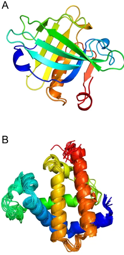
Figure 1.1. 3D protein structures. ... 6
Figure 1.2. Protein folding landscapes. ... 8
Figure 1.3. ESI Process. ... 18
Figure 1.4. Schematic of a Q-TOF... 22
Figure 1.5. Covalent labeling. ... 27
Figure 1.6. Photolysis experimental setup. ... 34
Chapter 2 Figure 2.1. Continuous-flow rapid mixing setup ... 58
Figure 2.2. pH independence of peptide labeling ... 60
Figure 2.3. ESI mass distributions of myoglobin control samples ... 63
Figure 2.4. Myoglobin folding studied by absorption spectroscopy ... 68
Figure 2.5. ESI mass distributions of oxidized myoglobin ... 70
Figure 2.6. Amino acid sequence of horse myoglobin ... 73
Figure 2.7. Relative ESI intensities of unoxidized myoglobin tryptic peptides ... 74
Figure 2.8. Normalized oxidation levels of myoglobin peptides ... 76
Figure 2.9. Crystal structure of native myoglobin ... 77
Chapter 3 Figure 3.1. ESI mass distributions of cytochrome c control samples ... 96
Figure 3.2. Cytochrome c folding studied by fluorescence spectroscopy ... 99
Figure 3.3. ESI mass distributions of oxidized cytochrome c ... 101
Figure 3.4. Amino acid sequence of horse cytochrome c ... 103
Figure 3.5. MS/MS spectra of selected oxidized cytochrome c peptides ... 104
xiii
Figure 4.1. Structure and sequence of S100A11... 126
Figure 4.2. ESI mass distributions of oxidized S100A11 ... 135
Figure 4.3. MS/MS spectrum of an oxidized S100A11 peptide ... 137
Figure 4.4. Normalized oxidation levels of S100A11 peptides ... 140
Figure 4.5. Structural changes of S100A11 during folding and dimerization ... 142
Chapter 5 Figure 5.1. Crystal structure of α1-antitrypsin in the metastable state ... 154
Figure 5.2. Amino acid sequence of α1-antitrypsin ... 163
Figure 5.3. MS/MS spectra of selected oxidized α1-antitrypsin peptides ... 164
Figure 5.4. Amino acid side chains of α1-antitrypsin determined to be oxidized ... 165
Figure 5.5. Normalized oxidation levels of α1-antitrypsin peptides ... 167
Figure 5.6. Structural changes during α1-antitrypsin folding ... 169
xv ∙OH – hydroxyl radical
A695 – absorbance at 695 nm
aMb – apomyoglobin
ANS – 1-anilino-8-naphthalene sulfonate
Atot– total peak area
Au – area of unmodified peak
CD – circular dichroism
CIDNP – chemically induced dynamic nuclear polarization
CRM – charged residue model
cyt c – cytochrome c
Da – Dalton
DI – dimeric protein folding intermediate
DN – native protein dimer
ESI – Electrospray Ionization
FRET - Fӧrster resonance energy transfer
Fu – fraction unmodified
fwhm – full width, half-maximum
GdnHCl – guanidinium hydrochloride
H2O2 – hydrogen peroxide
HDX – hydrogen/deuterium exchange
hMb – holomyoglobin
xvi MD – molecular dynamics
MF – folded protein monomer
MI – monomeric protein folding intermediate
MS – Mass Spectrometry
MS/MS – tandem mass spectrometry
MU – unfolded protein monomer
NMR – Nuclear Magnetic Resonance
NOL – normalized oxidation level
R – unoxidized peptide relative signal
Serpin – serine protease inhibitor
UPLC – ultra performance liquid chromatography
UV-Vis – ultraviolet-visible
Xaa – variable amino acid
α(t) – solvent accessibility as a function of folding time
α1AT – α1-antitrypsin
αrel – relative solvent accessibility
λem – emission wavelength
λmax – wavelength of maximal emission
Chapter 1 – Introduction
1.1 Protein Structure and Folding
While DNA encodes all of the information necessary for life, proteins are the
workhorse molecules of the cell. From cytoskeletal support and energy conversion to
transport and catalysis, the list of cellular tasks performed by proteins is seemingly
interminable. It is widely accepted that structure and function are intimately connected.
Protein functionality is achieved by utilizing the 20 naturally occurring amino acids
which represent a relatively small set of structural building blocks. All amino acids
(except for proline) have the same basic structure
CH COOH
H2N
R
where R is the side chain. Proline has a secondary amino nitrogen due to bonding with its
R-group and the resulting conformational strain is responsible for a great deal of
interesting structural effects (1). While all proteins are polymers of amino acids, most
must adopt a unique three dimensional structure in order to carry out their specific
functions (2). It still remains unclear how this protein structure is encoded within a linear
sequence (3). Four levels of protein structure can be distinguished.
Primary structure refers to the linear sequence of amino acids. This sequence is
encoded within DNA and it is assembled on ribosomes (4). With 20 naturally occurring
these sequences are encoded. Great diversity of function and regulation is obtained
through protein post-translational modifications (5). Secondary structure refers to the
organization of local contacts mediated by hydrogen bonds along the peptide backbone.
α-helices, β-sheets, and β-turns comprise the dominant secondary structural elements (4).
The three-dimensional orientation of such elements within a single protein chain
represents its tertiary structure. For many proteins the structural hierarchy stops there.
However, a considerable number of proteins exist as dimers, trimers, or larger oligomers
(6). Such association of distinct polypeptide chains, each with its own tertiary structure, is
referred to as quaternary structure.
1.1.1 Native Protein Structure
The native structure of a protein is only marginally stable compared to the
unfolded state. There is a large entropic penalty associated with the transition from a
disordered chain to one of highly defined structure, as well as an enthalpic penalty for
disrupting many interactions with the solvent. The protein must overcome these
unfavourable contributions through intramolecular contacts. Some important examples
are discussed below:
Hydrophobic interactions. Globular proteins exist in an aqueous environment however,
many amino acid side chains are hydrophobic. The clustering of said residues in the
protein interior results in a stabilizing contribution that is proportional to the buried
hydrophilic residues mostly on the surface where they interact with the solvent. Recent
experiments have demonstrated that for small-to-medium sized proteins, the hydrophobic
effect contributes approximately 60% to the overall stability (8).
Hydrogen bonding. Hydrogen bonds require both a donor group and an acceptor group.
Donors have a hydrogen atom bound to an electronegative heteroatom, eg. NH, OH,
-SH, and acceptors have lone electron pairs, eg. C=O. The peptide backbone, as well as
side chains, of an unfolded protein makes numerous hydrogen bonds with the solvent.
Upon folding, loss of those contacts must be compensated for with intramolecular
hydrogen bonds, most of which are found in α-helices and β-sheets. It has been estimated
that the average hydrogen bond stabilizes a protein by ~2-5 kJ mol-1 (9, 10), however this
bond energy is context sensitive (11).
Salt bridges. The positive charge on the side chains of Arg and Lys, as well as the
negative charge on those of Asp and Glu, often result in their localization on the protein
surface. Electrostatic pairing of these side chains is stabilizing. Occasionally such salt
bridges can also occur within the protein core. Charge-charge interactions on the surface
are weaker due to solvent screening. While perhaps not as important as other
intramolecular forces, it has been shown that optimizing the charged interactions on the
Cation-π interactions. While the positively-charged side chains of Arg and Lys can be
involved in salt bridges, they are also often found interacting with the π-electrons of the
aromatic amino acids Phe, Tyr, and Trp. A survey of the structures in the Protein Data
Bank revealed ~25% of all Trp residues involved in a significant cation-π interaction
(13). A bias for Arg-Tyr interactions has been shown to exist at protein-protein
interfaces, with the stabilizing electrostatic energy estimated to be ~10 kJ mol-1 (14). A
recent study found a specific cation-π interaction to be vital for the regulation of integrin
affinity and function (15). Similar interactions occur between aromatic residues. The
hydrogens on the aromatic rings are slightly positive. This allows the aromatic side
chains to interact favourably in an edge-to-face orientation, with the hydrogens of one in
close proximity to the π-electrons of another. Face-to-face side chain stacking, akin to
base stacking in nucleic acids, is also a possibility albeit with a slight offset to avoid
electrostatic clash between the electron clouds.
Disulfide bonds. The thiol side chains of cysteine residues can be oxidized to form a
disulfide (S-S) bond. These bonds can be intrachain as in lysozyme, or interchain as for
insulin. Disulfides serve to stabilize the native state by reducing the conformational
entropy of the unfolded state. Also, residue interactions required in the native state can be
promoted by disulfide linkages (16).
It has been over 50 years since the first X-ray structure of myoglobin was
determined (17). Currently the Protein Data Bank contains nearly 80000 protein
structures have been solved by X-ray crystallography. Solution NMR has been used for
~12% of structures. Crystallography is preferred for large proteins, and NMR is required
for those with highly dynamic regions. Static X-ray structures (Figure 1.1A) can be
helpful for visualizing binding interfaces, enzyme active sites, etc. but they are only
informative regarding native proteins. Partially folded states are often too flexible for a
detailed structural elucidation by X-ray methods. NMR can characterize these disordered
proteins, and it is capable of probing dynamic aspects of native proteins (Figure 1.1B).
1.1.2 Protein Folding
Anfinsen‟s experiments on ribonuclease showed that a denatured protein will
spontaneously refold to its correct native structure when placed in a suitable solvent
environment. Hence, the sequence of amino acids contains all of the information needed
to rearrange from a disordered polypeptide chain into a highly ordered native state (18).
This observation begs the question: How does a protein “know” its correct secondary,
tertiary and quaternary structure based only on primary structure? Much progress has
been made towards answering this question in the decades since Anfinsen‟s discovery
through experiment and simulation, although many details remain elusive.
In 1969 Cyrus Levinthal posited that it should take many times the age of the
universe for a polypeptide chain to find its native conformation based on a random search
(19). Proteins are continually synthesized in living organisms, and most fold within a few
seconds. This mismatch of timescales became known as the “Levinthal Paradox”, and it
Figure 1.1. 3D protein structures. (A) X-ray crystal structure of the bovine
β-lactoglobulin, a protein with a high β-sheet content (PDB ID: 1BEB). (B) Ensemble of
Levinthal assumed that each partially folded conformer was approximately degenerate in
energy with the unfolded state. The native conformation corresponds to a deep energy
well which can only be attained by an extensive random search (Figure 1.2A).
Levinthal‟s proposed solution involved the presence of defined pathways along which
proteins can fold on a biologically relevant timescale (20). Many different mechanisms
have been proposed to explain the properties of these putative pathways.
1.1.3 Protein Folding Mechanisms
The framework model suggests that local secondary structural elements can form
independently (21). These structural elements would then diffuse and collide, ultimately
coalescing into native tertiary structure. At the other end of the spectrum is the
hydrophobic collapse model. This proposal asserts that folding is initiated with an
extensive collapse of hydrophobic residues, followed by formation of secondary
structure. The nucleation model took up residence somewhere in the middle and stated
that partial native secondary structure would serve as a folding nucleus from which
further structure would propagate (22).
All three of the proposed models implied the population of semi-folded structures
(intermediates) along the pathway. Jackson and Fersht, however, reported that folding of
chymotrypsin inhibitor 2 occurred in a two-state manner, with no detectable
intermediates (23); a mechanism termed nucleation-condensation. Subsequent ϕ-value
Figure 1.2. Protein folding landscapes. (A) Levinthal folding funnel involves a random
conformational search for the native state. (B) A rugged free energy landscape that
confirmed the two-state folding. Recently Fersht and coworkers have proposed a unifying
mechanism (25, 26), that consists of three basic steps: 1) formation of a nucleus with
native-like topology, 2) polypeptide chain collapse, and 3) achievement of the native
conformation through structural consolidation. Under the posited unifying mechanism
proteins may appear to fold via the nucleation-condensation or framework models
depending on the stability of their relative folding intermediates and the height of the
corresponding transition barriers (27).
The current generally accepted view of protein folding extends Levinthal‟s
pathway notion by incorporating the presence of many possible routes to the native state
(28, 29). Support for this view came from studies on ultrafast folding proteins, and the
finding that the folding rate is proportional to the number of folding routes (30). The
presence of many possible pathways leading to the native structure forms the basis of the
folding funnel model (24, 31). This model envisions folding as a conformational search
on a rugged free energy landscape which is biased towards the native state (Figure 1.2B).
The population of folding intermediates, or lack thereof, can then be explained by the
degree of smoothness of the landscape.
The energetic bias of the funnel explains, at least partially, how proteins can
adopt complex structures on a biologically relevant timescale. Another factor that affects
the folding rate is the amount of native-like structure present in the unfolded state. The
denatured state ensemble (DSE) of a protein can be highly heterogeneous. However,
many recent accounts suggest the retention of some residual structure (32-34). This
would alleviate the difficulties envisioned by Levinthal by limiting the conformations
1.1.4 Folding Intermediates
Determining the mechanism by which a protein attains its unique native state
requires the structural characterization of the entire folding pathway. As mentioned
above, ϕ-value analysis has been useful for characterizing transition states. However, it is
critical to also assess the partially folded states that become populated en route to the
native state (35). The formation of intermediates for multidomain proteins is well
established (36) but the case for small proteins remains unclear (37).
Many intermediates are formed via productive folding events (38). In addition,
off-pathway species can become populated, which must then unfold before joining the
pathway to the native structure (39). The funnel model suggests that many parallel routes
are available to reach the native state, and that intermediate species may reflect kinetic
traps (Fig. 1.1B) (31). Wildegger and Kiefhaber have utilized double-jump folding
experiments to show that lysozyme refolding can proceed directly to the native state, or
via such a kinetic trap (40).
Many protein folding transitions are highly cooperative, implying that
intermediate species cannot be detected. Some proteins do populate equilibrium
intermediates that are similar to their kinetic counterparts (41), but this does not appear to
be universal. It is postulated that intermediates are always present (42). However, due to
their transient nature time-resolved experiments are required for their characterization.
Detection of folding intermediates has become a fairly common practice, but their
1.1.5 Misfolding
Elucidating protein folding mechanisms remains one of the most ardently pursued
goals in biology. Much of the interest in solving the protein folding problem stems from
the fact that many diseases result from misfolding (43). Some misfolding diseases, such
as cystic fibrosis, stem from a loss-of-function mutation which results in an altered
protein structure. The other main class of folding disorders involves protein aggregation.
Diseases of this type, i.e. Alzheimer‟s, Parkinson‟s, type 2 diabetes, and prion diseases,
have elicited intense research efforts from the medical community due to their prevalence
in the population and the resulting economic pressure (44). Misfolding intermediates can
be prone to aggregation and are potentially suitable drug targets. However, akin to their
correctly folded counterparts, the structural characterization of those species remains a
challenge. A recent combination of experimental and computational approaches allowed
for a detailed description of a misfolded intermediate in a small protein domain (45). A
thorough understanding of protein folding mechanisms will facilitate the development of
targeted therapeutics (46-48).
1.1.6 Structure Prediction and De Novo Protein Design
The ultimate goals of protein folding research are related but distinct: 1)
predicting the tertiary fold of a protein solely from its amino acid sequence, and 2)
synthesizing a protein to carry out a desired function. While some success has been
achieved in predicting folding rates from sequences (49), structure prediction is the
crucial objective. Good agreement between computational modeling and X-ray or NMR
challenge (50). Understanding amino acid positioning within a protein active site is
straightforward, but elucidating how the remainder of the sequence interacts to achieve
such positioning remains a mystery. Recently, Koder et al. designed a heme-binding
oxygen transport protein that exhibited many similar characteristics to natural proteins,
although the engineered molecules exhibited a higher affinity for oxygen than for carbon
monoxide (51). Successes in the design of proteins with improved or novel functions will
likely become more common as new aspects of protein folding are uncovered.
1.2 Studying Protein Folding
Protein folding is an extremely rapid process, in most cases complete after only a
few seconds, with some ultra-fast domains requiring only a few microseconds (52). This
short timescale can complicate measurements, and thus equilibrium studies are often used
as a substitute. Much information about protein folding pathways has been garnered from
equilibrium experiments, which monitor protein structure as a function of denaturant;
frequently used denaturants include pH, chaotropic agents, and temperature. Many salient
details regarding in vivo protein folding can be uncovered through in vitro kinetic
experiments. These studies begin with a denatured protein which is exposed to a rapid
change in environment to trigger folding. Conventional triggers include rapid mixing
steps, most commonly stopped-flow, to introduce a pH jump or denaturant dilution (53).
Photochemical and temperature-jump methods have also been described (54). In addition
to a folding trigger, kinetic studies also require a suitable detection method, some
1.2.1 Optical Methods
Absorption spectroscopy. While UV-Vis absorption spectroscopy is most commonly used
for protein concentration measurements, it can also be used to monitor time-resolved
conformational changes. Proteins themselves do not absorb much light in the UV/visible
range so this is typically only used for those with external chromophores incorporated
into their structure. Changes in absorption result from alterations to the environment of a
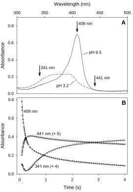
chromophore. Two intermediates in the folding of cytochrome c after a pH jump were
detected with absorption spectroscopy, providing evidence for the proposal of a four-state
mechanism (53).
Circular dichroism. CD refers to the differential absorption of left- and right-handed
circularly polarized light within a chiral molecule. CD is most often utilized to investigate
the protein backbone (55). Characteristic CD profiles are obtained for α-helices, β-sheets,
and random coils. Thus, the secondary structure of a protein can be interrogated in a
straightforward manner.
Fluorescence spectroscopy. Aromatic amino acid residues are intrinsically fluorescent,
although usually only tryptophan fluorescence is measured. The emission properties of
the Trp side chain are highly sensitive to the solvent environment. The λmax in water is
around 355 nm, while in a hydrophobic environment (such as the protein core) it is
around 335 nm (56). Interpretation of fluorescence data is greatly simplified in proteins
residues can be complicated but single Trp mutants can be used to deconvolute the
fluorescence signal and localize the structural changes taking place during folding (57).
Fӧrster resonance energy transfer. FRET involves energy transfer between a donor and
an acceptor. The emission spectrum of the donor must partially overlap with the
absorption spectrum of the acceptor. After excitation of the donor, fluorescence intensity
measured from the acceptor reports on their relative distance. FRET is often termed a
„molecular ruler‟ for this reason. Care must be taken when choosing the fluorophores for
protein studies, as large bulky dyes can sometimes interfere with folding. The
incorporation of fluorescent unnatural amino acids represents an interesting alternative
(58). FRET studies are particularly intriguing as they offer the possibility of monitoring
the dynamics of single protein molecules (59).
1.2.2 Nuclear Magnetic Resonance Spectroscopy
NMR spectroscopy can provide a comprehensive portrayal of protein dynamics at
the atomic level. NMR active nuclei, typically 15N, 13C and 19F, can be incorporated into
protein sequences to act as molecular reporters, along with naturally occurring 1H,
through the use of myriad dynamics techniques (60). 1D 19F NMR combined with
stopped-flow mixing allowed the folding of E. coli DFTR, with engineered fluorinated
tryptophans, to be followed on the order of seconds (61), however 2D experiments are
more common. Relaxation dispersion measurements have been used to uncover
unfolding/refolding events in aMb (62) and a transient folding intermediate in a small
photo-CIDNP spectroscopy allows for structural transitions on the order of tens of
milliseconds to be characterized (64). This method, which measures the
cross-polarization of amino acid side chains from an activated aromatic residue, has revealed a
preformed hydrophobic cluster in the unfolded state of a small Trp-cage protein (65).
Because 1H nuclei result in an NMR signal but 2H nuclei do not, NMR is a suitable
detection method for hydrogen/deuterium exchange experiments (see section 1.4.1) and
has been used to characterize folding intermediates populated as early as ~0.5
milliseconds (66).
1.2.3 Computer Simulations
While experimental methods routinely interrogate folding processes on the
(sub)millisecond timescale, they struggle to access faster regimes. Computer simulations
historically have had the opposite problem; accession of biologically relevant timescales
requires an enormous amount of computing power and time. This is troublesome because
experimental validation of computational outputs is required. Since the advent of
molecular dynamics (MD) in the late 1970s (67) much improvement has been made in
the time resolution of simulated folding studies (68). Recent work from Shaw and
coworkers has caused a paradigm shift by extending MD timescales by over two orders
of magnitude (69). A new supercomputer, ANTON, has allowed for a greater overlap
between simulation and experiment. Folding trajectories for many small protein domains
have been simulated for upwards of 1.1 milliseconds, producing structures that are in
1.3 Mass Spectrometry
MS measures the mass-to-charge ratio (m/z) of gas phase ions. MS was
introduced nearly a century ago. Unfortunately, traditional ionization sources were too
harsh for the transfer of large, intact biomolecules into the gas phase. This feat was first
accomplished nearly 25 years ago with the advent of two „soft‟ ionization techniques.
The 2002 Nobel Prize in Chemistry was awarded for techniques to identify and
structurally characterize biomolecules of which Koichi Tanaka and John Fenn shared one
half for their development of MALDI and ESI techniques, respectively. Over the past 25
years MS has become a widely used technique for the analysis of biological molecules
(70). A typical mass spectrometer comprises an ion source, a mass analyzer, and a
detector.
1.3.1 Ionization Techniques
1.3.1.1 Matrix-Assisted Laser Desorption/Ionization
MALDI utilizes a pulsed laser to desorb biomolecular analytes from a solid
matrix. Organic molecules in the matrix absorb UV photons and desorb from the surface
taking proteins with them. Charge is then transferred from the matrix molecules to the
gaseous proteins. Because MALDI is a pulsed ionization method and produces very low
charge states, it is most often interfaced with a time-of-flight analyzer (71). Tanaka et al.
were able to detect singly charged proteins upwards of 25 kDa along with multimers up
to 100 kDa, albeit with greatly reduced sensitivity (72). Around the same time, Karas and
could be detected (73). MALDI is still routinely used for protein identification, and it has
also found applications for tissue imaging (74).
1.3.1.2 Electrospray Ionization
ESI was first conceived by Dole et al. over 40 years ago (75). An explosion in the
usage of ESI-MS occurred after Fenn and coworkers demonstrated its utility for studying
biological macromolecules (76). ESI introduces analyte ions into the MS directly from
solution and is thus commonly interfaced with liquid chromatography. Analyte solution is
pumped through a spray needle which is held at a high potential, ~3-5 kV. In positive ion
mode, enrichment of positive charges occurs at the needle tip resulting in a Taylor cone
(Figure 1.3). With the help of a heated, concentric nebulizing gas flow, a mist of solvent
droplets is emitted toward the MS. The droplets shrink through solvent evaporation until
they reach the Rayleigh limit (77) at which point Coulombic fission occurs. This process
of evaporation and fission is repeated until naked gas-phase ions are produced (78). Two
mechanisms have been proposed for this final step.
Ion evaporation model. The IEM was first put forth by Iribarne and Thompson to
describe how small molecules became ionized from droplets close to the Rayleigh limit
(79). In this model, solvated ions are ejected from the droplet surface. Computer
simulations have shown this to be the likely ionization mechanism for small molecules
and salt clusters (80, 81). Ahadi and Konermann recently utilized a simple MD model to
show that the ionization of unfolded protein chains likely proceeds via the IEM as well
+ + + + + + + ++ + + + + + + + -+ + + ++ + + + + + + ++ + + + + + + + + + + + + + + + + + + + + + + + + + + + + + + + ++ ++ + + 3-5 kV e -e
-MS
spray needle solvent evaporation + droplet fission
Figure 1.3. ESI Process. Simplified schematic representation of the electrospray
Charged residue model. The CRM posits that gas phase ions are produced via complete
solvent evaporation. Folded globular proteins are thought to become ionized by this
mechanism, as the maximum ESI-MS charge state for a given protein shows good
agreement with the Rayleigh charge for a water droplet of the same radius (83). Recent
MD simulations have provided further evidence supporting the notion that ionization of
folded proteins proceeds via the CRM (82).
1.3.2 Mass Analyzers
Once gas-phase ions have been produced and introduced into the MS, they must
be separated and detected based on their m/z. Various mass analyzers are available,
including quadrupole ion traps and ion cyclotron resonance cells. The two most common
are linear quadrupoles and time-of-flight analyzers.
1.3.2.1 Quadrupole Mass Analyzer
A quadrupole is a set of 4 parallel cylindrical rods. An RF voltage and a
superimposed DC voltage are applied to each opposite rod pair (84). For a given voltage
setting only ions of a certain m/z will be transmitted through the quadrupole. All other
ions will collide with the rods due to their unstable trajectories. Sequential transmittance
of all ions in a given m/z range is accomplished by changing the voltages in small steps.
Quadrupoles exhibit limited resolution and mass range. Therefore, they are often coupled
to other mass analyzers, such as a time-of-flight unit. In such a situation, the quadrupole
can be operated in RF-only mode (with no DC voltage). In this case the quadrupole acts
A benefit of linking two analyzers in tandem is the possibility to carry out
MS/MS, or tandem-MS, measurements. In these experiments, the first quadrupole
voltages are „parked‟ to allow transmittance of ions of a certain m/z. These selected ions
are then fragmented in a collision cell, and the resulting fragments are detected in the
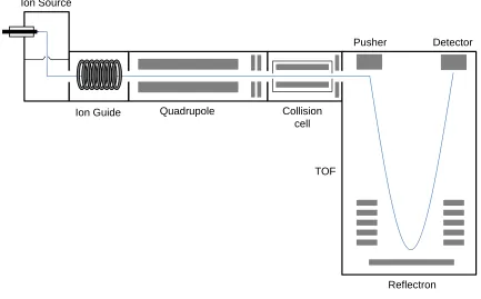
second mass analyzer. Quadrupole time-of-fight (Q-TOF) mass spectrometers can easily
be interfaced with liquid chromatography systems and have thus become the workhorses
of the proteomics and pharmaceutical industries.
1.3.2.2 Time-of-Flight Mass Analyzer
Ions are separated in a TOF analyzer based on their m/z. An electric field is
applied to a packet of ions and this potential energy is transformed into kinetic energy as
the ions are accelerated through the TOF tube.
2
2 1 )
(ze U mv (1-1)
where ze is the ion charge and ΔU is the applied potential gradient. Rearrangement of Eq.
1-1 yields
m U ze
v 2 (1-2)
The flight time on an ion is hence given by
2 / 1
2
z m U e L v L
where L is the path length. A time-to-digital converter is used to transform the measured
drift times into a mass spectrum.
The applied electric voltage in the TOF may not affect all ions of a given m/z
equally, resulting in a distribution of kinetic energies and thus flight times (85). This is
seen as peak broadening that decreases resolution in the mass spectrum. The issue can be
addressed by incorporating a reflectron (Fig. 1.3). This device acts as an ion mirror and
consists of a stack of rings with increasing potential. Ions with larger velocities will
penetrate deeper into the reflectron, while slower ones will follow a shallower trajectory.
This change in path length compensates for slight differences in kinetic energy and
allows all ions of a given m/z to be detected simultaneously, improving peak resolution
by a factor of ~5.
1.3.3 Protein Folding Studied by Electrospray Mass Spectrometry
The protein charge state distribution seen in an ESI mass spectrum has been
shown to be dependent on its solution phase conformation (86) and this feature has been
exploited in protein folding studies (87). While equilibrium studies have been performed
in this way (88), the fast detection of MS provides the opportunity to investigate kinetic
folding processes (89) and even enzymatic reactions (90). Such time-resolved
experiments utilize solution-phase mixing interfaced directly with a mass spectrometer
(91, 92). Additional structural information can be obtained through the incorporation of a
conformationally sensitive labeling step prior to MS injection. Various such labeling
Ion Source
Reflectron TOF
Collision cell Quadrupole
Ion Guide
Pusher Detector
Figure 1.4. Schematic of a Q-TOF. Simple representation of the major components of a
quadrupole time-of-flight mass spectrometer. The blue line indicates the path travelled by
1.4 Protein Labeling
Most optical techniques only report on global structural features of proteins. To
garner detailed mechanistic insights into protein structure, dynamics, and folding,
spatially resolved information is required. There are two general methods that can be
coupled to MS for such a purpose: hydrogen/deuterium exchange (HDX) and covalent
labeling. In broad terms, HDX monitors hydrogen bonding patterns in a polypeptide
backbone and covalent labeling probes the solvent accessibility of amino acid side
chains. Thus, results obtained from these two techniques are complementary and can
provide a comprehensive picture of the protein of interest (93).
1.4.1 Hydrogen/Deuterium Exchange
The exchange of hydrogens attached to electronegative heteroatoms in proteins
with deuterium from the solvent was first described by Linderstrøm-Lang in the early
1950s (94). The kinetics of HDX, and their dependence on temperature and pH have
since been characterized (95) as well as the effects of neighboring amino acid residues
(96). The main principle of HDX is that exposed hydrogens will exchange with solvent
quite rapidly while those involved in hydrogen bonds, as in the case of α-helices and
β-sheets, will exchange much more slowly. Significant mechanistic understanding of HDX
has come from NMR experiments conducted by Englander and coworkers (97). The
nuclear spin of hydrogen (1H) gives rise to an NMR signal while that of deuterium (2H)
does not, making NMR a preferred readout for hydrogen exchange experiments. Many
pathways for model proteins such as cytochrome c (99) and apomyoglobin (66) have
been characterized.
In addition to altering nuclear spin, the exchange of hydrogen for deuterium also
increases the mass of an analyte. This effect can be exploited for MS experiments (100).
The classic “bottom-up” approach was first described by Zhang and Smith nearly 20
years ago (101) and it involves monitoring the mass increase of proteolytic peptides, as a
function of deuteration time. Bottom-up HDX-MS has been utilized extensively ever
since (102), including the study of changes in receptor dynamics after drug binding
protein-interactions (103), the thermodynamic stability of proteins (104), and the cyclic
motions of molecular machines (105). Pulsed labeling methods have been described
whereby the hydrogen bonding status of folding intermediates can be reported, often with
single-residue resolution (106), however structural data should be interpreted with
caution due to the basic pH required for labeling (107). Despite being widely used,
HDX-MS has two main drawbacks that must be addressed: 1) deuterium scrambling and 2)
back-exchange.
Akin to the mechanism of peptide fragmentation (108), deuterium scrambling is
the process by which protons and deuterons can migrate along the backbone of a
collisionally activated peptide, thus reducing the measured spatial resolution (109, 110).
Radical-based fragmentation techniques such as electron capture dissociation (ECD) and
electron transfer dissociation (ETD) have been described as alternatives to classical
collision techniques. They have been shown to be less sensitive to neighboring amino
acids, resulting in a higher sequence coverage (111) as well as having the capability to
intact proteins have shown good agreement with NMR measurements with near single
amino acid resolution (113, 114). These “top-down“ approaches are conducted by
fragmentation of intact proteins, and hence they do not require proteolytic digestion. A
combination of proteolytic digestion and electron-based fragmentation has been termed
“middle-down“. This approach is capable of measuring accurate deuteration levels with
amino acid resolution as in “top-down“ experiments but with simplified data analysis due
to peptide, rather than protein, precursor ions (115).
Back exchange is another issue that must be considered. In experiments involving
proteolytic digestion, sample clean-up and peptide separation is performed via liquid
chromatography (LC) prior to MS analysis. LC is performed with a gradient of aqueous
and organic solvents which implies that deuterated peptides are in contact with protiated
water for much of the analysis. This allows deuterium to exchange with hydrogens from
the LC solvent, thereby diminishing the amount of structural information. The inclusion
of fully-labeled control samples can correct back-exchange to some extent.
1.4.2 Covalent Labeling
An entirely different MS-based structural approach follows a strategy where the
target protein is modified with covalent labels. This provides the opportunity for
extensive downstream sample handling such as dialysis, lyophilization, and
chromatography. The amount of labeling achieved at any given site is modulated by both
the intrinsic reactivity of the amino acid with the label and the solvent accessibility of the
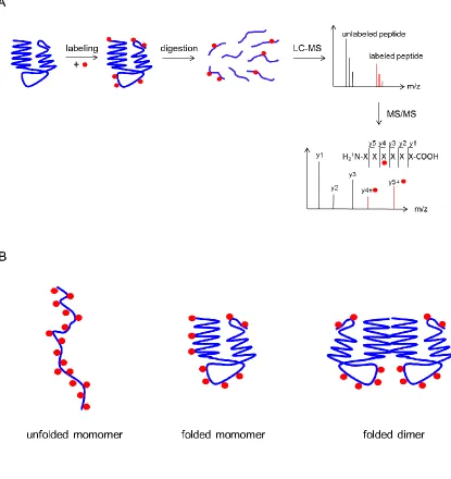
side chain. Figure 1.5A depicts the typical workflow in a covalent labeling experiment.
Extensive labeling is seen for unfolded proteins, whereas the decreased solvent
accessibility of the folded state results in fewer modifications. Side chain protection at
protein-protein binding interfaces results in protection as well. Covalent labels fall into
two general categories, those that react with specific amino acids and those that exhibit
more promiscuous reactivity.
1.4.2.1 Specific Labeling
Specific labeling reports directly on solvent accessibility since all labeled sites
will have identical reactivities. Common targets are hydrophilic residues on the protein
surface such as Lys, Arg, Asp, Glu, and Cys. Predictable mass increases at labeled
residues can be easily tracked by MS. Many different labeling agents targeting various
amino acids have been described in the literature (116) but discussion will be limited to a
select few.
Lysine modification and cross-linking. The positively charged side chain of lysine
provides an ideal modification target due to its propensity to reside on protein surfaces.
Acetylation of exposed lysines with MS detection dates back 20 years (117). Most often
lysine modification is utilized to map protein binding interfaces such as HIV-1 reverse
transcriptase contacts within the viral RNA:tRNA complex (118). While many lysines are
solvent exposed, some are globally protected within the protein core. Fitzgerald and
coworkers monitored the modification of buried lysines as a function of denaturant
concentration to investigate the thermodynamic stability of proteins in a multicomponent
Figure 1.5. Covalent labeling. (A) Typical workflow of a covalent labeling experiment.
Proteins are labeled in solution and digested. Resultant peptides are separated and
detected with LC-MS. Label site determination is achieved through MS/MS experiments.
to their being used in crosslinking experiments. These studies use bifunctional molecules
of varying lengths to determine distances between lysines on a protein or within
interacting proteins (120). Crosslinking data recently provided distance constraints for
computational modeling that determined the subunit arrangement of a eukaryotic 16-mer
chaperonin (121).
Carboxylate labeling. Similar to lysine, aspartic and glutamic acid are abundant in
proteins and most often found on the surface. Mapping protein solvent accessible surface
area via modification of acidic residues has forged ahead through the efforts of Gross and
coworkers. The original study used glycine ethyl ester and MS to determine the
orientation of the FMO antenna protein on a bacterial membrane (122). Recently the
method has been extended to monitor differences in solvent accessibility of multiple
conformational states of calmodulin (123).
Thiol labeling. Cysteine residues not involved in disulfide bridges contain a free –SH
group that is available for modification. Such free Cys residues are rare in most proteins.
Genetic engineering, however, allows the introduction of additional Cys residues. Due to
the small, polar nature of the side chain these mutations can be tolerated to a far greater
extent than Lys, Glu, or Asp (124). While the solvent accessibility of Cys residues as a
function of denaturant has led to structural characterization of protein folding
intermediates under equilibrium conditions (125), thiol labeling has predominantly been
used in kinetic experiments. While many amino acids require extended incubation with a
milliseconds. As a result, the temporal changes in Cys labeling have provided structural
information on the folding pathways of proteins such as barnase and monellin (126-128).
1.4.2.2 Non-Specific Labeling
While residue-specific labeling can simplify data analysis the spatial resolution
often suffers. Increased sequence coverage can be attained through the combination of
multiple labeling reagents (129). A conceptually simpler method is the use of semi- or
non-specific labels. Vachet and coworkers have shown that diethylpyrocarbonate (DEPC)
can modify Ser, Thr, His, and Tyr residues resulting in ca. 25% coverage of an average
protein. Labeling times as short as a few minutes provide significant modification levels
with minimal perturbation to protein structure (130). Jumper and Schriemer described a
method for interrogating protein topography via production of carbene labels by
photolysis of a diazarine-modified precursor (131). This carbene method provided good
temporal resolution, as well as the potential to probe every amino acid in a protein
sequence. By far however, the preferred non-specific labeling method for biomolecular
structure analysis has been the production of hydroxyl radicals (·OH) (132).
1.4.3 Hydroxyl Radical Labeling
In 1978 Galas and Schmitz introduced the term footprinting to describe
experiments that sought to determine regions of nucleic acids that were protected from
nucleases by proteins (133); the extension to proteolytic cleavage of proteins came much
than enzymes to cleave nucleic acids (135). Hydroxyl radical footprinting continues to be
used to study dynamics of nucleic acids in both equilibrium and time-resolved
experiments (136, 137),
The pioneering work of Chance and coworkers showed that ·OH could modify
solvent exposed amino acid side chains of a protein without backbone cleavage (138).
The term “footprinting” is now commonly used for hydroxyl radical labeling of protein
surfaces (139), binding interfaces (140), and folding mechanisms (141). While hydroxyl
radicals have the potential to modify any of the 20 amino acids, only ~50% are typically
seen in a protein labeling experiment. The most reactive residues tend to be the
sulfur-containing side chains (Cys and Met), followed by the aromatics (Trp, Tyr, Phe). Other
commonly oxidized side chains are those of His, Leu, Ile, Val, Pro, Glu, and Arg. The
oxidation of these side chains can progress through complex mechanisms which have
been well described (142, 143). The most frequently observed oxidation products involve
the incorporation of an oxygen atom, resulting in a +16 Da mass shift, although other
There are several radical generation approaches for protein labeling studies, such as
Fenton chemistry (144), electrochemistry (145), and corona discharge methods (146,
147). Labeling has been shown to be rather insensitive to the method used, however only
two have really gained a foothold in the field.
1.4.3.1 Radiolysis of Water
The generation of ·OH directly from water via synchrotron radiation for protein
structural studies was pioneered by Chance when he utilized the amount of labeling of
aMb as a function of denaturant to probe its unfolding pathway (148). A series of articles
by Xu and Chance outlined much of the chemistry involved in ·OH labeling (149-151).
accord with published rate constants for the free amino acids (152). ·OH labeling via
millisecond X-ray radiolysis of water has been used by Chance and coworkers to gain
insights into enzyme mechanisms (153) and the structure/function relationship of integral
membrane proteins (154). Since access to synchrotron light sources is limited, the Chance
group has maintained a near monopoly in this field.
A water radiolysis method with broader accessibility uses γ-rays to generate ·OH.
Because of the relatively long labeling time required (minutes), γ-ray radiolysis is well
suited for equilibrium experiments and has been exploited mainly in differential studies.
Experiments of this type seek to gain topological information on discrete conformational
states of a protein. Examples include the calcium induced conformational change of
calmodulin (155), the effect of heme removal from myoglobin (156), and the prepore to
pore transition of the protective antigen from Bacillus anthracis (157).
To overcome the deficiencies of limited synchrotron access and long labeling
times in γ-ray studies, Sharp and coworkers recently used a pulsed electron gun to
generate ·OH from water on the submicrosecond timescale (158). This proof-of-principle
experiment monitored the solvent accessibility of residues within model proteins
ubiquitin and β-lactoglobulin. The extremely fast labeling time achieved with this method
is highly desirable for the study of protein folding in a time-resolved manner, however
1.4.3.2 Photolysis of Hydrogen Peroxide
Early studies mapped protein surfaces using concentrated (~15% v/v) H2O2 and
prolonged exposure to low intensity UV light (159). While this method produces
modified proteins, spurious „background‟ oxidation can occur from the presence of the
peroxide. More recently, a photolytic approach has evolved for generating radicals from
dilute (~0.1% v/v) solutions of peroxide via homolytic cleavage with a pulsed UV laser
(160, 161). Non-irradiated control samples have shown that incubation of the protein with
such low concentrations of peroxide exhibit very little background oxidation. A seminal
paper by Hambly and Gross described the pulse-labeling approach in combination with a
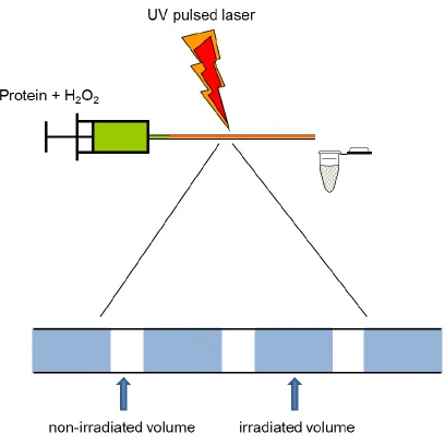
continuous-flow reaction setup (160). This continuous-flow system is preferable to the
static system of Aye et al. (161)because with a simple plug flow approximation,
„single-hit‟ conditions can be attained. „Single-„single-hit‟ refers to the regime in which each protein
molecule only “sees” one labeling pulse. This can be achieved by adjusting the flow rate
and laser frequency such that two adjacent irradiated volumes are separated by a
non-irradiated volume (Figure 1.6). Oxidation has been shown to disrupt protein structure
(162). A second labeling pulse may therefore modify side chains that were not initially
solvent exposed, resulting in artifactual data. Plug flow constitutes a useful zero-order
approximation. However, a more accurate picture of the dynamics within the reaction
chamber takes into account laminar flow (163). Hambly and Gross also showed that the
·OH lifetime in solution can be tuned via the addition of radical scavengers, such as
glutamine. This results in a labeling pulse as short as ~1 µs (160) which is shorter than
oxidation-induced protein unfolding (164).
Figure 1.6. Photolysis experimental setup. Protein and peroxide are mixed and pumped
through a UV transparent capillary onto which the laser is focused. Proper adjustment of
flow rate and laser frequency leads to non-irradiated solution separating adjacent
·OH labeling by laser photolysis of peroxide has been used in many recent protein
structural studies. Most seek to identify differences in solvent accessibility between
multiple states of a protein. The interface of the carbohydrate-binding, homodimeric
protein galectin-1 was characterized through comparison of labeling levels for the
monomers and for the dimer. Results were shown to be in accord with theoretical solvent
accessibilities calculated from MD simulations (140). Similarly, the conformational
changes in calmodulin concomitant with peptide binding have been described (165). Pan
et al. extended the methodology to look at exposed Met residues of the integral
membrane protein bacteriorhodopsin (BR) (166).
The very short labeling pulse achieved in laser photolysis makes it ideally suited
to investigate rapid, time-resolved changes in protein structure however there is a dearth
of such studies in the literature. Following work that will be presented later in this thesis,
Pan et al. used rapid mixing followed by oxidative labeling to explore the BR folding
mechanism (141). A temperature-jump method has also recently been described that is
1.5 Scope of Thesis
This work extends the photochemical oxidation method developed by Hambly
and Gross (160) to study protein folding transitions through the combination of
continuous-flow rapid mixing, pulsed labeling, and MS detection. The extremely short
radical lifetime makes such a method ideally suited to study rapid structural changes.
Prior to the work described here, ·OH labeling had only been utilized for equilibrium
measurements.
We will describe the first application of pulsed oxidative labeling to characterize
transient protein unfolding intermediates, along with a comprehensive data analysis
strategy to deal with differing peptide reactivities (Chapter 2). The folding pathway of a
small model protein is then characterized (Chapter 3). In both cases, the labeling data are
correlated with optical measurements. The approach is extended to monitor the folding
and assembly of a protein complex (Chapter 4). Finally, we apply oxidative labeling
method to explore the folding of a large protein with direct implications in human disease
(Chapter 5). These results are explicitly compared with hydrogen bonding data from
HDX experiments.
The labeling approach introduced here provides an avenue for obtaining
residue-resolved structural information regarding side-chain accessibility during rapid protein
folding processes, with application to other biomolecular events as well. The combination
of the resultant labeling data with those obtained from HDX allows for a comprehensive