Correspondence
The spectrum of presentations of venous infarction caused by deep cerebral vein thrombosis
To the Editor: Van den Bergh et al.1 illustrated important
features related to cerebral venous thrombosis (CVT) and spe-cifically deep cerebral venous thrombosis (DCVT). Their four cases occurred in women. CVT is more common in women, and DCVT has a female to male ratio of 8.3:1. All women were taking oral contraceptives (OCP). It has been shown that OCP, even at low doses, increases the risk of CVT. In the presence of congenital thrombophilia, this risk is 100 times higher.2
Diag-nosis is difficult in DCVT because there are many clinical and radiologic presentations. DCVT should be considered in a pa-tient presenting with headache, altered mental status, neuro-logic symptoms not easily localized to an arterial supply, and imaging abnormalities seen bilaterally in thalamus and basal ganglia. These areas are well perfused via small branches from both anterior and posterior circulations, so an arterial stroke is not the culprit.3-5
Zeyad Morcos,Granad Island, NE
Reply from the Authors:We thank Dr. Morcos for his interest in our article. We agree that DCVT is a diagnostic challenge, especially in the case of partial thrombosis or sufficient collater-als.1 The recommendations made by Dr. Morcos are
appropri-ate—to consider DCVT in case of headache, altered mental status, neurologic symptoms not easily localized to an arterial supply accompanied by thalamus and basal ganglia abnormalities, espe-cially if bilateral.
It is unclear whether known risk factors for CVT in general can be directly applied to DCVT, but it seems to be more common in women. However, it is difficult to establish the role of OCP usage as an additional risk factor since its use is extremely com-mon acom-mong women in the reproductive age.6-8The change in the
sex ratio of cases of sinus thrombosis over time provides indirect evidence.
Until the mid-1970s, men and women were affected in equal proportions. More recently, there has been a significant female predominance among young adults with sinus thrombosis (70 to 80% of cases are in women of childbearing age) but not among children or elderly persons.9 This might be caused by the
in-creased use of OCP, in particular third-generation contraceptives that contain gestodene or desogestrel.8
Walter M. van den Bergh, MD, Irene van der Schaaf, Jan van Gijn,Utrecht, The Netherlands
Copyright © 2006 by AAN Enterprises, Inc.
References
1. Van den Bergh WM, Van der Schaaf I, Van Gijn J. The spectrum of presentations of venous infarction caused by deep cerebral vein thrombo-sis. Neurology 2005;65:192–196.
2. Bousser M, Kittner S. Oral contraceptive and stroke. Cephalalgia 2000; 20:183–189.
3. Baumgartner R, Landis T. Venous thalamic infarction. Cerebrovasc Dis 1992;2:353–358.
4. Ur Rahman N, Al Tahan A. Computed tomographic evidence of an exten-sive thrombosis and infarction of the deep venous system. Stroke 1993; 24:744 –746.
5. Morcos Z, Sundararajan S. Cerebral venous thrombosis. In: Suarez JI, ed. Critical care neurology and neurosurgery. Totowa, NJ: The Humana Press, 2004;379 –393.
6. Martinelli I, Sacchi E, Landi G, Taioli E, Duca F, Mannucci PM. High risk of cerebral-vein thrombosis in carriers of a prothrombin-gene muta-tion and in users of oral contraceptives. N Engl J Med 1998;338:1793– 1797.
7. de Bruijn SF, Stam J, Koopman MM, Vandenbroucke JP. Case-control study of risk of cerebral sinus thrombosis in oral contraceptive users and in carriers of hereditary prothrombotic conditions. BMJ 1998;316:589 – 592.
8. de Bruijn SF, Stam J, Vandenbroucke JP. Increased risk of cerebral venous sinus thrombosis with third-generation oral contraceptives. Lan-cet 1998;351:1404 –1404.
9. Stam J. Thrombosis of the cerebral veins and sinuses. N Engl J Med 2005;352:1791–1798.
Bilateral involvement of a single cranial nerve: Analysis of 578 cases
To the Editor: We read Dr. Keane’s article with interest.1Dr.
Keane’s analysis of the inpatients he personally examined over a 34-year period at the Los Angeles County/University of Southern California Medical Center suggested that bilateral involvement of the same cranial nerve is uncommon and is usually associated with cranial nerve VI damage.
However, the most rostral of cranial nerves, the olfactory nerve, was not mentioned. Inclusion of this cranial nerve may have substantially changed his results. Bilateral cranial nerve I damage is frequently seen associated with head trauma, including up to 60% of those with severe head injury. Likewise, a variety of other neurologic conditions induce bilateral olfactory deficits, in-cluding tumors, epilepsy, demyelinating disorder, degenerative disorders including Parkinson’s disease and senile dementia of the Alzheimer’s type, and migraine.2,3
Furthermore, inclusion of this cranial nerve would have mark-edly changed his results because, based on demographics alone, cranial nerve I dysfunction would have been substantial. It is estimated that half of those over the age of 65 and three-quarters of those over the age of 80 have a reduced ability to smell. Fur-thermore, many medications used in the treatment of neurologic conditions may have also been anticipated to impair his patients’ olfactory ability.
The lack of inclusion of cranial nerve I in Dr. Keane’s report highlights the widespread practice of overlooking cranial nerve I in the neurologic examination. In a retrospective study of 94
neu-rology inpatient consultations recorded in the hospital charts, only four had cranial nerve I testing (and those were described as “WNL”).4A myriad of neurologic conditions manifest bilateral
cra-nial nerve I dysfunction, and discovery of such loss is important for safety and quality of life.5
Alan R. Hirsch, MD, Stephanie Fulton, Thomas L. Wilding, Frances Groen,Chicago, IL
Reply from the Author:I agree with the authors on the impor-tance of testing the olfactory nerve. I hope that their letter will be more persuasive than my harangues to the residents that the cranial nerves do not begin with II.
James R. Keane,Los Angeles, CA
Copyright © 2006 by AAN Enterprises, Inc.
References
1. Keane JR. Bilateral involvement of a single cranial nerve: analysis of 578 cases. Neurology 2005;65:950 –952.
2. Murphy C, Doty RL, Duncan HL. Clinical disorders of olfaction. In: Doty RL, ed. Handbook of olfaction and gustation, second edition, revised and expanded. New York: Marcel Dekker, Inc., 2003.
3. Hirsch AR. Olfaction in migraineurs. Headache 1992;32:233–236. 4. Hirsch AR, Colavincenzo ML. Failure of physicians to assess olfactory
ability in neurologic inpatients. Chemical Senses 1999;24:607– 608. 5. Hirsch AR. Listening to patients with chemosensory dysfunction.
Addendum to assessment: Prevention of
post–lumbar puncture headaches: Report of the TTAS of the AAN
To the Editor:We read with interest the report of the Therapeu-tics and Technology Assessment (TTA) Subcommittee of the AAN providing an addendum to the assessment on the prevention of post-lumbar puncture headaches (PLPHAs) following diagnostic LPs.1We were surprised that a consensus was reached for a Level
A recommendation. Only one Class I article was cited.2
In the Strupp et al. study, 306 patients were allocated ran-domly to the “atraumatic” vs “traumatic” needle, yet 230 are eval-uated, representing a drop-out rate of 25%. Of the 76 drop-outs, 25 did not return the evaluation sheet. Since the Subcommittee’s recommendation includes a mandate to enact widespread educa-tional strategies to impact neurologic practice, we think an intention-to-treat analysis that incorporates all randomized pa-tients would be important.
When further assessing the Strupp et al. article,2the control
event rate of 24% vs experimental event rate of 12% translates to a relative risk reduction of 50% and absolute risk reduction of 12% (NNT⫽ 8.3). However, the CI calculates to 0.12⫾ 0.1 (i.e., an absolute risk reduction of 0.02 to 0.22).
The new conclusion—“now also one study providing Class I evidence in a patient population undergoing diagnostic LPs with a 22-guage needle supports the use of an atraumatic spinal needle to reduce the frequency of PLPHA”—is unlikely. Additionally, the studies leading to the addendum do not provide data higher than Class IV evidence addressing other relevant primary outcomes (e.g., occurrence of back pain, technical variables between the two procedures).
We question the new conclusion that supports the use of an atraumatic spinal needle to reduce the frequency of PLPHA, and the recommendations to develop and disseminate standardized training materials for practitioners, and to track acceptance and implementation within the neurologic community.
Phillip L. Pearl, William M. McClintock,Washington, DC
Reply from the Authors:We thank Drs. Pearl and McClintock for their comments and appreciate this opportunity to respond.
Pearl and McClintock state that 306 patients were random-ized. The authors of the original article2state that 51 patients did
not meet the inclusion criteria and were not randomized. There-fore, 255 subjects were randomized. Of the randomized patients, 25 did not return the evaluation sheet—12 from the traumatic group and 13 from the atraumatic group. The 230 patients who returned the evaluation sheets were equally divided and the re-sults for them are reported using intention-to-treat analysis. A 10% drop-out rate is acceptable and does not change the evidence class.
We agree with Pearl and McClintock that the number needed
to treat to prevent one PLPHA by using a 22-G atraumatic needle rather than a 22-G traumatic needle, based on the article in ques-tion,2is eight, and indicated this in the Discussion. We agree also
that a reduction from 24% to 12% represents a relative risk reduc-tion of 50% and an absolute risk reducreduc-tion of 12%. We elected to present the information only in terms of numbers needed to treat, because we consider that number to be easier to understand than RRR or ARR.
Pearl and McClintock state that the CI on the absolute risk reduction of 12% is 2% to 22%, without providing the basis for their statement. If they are correct, the best estimate for the absolute risk reduction remains 12%; eight is the most likely estimate of the “number needed to treat,” and the likelihood that the true number is greater than eight is the same as the likelihood that it is smaller than eight.
The original assessment and the addendum discussed techni-cal aspects within the Discussion. The presence of a learning curve to the use of a new technology is not considered, in general, a “primary outcome” of the use of that technology. Pearl and McClintock suggest also that the occurrence of back pain should be considered an additional relevant primary outcome. We point out that there can be only one primary outcome, while recognizing the need to be aware of secondary outcomes that might detract from the value of benefits measured within the primary outcome. The decision for a Level A recommendation was based on avail-able evidence for post-LP headache. It took into consideration not only the new (2001) Class I evidence in diagnostic LPs, but also the Class I evidence in spinal anesthesia studies, all pointing to the reduction in the frequency of PLPHA by using non-cutting needles.
The recommendation to develop training materials and track acceptance are consistent with the mission of the AAN to serve its constituent members as they, in turn, serve patients with neurologic diseases. We stand by our original conclusions and recommendations.
Considering Drs. Pearl and McClintock’s affiliation, we point out that our recommendations pertain to the adult population, in which there are high quality data, and make no comments about the pediatric population.
Carmel Armon, Randolph W. Evans, MD, for the Therapeutics and Technology Assessment Subcommittee,Springfield, MA
Copyright © 2006 by AAN Enterprises, Inc.
References
1. Armon C, Evans RW. Addendum to assessment: Prevention of post-lumbar puncture headaches: Report of the TTAS of the AAN. Neurology 2005;65:510 –512.
2. Strupp M, Schueler O, Straube A, Von Stuckard-Barre S, Brandt T. “Atraumatic” Sprotte needle reduces the incidence of post-lumbar punc-ture headaches. Neurology 2001;57:2310 –2312.
Camptocormia: Pathogenesis, classification, and response to therapy
To the Editor: Azher et al. concluded that of 16 patients diag-nosed with camptocormia, 11 developed it in association with Par-kinson disease (PD). Four had dystonia and one had Tourette syndrome.1However, they did not report or perhaps did not
per-form electrodiagnostic testing and more specifically needle electro-myography (EMG) examination of the thoracic paraspinal muscles in the individuals with isolated camptocormia and cervical paraspinal muscles in those who also manifested head drop.
The authors noted that no specific neuroimaging abnormalities were seen except for a thoracic syrinx in one patient. They did not report or did not perform thoracic or cervical spine MRI or CT imaging in each of the 16 patients and did not specifically remark about the signal characteristics of the extensor muscles in any case. These are significant limitations of their study.
The authors do reference in their Discussion previous studies demonstrating that thoracic extensor myopathy can cause campto-cormia and one study of four patients with PD and camptocampto-cormia
disorders. A thorough evaluation that includes nerve conduction study, repetitive stimulation, EMG of limbs and involved extensor muscles, MR imaging of the spine with attention not only to bony and neural elements but also the signal characteristics of the paravertebral muscles, and possibly muscle biopsy of limb or para-vertebral muscle often uncovers a neuromuscular cause.
With isolated head drop, the most common neuromuscular causes we have encountered are isolated cervical extensor myop-athy, inclusion body myopmyop-athy, myasthenia gravis, and amyotro-phic lateral sclerosis (ALS). For those with isolated camptocormia, the most common neuromuscular causes we have encountered are isolated thoracic extensor myopathy and ALS. For those with both the most common causes we have encountered are isolated exten-sor myopathy (cervical and thoracic) and ALS.
Reply from the Authors:We appreciate Dr. McCluskey’s inter-est in our review article and his comments. Although he provides no information on his cases, he argues that extensive neurophysi-ologic and neuroimaging evaluations may uncover neuromuscular etiologies for camptocormia.
As stated in the first paragraph of the Discussion portion of our article,1 our series was based on a population of patients
evaluated in a movement disorders clinic, hence the preponder-ance of movement disorders. Although a few reports suggest that extensor thoracic myopathies may be responsible for some cases of camptocormia, the interpretation of the muscle biopsies and EMG in those cases has been controversial and many experts have attributed those changes to chronic contractions of the antigravity muscles involved in compensatory trunk extension rather than to primary myopathies.2
In a study of 27 patients with camptocormia, EMG of the paravertebral muscles was difficult since the patients were unable to extend their trunks, and the recordings were uninterpretable in 16 cases. Other studies showed nonspecific changes in muscle biopsies without any evidence of myopathy.3 Likewise, imaging
studies often reveal changes that are likely to be secondary to rather than the primary cause of camptocormia.
Even though imaging studies were not done in all our patients, in those who had X-rays or MRIs of the spine, we could not find any radiologic features that differentiated patients with PD and camptocormia from those without camptocormia. We believe that our Discussion adequately addresses these concerns, although be-cause of space limitations we were not able to review all the relevant studies in detail.
Finally, the primary aim of our article was to draw attention to the heterogeneous etiologies of camptocormia, summarized in sup-plementary table E-1 on theNeurology Web site (www.neurology. org).
Joseph Jankovic, MD, Shaheda Azher, MD,Houston, TX
Copyright © 2006 by AAN Enterprises, Inc.
References
1. Azher SN, Jankovic J. Camptocormia: pathogenesis, classification, and response to therapy. Neurology 2005;65:355–359.
2. Laroche M, Delisle MB, Aziza R, et al. Is camptocormia a primary mus-cular disease? Spine 1995;20:1011–1016.
3. Djaldetti R, Mosberg-Galili R, Sroka H, et al. Camptocormia (bent spine) in patients with Parkinson’s disease— characterization and possible pathogenesis of an unusual phenomenon. Mov Disord 1999;14:443– 447.
Marchiafava–Bignami disease: Diffusion-weighted MRI in corpus callosum and cortical lesions
To the Editor:Me´ne´gon et al. recently reported abnormalities on diffusion-weighted MRI (DWI) in a series of six patients with Marchiafava-Bignami disease (MBD).1
Partial callosal signal abnormality is common on FLAIR and T2-weighted MRI in past reports.2,3In this study, DWI lesions
involved the entire corpus callosum (CC) in all patients, prompting the authors to suggest that complete affection of the CC may be obligatory in MBD. Although sensitivity of DWI is likely superior to conventional MRI, this hypothesis appears questionable on the background of histopathologic results re-porting the sparing of callosal areas from demyelination or necrosis4 and recent studies showing partial involvement on
both conventional MRI and DWI.3,5 Moreover, Me´ne´gon et al.
reportedly acquired conventional and DWI images in axial slices only, an important limitation for comprehensive evalua-tion of the entire CC.
The authors proposed that markedly reduced callosal apparent diffusion coefficient (ADC) may indicate poor prognosis, which contradicts recent reports.3A prospective, multicenter MRI study
including sagittal imaging may provide the most comprehensive results on the prognostic value of DWI in MBD. Correlative stud-ies establishing a link between ADC changes and histologic find-ings in MBD would be important but difficult due to the lack of an appropriate animal model and decreasing autopsy rates in this rare disorder.
The lateral-frontal predilection of cortical diffusion abnor-malities1,5is consistent with the autopsy finding of Morel’s
lam-inar sclerosis in the same areas.4,5Nevertheless, evidence that
diffusion abnormalities correspond to this highly specific form of cortical injury is insufficient as comparable DWI lesions con-sistent with cytotoxic edema have also been demonstrated fol-lowing cerebral hypoxia, prolonged seizure activity, or in Wernicke-Korsakoff disease, which may accompany severe cases of MBD.1,3
The discrepancy between MRI studies suggesting poor prog-nosis in cases with cortical involvement1,5 and autopsy series
reporting Morel’s laminar sclerosis predominantly in cases with a mild course4also stresses the need for direct comparison of
cortical diffusion abnormalities with histopathologic findings. Assuming that the observed cortical MRI lesions reflect a pathology corresponding to Morel’s laminar sclerosis, demon-stration of the latter in the acute stage would indicate that cortical pathology in MBD may not result from secondary corti-cal neuronal degeneration due to corti-callosal lesions as proposed earlier.4
Alexander V. Khaw, Alexander Heinrich,Greifswald, Germany
Reply from the Authors:We thank Drs. Khaw et al. for their comments on our article where we describe MRI abnormalities in the sub-acute phase of MBD using conventional and DWI in axial and sagittal planes.1We observed diffuse corpus callosum
abnor-malities in all the cases but never suggested the complete lesion of the CC as a new mandatory diagnosis criteria of MBD.
The low number of subjects, the anatomo-pathologic data, and the recent descriptions of partial corpus callosum diffusion hyper-intensities in MBD do not allow us to draw this conclusion.2Our
results also suggested a relationship between the values of the ADC/NAWM ratio and the long-term prognosis but with possible bias related to the low number of subjects.
The gold standard for such a study would be to compare the MRI data to the brain anatomo-pathologic lesions observed in subjects who died during follow-up. This would also answer ques-tions concerning the molecular basis of the cortical lesions ob-served in our patients. Similar cortical lesions to those obob-served in our study have been reported in one case without history of sei-zure or cerebral anoxia. As in our report, this suggests a possible primary cortical involvement in MBD.4
Two arguments promote the hypothesis of poor prognosis asso-ciated with cortical lesions: the poor clinical outcome of patients in Johkura et al. study5and of the three patients in our study and
the high frequency of cortical lesions in autopsy series.6
We agree with the authors that a multicenter MRI study using the uniform modalities of DWI acquisition and analysis would provide more comprehensive results on the prognosis value of MRI in the earlier phase of MBD.
P. Me´ne´gon, I. Sibon, C. Pachai, J.M. Orgogozo, V. Dousset,
Bordeaux, France
Copyright © 2006 by AAN Enterprises, Inc.
References
1. Menegon P, Sibon I, Pachai C, Orgogozo JM, Dousset V. Marchiafava-Bignami disease: diffusion-weighted MRI in corpus callosum and cortical lesions. Neurology 2005;65:475– 477.
2. Heinrich A, Runge U, Khaw AV. Clinicoradiologic subtypes of Marchiafava-Bignami disease. J Neurol 2004;251:1050 –1059.
3. Hlaihel C, Gonnaud PM, Champin S, Rousset H, Tran-Minh VA, Cotton F. Diffusion-weighted magnetic resonance imaging in Marchiafava-Bignami disease: follow-up studies. Neuroradiology 2005;47:520 –524. 4. Brion S. Marchiafava-Bignami disease. In: Vinken PJ, Bruyn GW, eds.
Handbook of clinical neurology. Amsterdam: North Holland, 1977;317– 329.
5. Johkura K, Naito M, Naka T. Cortical involvement in Marchiafava-Bignami disease. Am J Neuroradiol 2005;26:670 – 673.
Primary central nervous system lymphomas (PCNSL): MRI response criteria revised
To the Editor:Ku¨ ker et al. propose revised response criteria for primary CNS lymphoma (PCNSL) after completion of tumor-directed therapy.1Of 68 patients with contrast enhancing lesion
after methotrexate (MTX) therapy, they identified four with small lesions (5 mm in diameter or “bandlike”) in the area of primary tumor, hemorrhage, biopsy, or infection. The lesions were not further treated and did not show any change at 9, 24, 32, and 54 months follow-up.
Such lesions may reflect “unconfirmed” complete remissions (CRu) and not residual tumor, as proposed by an international workshop on standards of baseline evaluation and response crite-ria for PCNSL.2The limits of residual lesion size as proposed by
Ku¨ ker et al. are arbitrary. In their retrospective analysis, they do not include information on the 64/111 cases who met the criteria of partial remission (PR) and how decisions for either salvage therapy vs waiting were made in these cases.
In a series of 65 patients treated with a combined systemic and intraventricular chemotherapy, patients who met the criteria of a PR were not treated with salvage therapy irrespective of residual tumor size.3From these cases (plus another 23 patients treated
with the same regimen), we identified seven cases who met the following criteria: completion of therapy, residual contrast en-hancing lesion, no low grade lymphoma, and follow-up for at least 1 year. Among these, four matched the revised response criteria as proposed by Ku¨ ker et al. and two of them showed an early relapse 3 and 6 months after completion of therapy. The other three showed residual contrast enhancing lesions, all located within the primary tumor region, measuring 8 mm. These pa-tients showed a progression free survival of 20⫹, 29, and 88⫹ months. Two of the patients showed spontaneous disappearance of the lesions and the other a partial involution.
We conclude that categorizing patients’ responses according to these revised criteria is not predictive of their individual course after therapy. Therefore, patients with CRu should be carefully observed with serial scans.2
Uwe Schlegel, Annika Ju¨ rgens, Hendrik Pels,Bochum, Germany
Reply from the Authors: We read Schlegel et al.’s letter with interest and thank them for the additional data. We agree with their final statement that all patients with PCNSL should be closely monitored. However, surveillance scanning cannot be re-stricted to patients classified as complete response (CR) with
small residual lesions but has to include patients with CR as defined by the original MacDonald criteria. These patients have also been shown to relapse early after CR.
In the NOA-03 trial4 1 of 11 patients without any residual
contrast enhancing lesion relapsed within the first 6 months after the completion of therapy. It is plausible that Schlegel et al.’s patients who relapsed early after unproven CR did so at the site of the contrast-enhancing residual. It is not surprising that relapses may occur in patients with residual lesions, as in those without. The crucial point about the new classification scheme is that this is not even most frequently the case. In our treatment trial, all patients with residual contrast uptake were supposed to be treated as partial response (PR) and hence submitted to further therapy.
The four patients we reported who were not receiving salvage therapy were an exception and protocol violation. We may assume that among the remaining 64 patients with residual contrast-enhancing lesions who received further therapy several were treated unnecessarily.
This point is confirmed by Schlegel et al., who describe three patients with even larger lesions that did not progress over a long time in spite of a lack of treatment. The suggested new criteria for modified CR may even have to be expanded if more data become available.
As in all classifications, sensitivity has to be balanced against specificity, which in this case does seem to advocate a careful approach. Further work and the application of more sophisticated imaging modalities such as PET may be necessary to better define the presence of residual viable tumor tissue.
W. Kuker, Ulrich Herrlinger,Oxford, UK
Copyright © 2006 by AAN Enterprises, Inc.
References
1. Ku¨ ker W, Na¨gele T, Thiel E, et al. Primary central nervous system lymphomas (PCNSL): MRI response criteria revised. Neurology 2005;65: 1129 –1131.
2. Abrey LE, Batchelor TT, Ferreri AJM, et al. Report of an International Workshop to Standardise Baseline Evaluation and Response Criteria for Primary CNS Lymphoma. J Clin Oncol 2005;23:5034 –5043.
3. Pels H, Schmidt-Wolf IGH, Glasmacher A, et al. Primary central nervous system lymphoma: results of pilot and phase ii study of systemic and intraventricular chemotherapy with deferred radiotherapy. J Clin Oncol 2003;21:4489 – 4495.
4. Herrlinger U, Kuker W, Uhl M, et al. Neuro-Oncology Working Group of the German Society. NOA-03 trial of high-dose methotrexate in primary central nervous system lymphoma: final report. Ann Neurol 2005;57:843– 847.
Use of serum prolactin in diagnosing epileptic seizures: Report of the Therapeutics and Technology Assessment Subcommittee of the American Academy of Neurology
To the Editor:The AAN Therapeutics and Technology Assess-ment Subcommittee recently reported on the use of serum prolac-tin (PRL) in differentiaprolac-ting epileptic seizures from nonepileptic seizures (NES).1 The authors addressed two main concerns:
whether serum PRL is useful in distinguishing individual epilep-tic seizures from psychological NES; and whether serum PRL is useful in distinguishing individual epileptic seizures from other paroxysmal neurologic conditions.
The diagnosis and treatment of patients with psychological NES has long confounded neurologists, psychiatrists, and emer-gency department physicians. Currently, no randomized double blind, placebo controlled trial (RCT) has been completed for NES.2A common concern with diagnoses in the Diagnostic and
Statistical Manual of Mental Disorders–IV is that psychiatric diagnoses have no physiologic correlates. While aggregate data on depression and anxiety states have revealed alterations in the HPA axis, these findings are not applicable to the diagnosis of those with major depressive disorders or post traumatic stress disorders. NES are the exception to this rule, with diag-nosis validated by a physiologic measure—the gold standard,
Trimble first showed that generalized tonic clonic seizures (GTC), but not NES, raised serum PRL.3 Pooling the available
data of the 10 studies meeting inclusion criteria, the subcommit-tee authors found a sensitivity of 60% for GTC and 46% for com-plex partial seizures (CPS), and a specificity of ⬃96% for both. They found a positive predictive value of 93 to 99%.
Cragar et al. similarly found lack of PRL elevation has an average 89% sensitivity to psychological NES.4 Clinically, this
translates into a strong confirmation of a diagnosis of epileptic seizures when an elevated PRL is found in patients with GTC or CPS-like events suspected of being NES. The authors concluded that serum PRL rise is probably a useful adjunct to differentiate GTC or CPS from NES.
This report is timely in light of the difficulty in management of patients with NES. Harden et al. found that neurologists and psychiatrists differ significantly in their opinion, with psychia-trists believing video-EEG was inaccurate in NES diagnosis, com-pared to neurologists.5
Reply from the Authors: We are grateful to Dr. LaFrance for his comments about our prolactin therapeutics and technology assessment article. As he points out, it was a critical compendium of prior work, and we prepared it in part because this potentially useful physiologic marker has never really “caught on” in clinical practice. Although the test can have excellent specificity and pos-itive predictive value, this is true only in a setting of high suspi-cion for epileptic generalized tonic-clonic or complex partial seizures, and in the absence of several confounding factors.
Interpretation of the assay therefore is far from automatic, and still requires keen clinical judgment.
David Chen, Yuen T. So, MD, PhD, Robert S. Fisher, MD, PhD, Stanford, CA
Copyright © 2006 by AAN Enterprises, Inc.
References
1. Chen DK, So YT, Fisher RS. Use of serum prolactin in diagnosing epilep-tic seizures: report of the Therapeuepilep-tics and Technology Assessment Sub-committee of the American Academy of Neurology. Neurology 2005;65: 668 – 675.
2. LaFrance WC Jr., Devinsky O. The treatment of nonepileptic seizures: historical perspectives and future directions. Epilepsia 2004;45(suppl 2):15–21.
3. Trimble MR. Serum prolactin in epilepsy and hysteria. Br Med J 1978;2: 1682.
4. Cragar DE, Berry DT, Fakhoury TA, Cibula JE, Schmitt FA. A review of diagnostic techniques in the differential diagnosis of epileptic and non-epileptic seizures. Neuropsychol Rev 2002;12:31– 64.
5. Harden CL, Burgut FT, Kanner AM. The diagnostic significance of video-EEG monitoring findings on pseudoseizure patients differs between neu-rologists and psychiatrists. Epilepsia 2003;44:453– 456.
Correction
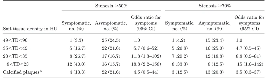
Plaque density on CT, a potential marker of ischemic stroke
In the article, “Plaque density on CT, a potential marker of ischemic stroke” (Neurology2006;66:118 –120) by J.-M. Serfaty et al., table 2 contains significant errors. The accurate table is as follows:
Table 2Association between symptoms and plaque characteristics
Stenosisⱖ50% Stenosisⱖ70%
Soft-tissue density in HU
Symptomatic, no. (%)
Asymptomatic, no. (%)
Odds ratio for symptoms
(95% CI)
Symptomatic, no. (%)
Asymptomatic, no. (%)
Odds ratio for symptoms
(95% CI)
49⬍TD⬍96 1 (3.3) 25 (24.5) 1.0 1 (4.2) 15 (23.4) 1.0
35⬍TD⬍49 5 (16.7) 22 (21.6) 5.7 (0.6–52) 5 (20.8) 16 (25.0) 4.7 (0.5–45) 23⬍TD⬍35 8 (26.7) 17 (16.7) 11.8 (1.3–102) 7 (29.2) 12 (18.8) 8.8 (0.9–81)
⫺8⬍TD⬍23 12 (40.0) 16 (15.7) 18.8 (2.2–158) 8 (33.3) 8 (12.5) 15 (1.6–142) Calcified plaques* 4 (13.3) 22 (21.6) 4.5 (0.5–44) 3 (12.5) 13 (20.3) 3.5 (0.3–37)
Each quartile of soft-tissue density and the excluded calcified carotid group (Reader 1):p⫽0.004 (stenosis⬎50%) and
p⫽0.04 (stenosis⬎70%). Similar results were obtained for Reader 2.
* Two patients could not be included in this group as no reliable clinical information could be retrieved.