ABSTRACT
CHERRY, NATALIE ANNE. Ecological Diversity of Bartonella. (Under the direction of Edward B. Breitschwerdt and Sam L. Jones.)
The Bartonella genus is composed of over 22 species or subspecies of gram-negative, fastidious, facultative intracellular bacteria that have been identified as the cause of, or
associated with, numerous clinical manifestations in humans and dogs. Bartonella species have also been detected in a wide range of other mammalian species, including ruminants,
horses, rodents, and sea mammals. Individual Bartonella spp. have co-evolved with specific mammalian hosts, such as B. bacilliformis and B. quintana with humans, B. henselae with domestic cats, B. bovis with cattle, and presumably, B. vinsonii subsp. berkhoffii with canids. When an organism infects a host without causing clinical pathology, the host species is
considered a reservoir, a mammalian niche that allows the pathogen to survive, propagate,
and facilitate dispersal to other hosts. The adaptation of Bartonella spp. to a broad range of mammalian hosts, as well as vectors which transmit these bacteria, is representative of the
complexity of the ecological diversity of members of the Bartonella genus.
To further define this ecological diversity and implications of this diversity to human
and veterinary medicine, a detailed review of the literature is provided relative to the host,
vector, physiological, geographic, and genetic ecology. Furthermore, three studies were
conducted to determine the prevalence of Bartonella in various mammalian populations, including cattle, dogs, humans, and horses.
host of this Bartonella sp., from North Carolina, with emphasis on development of new PCR primers to enhance detection of B. bovis in these ruminants (Cherry et al., 2009). Results of this study revealed a molecular prevalence of Bartonella in over 80% of cattle tested and reported the first detection of B. henselae in cattle. Next, a study was conducted to determine if Bartonella infection was the cause or a co-factor in illness in a veterinarian/dog breeder in Virginia and her sick Doberman pinschers (Cherry et al., 2011a). Evidence of Bartonella
infection and/or exposure was detected by serological, molecular, and/ or microbiological
methods in the breeder and five of her dogs. These results, together with the clinical
manifestations and environmental factors described, indicated Bartonella spp. likely played a primary or secondary role in the breeder and dogs’ illnesses. Lastly, a study was performed
to identify Bartonella infection in four horse populations, healthy adults (49), sick foals (15), adults with musculoskeletal manifestations (26), and adults presenting with colic (96), by
serological, molecular, and microbiological methods which is the first Bartonella prevalence study conducted in horses to our knowledge. Bartonella DNA was detected in the healthy (1/49), sick foal (3/15), and musculoskeletal disease (5/26) populations, though there was no
evidence of Bartonella infection in any horses within the colic population. By statistical analysis, Bartonella infection was most frequent in sick foals and adult horses with musculoskeletal manifestations, indicating further studies are warranted to determine the
pathogenic potential of bartonellosis in equids.
Development of detection methods for diagnosis of Bartonella infection and
has demonstrated the ubiquitous nature of Bartonella spp. Without these developments, researchers, clinicians, veterinarians, and the general public would not be aware of the
ecological diversity, pathogenic potential, or risk factors of Bartonella infection in humans or animals. Further investigation is warranted into the mechanisms of transmission from vectors
Ecological Diversity of Bartonella
by
Natalie Anne Cherry
A dissertation submitted to the Graduate Faculty of North Carolina State University
in partial fulfillment of the requirements for the degree of
Doctor of Philosophy
Comparative Biomedical Sciences
Raleigh, North Carolina
2012
APPROVED BY:
_______________________________ ______________________________
Edward B. Breitschwerdt Sam L. Jones
Committee Chair Committee Co-chair
DEDICATION
This dissertation is dedicated to my parents, Elwood and DiAnna Cherry, and my entire
BIOGRAPHY
ACKNOWLEDGMENTS
My sincerest gratitude goes out to all of my committee members for their kindness,
support, and encouragement throughout my PhD career at NC State. I would like to thank Dr.
Ed Breitschwerdt, my mentor and advisor, whose unwavering support has allowed me to
grow as a student, a scientist, and a person. I would also like to thank my other committee
members, Dr. Sam Jones, my co-advisor, Dr. Ricardo Maggi, and Dr. Jennifer Davis, for
their advice, encouragement, technical expertise, and clinical knowledge.
Thank you to the former and current faculty and staff of the Intracellular Pathogens
Research Laboratory and Vector Borne Disease Diagnostic Laboratory for their belief in me
and technical assistance, and for an enjoyable experience while working on my degree,
including Dr. Pedro Diniz, Dr. Belen Cadenas, Dr. Nandhakumar Balakrishnan, Dr. Patricia
Mascarelli, Julie Bradley, and Barbara Hegarty. I was very blessed to also have the support
of the graduate students in the CBS program, including Mrudula Varanat, Jingjing Li, Rita
Simoes, and Elizabeth Pultorak. An additional thank you to Dr. Shila Nordone, Melissa
TABLE OF CONTENTS
List of Tables ... vii
List of Figures ... viii
List of Abbreviations ... ix
Chapter 1. Ecological diversity of Bartonella ...1
Introduction ...2
Host ecology ...3
Reservoir hosts ...4
Humans ...4
Cats ...6
Canids ...10
Cattle ...14
Rodents ...15
Accidental hosts ...16
Humans ...17
Cats ...21
Domestic dogs ...22
Horses ...25
Summary ...26
Vector ecology ...26
Sandflies ...27
Lice ...28
Fleas ...29
Biting flies ...30
Ticks ...31
Summary ...33
Virulence factors and pathogenesis ...34
Erythrocytes ...35
Endothelial cells ...38
Epithelial cells ...40
Other cell types ...41
Geographic distribution and genetic diversity ...43
Summary ...49
References ...50
Chapter 2. PCR detection of Bartonella bovis and Bartonella henselae in the blood of beef cattle ...79
Abstract ...81
Introduction ...82
Materials and methods ...84
Results ...87
Discussion ...89
References ...92
Chapter 3. Ecological diversity of Bartonella species among dogs and their owner in Virginia ...99
Abstract ...101
Introduction ...101
Materials and methods ...104
Results ...108
Discussion ...116
References ...121
Chapter 4. Serological and molecular prevalence of Bartonella spp. infection in healthy and sick horses and foals from the southeastern United States ...135
Abstract ...138
Introduction ...139
Materials and methods ...141
Results ...143
Discussion ...146
Footnotes ...153
References ...153
LIST OF TABLES Chapter 1. Ecological diversity of Bartonella
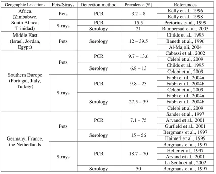
Table 1. Geographic locations, populations tested, detection methods, and prevalence of
Bartonella henselae in domestic cats worldwide ...77
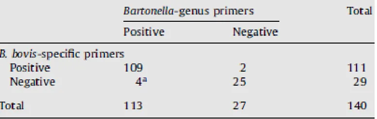
Chapter 2. PCR detection of Bartonella bovis and Bartonella henselae in the blood of beef cattle
Table 1. Comparative efficiency of Bartonella-genus primers and B. bovis-specific primers for the detection of B. bovis ...98
Chapter 3. Ecological diversity of Bartonella species among dogs and their owner in Virginia
Table 1. Signalment, year of illness onset and Bartonella detection, primary clinical
abnormalities, Bartonella species identified by DNA sequencing, and the clinical status at the conclusion of the study for five Doberman pinscher dogs kennel and the kennel
LIST OF FIGURES
Chapter 3. Ecological diversity of Bartonella species among dogs and their owner in Virginia
Figure 1. Agarose gel electrophoresis illustrating Bartonella genus intergenic spacer (ITS) polymerase chain reaction (PCR) amplicons (lane 2) obtained from the paraffin-embedded histocytic sarcoma from Dog 2 ...133
LIST OF ABBREVIATIONS
PCR – polymerase chain reaction
CSD – Cat Scratch Disease
BAPGM – Bartonella alpha-Proteobacteria growth medium T4SS – Type IV secretion system
BEP – Bartonella effector protein BadA – Bartonella adhesin protein A TAA – trimeric autotransporter adhesin
HPC – hematopoietic progenitor cells
TLR – toll-like receptor
LPS – lipopolysaccharide
PFGE – pulse-field gel electrophoresis
MLTS – multi-locus type sequencing
Vomp – variably-expressed outer membrane proteins
Ecological Diversity of Bartonella
Introduction
Microbial ecology is the study of the relationships of microorganisms with each other
and their environment. The genus Bartonella belongs to the class alpha-Proteobacteria and is comprised of pleomorphic, gram-negative, fastidious, facultative intracellular bacteria which
are primarily vector-borne. Research over the past two decades has elucidated the vast
ecological diversity of this genus with respect to host, vector, geographic, and genetic
ecology.
Over 22 species, subspecies, or Bartonella with Candidatus status have been
identified worldwide in a variety of hosts, including humans and domestic and wild animals,
including dogs, cats, deer, horses, cows, marine mammals, feral swine, and numerous rodent
species (Breitschwerdt and Kordick, 2000; Boulouis et al., 2005; Maggi et al., 2005, 2008;
Chomel et al., 2006; Beard et al., 2010). Many Bartonella species have been shown to cause, or be associated with, clinical pathology in some of these hosts.
The symbiotic relationships of Bartonella species with their respective mammalian hosts is complex. This is due to their promiscuous, fastidious nature and the numerous
clinical manifestations they can induce upon infection. Therefore, the study of Bartonella
species in an experimental setting, in vitro and in vivo, does not typically mimic natural infection. Advancements in serological, molecular, and microbiological techniques has
Host Ecology
With the exception of B. bacilliformis and B. quintana, which are classified as “specialists” because they generally only utilize humans as reservoirs, most Bartonella
species are referred to as “generalists” because they are able to infect and persist in numerous
animal hosts. Although B. quintana has been categorized as a “specialist”, there is evidence to support the possibility that this Bartonella sp. may have adapted the means to utilize another mammalian species, the domestic cat, as a reservoir host (Drancourt et al., 1996; La
et al., 2005; Breitschwerdt et al, 2007a). For example, B. henselae is classified as a “generalist” because though the cat is considered the primary reservoir of this species, B.
henselae DNA has been identified in many other mammalian species (Breitschwerdt and Kordick, 2000; Maggi et al., 2005, 2008; Valentine et al., 2007; Harms et al., 2008; Jones et
al., 2008; Chomel et al., 2009; Beard et al., 2010; Breitschwerdt et al., 2010a).
In contrast to other vector-borne diseases which are typically limited to one host,
many Bartonella species have the ability to be transmitted from a reservoir host to an accidental host. Interestingly, the reservoir host of one Bartonella sp. may be infected with other Bartonella spp., resulting in its utilization by Bartonella as an accidental host as well as a reservoir. Bartonella infection may at times be readily cleared by these hosts, but it has demonstrated the ability to evade some host immune systems (Vermi et al., 2006; Popa et al.,
2007). In cases where the bacteria are able to accomplish this, chronic infection may result,
leading to pathophysiological complications in accidental hosts by inadvertent transmission.
hosts resulting in chronic bacteremia and further evasion of the adaptive immune system
(Merrell and Falkow, 2004). This strategy is useful for Bartonella as a vector-borne blood pathogen because in order for a particular bacterium to be successful, it must evolve with a
primary mammalian source that is able to sustain the bacteria, essentially forming a
“commensal” relationship with its host (Dale and Moran, 2006). The establishment of a
primary reservoir host for Bartonella spp. is key because it allows for persistent infection at a high level of bacteremia for a prolonged period of time in a host, permitting uptake by
blood-feeding arthropods and subsequent transmission to other hosts.
Reservoir hosts Humans
Humans are the known reservoir for two species of Bartonella, B. bacilliformis and B.
quintana. B. bacilliformis is the causative agent of Carrion’s disease, characterized by massive hemolytic anemia, which is found primarily in the Andes region of Peru, Columbia,
and Ecuador (Ihler, 1996). It was the only member of the genus Bartonella until 1993, when the Rochalimaea and Bartonella genera were merged (Brenner et al., 1993). Alberto Barton first identified this bacteria in erythrocytes in 1905, and Towsend first documented the
sandfly, Lutzomyia verrucarum, as the natural vector of B. bacilliformis in 1913. Before the etiologic agent of this human bartonellosis agent was identified, there is evidence that pre-
Columbian natives of South America were affected by disease caused by B. bacilliformis
identified in the Andes region of South America (Gray et al., 1990; Cooper et al., 1997; Ellis
et al., 1999a; Kosek et al., 2000; Maco et al., 2004), despite medical advances.
Bartonella quintana is the causative agent of trench fever which is transmitted by the human body louse (Pediculus humanus humanus). Trench fever, also known as five day fever characterized by recurrent, cyclical fever, headaches, dizziness, and leg pain, was first
documented in soldiers during World War I with an estimate of more than one million
affected individuals at that time (Maurin and Raoult, 1996). Cases were again reported
during World War II (Kostrzewski, 1949).
Interestingly, it appears that the clinical manifestations of B. quintana infection are quite variable, presumably due to individual host responses. McNee et al. documented two
different clinical presentations in individuals with trench fever; one group presented with
classic characteristics of trench fever, whereas the other group presented with shorter initial
illnesses but with frequent relapses (McNee et al., 1916). Other pathophysiological
consequences of B. quintana infection in humans include chronic bacteremia (Kostrzewski, 1949; Foucault et al., 2002; Foucault et al., 2003; Capo et al., 2003), endocarditis (Houpikian
and Raoult, 2005), bacillary angiomatosis (Koehler et al., 1992; Santos et al., 2000; Sala et
al., 2005), and lymphadenopathy (Raoult et al., 1994; Drancourt et al., 1996). B. quintana has re-emerged as a human pathogen in recent years with risk factors including homelessness,
low socioeconomic status, alcoholism, body lice infestation, and, potentially, exposure to cats
(Spach et al., 1995; Jackson et al., 1996; Koehler et al., 1997; Spack and Koehler, 1998;
Cats
The domestic cat is the known reservoir host of B. henselae, the agent of CSD, but cats can also be infected with B. quintana (Drancourt et al., 1996; Parrot et al., 1997; Scully et al., 1998; La et al., 2005; Breitschwerdt et al., 2007a) and potentially serve as reservoirs
for B. clarridgeiae (Clarridge et al., 1995; Kordick et al., 1999) and B. koehlerae (Droz et al., 1999; Rolain et al., 2003a; Avidor et al., 2004). A high prevalence of chronic bacteremia in
reservoir populations allows Bartonella spp. to be transmitted more effectively from host to host, as demonstrated in cat populations worldwide.
B. henselae was first isolated from cats in the early 1990s (Regnery et al., 1992a; Koehler et al., 1994). Since that time, further research, including epidemiological data and
experimental infection and transmission studies, have proven the cat as a competent
mammalian reservoir host of B. henselae. A study by Koehler et al. in 1994 demonstrated cats were able to remain Bartonella bacteremic for months at a time, and the authors also isolated B. henselae from 41% of 61 cats in the San Francisco Bay region of California. Another study in northern California documented B. henselae bacteremia in 39.5% of cats with an overall seroreactivity of 81% against B. henselae antigens (La Scola et al., 2002); additionally, the B. henselae antibody titers were higher in the bacteremic cats than in the nonbacteremic ones. Authors of this study also evaluated the population of cats tested for risk
factors for B. henselae infection, and they found stray or impounded cats, younger cats, and flea-infested cats were more likely than pet cats to be bacteremic.
A study in France documented 53% of stray cats as bacteremic with either B.
seroprevalence in pet and feral cats in the United Kingdom was reported as 40.6 and 41.8%,
respectively (Barnes et al., 2000). More recently, B. henselae or B. clarridgeiae DNA was amplified from 47.8% of cats from Alabama and Virginia and from 59.8% of the fleas
collected from those cats (Lappin et al., 2006).
Since Bartonella henselae can be transmitted from cats to humans by bite or scratch, potentially causing clinical illness in the non-reservoir human host, other groups have used
molecular methods to test for Bartonella DNA in cat samples other than blood. Recently, Kim et al. documented PCR evidence of B. henselae from more than 40% of the saliva, nails, and blood samples of feral cats in Korea (Kim et al., 2009). In the US, blood and tissue
samples from feral cats were tested for Bartonella DNA, and either B. henselae or B.
clarridgeiae was amplified from 31.4% of skin biopsies, 17.6% of both gingival and claw bed swabs, and 56.9% of blood samples (Lappin and Hawley, 2009).
Historically, studies involving cats experimentally-infected with B. henselae have yielded comparable results in regard to bacteremia but more contradictory results in clinical
presentation. Two studies documented no signs of clinical illness in cats inoculated with B.
henselae (Regnery et al., 1996; Abbott et al., 1997), similar to cats naturally-infected with B.
henselae (Regnery et al., 1992a; Kordick et al., 1995). Other studies resulted in febrile, self- limiting clinical illness in experimentally-infected cats. One study demonstrated bacteremia
in young cats via intravenous inoculation of B. henselae as early as 2 weeks post-infection, persisting up to 32 weeks (Guptill et al., 1997). B. henselae-inoculated cats developed fever and anorexia, though these symptoms were typically self-limiting, lasting only 2-7 days.
even though they recovered from initial infection and were outwardly healthy. Though this
experiment does not mimic the natural route of infection of cats with B. henselae by cat fleas, these results show that cats are able to recover from initial infection and remain clinically
healthy while sustaining a high level of bacteremia, as these authors concluded. Another
study by Guptill et al. in 1998 sought to determine if B. henselae infection in female cats resulted in perinatal transmission and/or reproductive effects. These authors found that
bacteremic mother cats inoculated with B. henselae intradermally did not transfer the bacteria to their offspring but did report reproductive failure, including delayed conception and failure
to conceive.
Kordick et al. inoculated specific-pathogen free cats with B. henselae and/or B.
clarridgeiae in 1999 by blood transfusion which produced similar results to the previous study, including acute, self-limiting illness a few days post-B. henselae or B. clarridgeiae
inoculation and histopathological lesions in major organs. Authors of another study
documented similar but more severe clinical illness in cats inoculated intradermally with a
known pathogenic strain of B. henselae (O’Reilly et al., 1999). These authors concluded the variable results between the studies may have resulted from hereditary components,
inoculation routes, or, more likely, virulence of the B. henselae strain used as the inoculum. Inoculation route is a critical variable when evaluating pathogenesis of a bacteria in a
mammalian host experimentally, and studies delivering B. henselae intradermally have been the most effective, yielding a larger percentage of cats infected as well as a higher level of B.
considering B. henselae transmission to cats is essentially intradermal through the bite of cat fleas.
B. clarridgeiae was first isolated from the pet cat of an HIV-positive patient
(Clarridge et al., 1995). The worldwide distribution of B. clarridgeiae isolates is inconsistent, ranging from no greater than 10% in some studies (southeastern United States, Japan,
Taiwan) up to 17 to 36% in some regions (France, Philippines, the Netherlands, Thailand).
The high prevelance of B. clarridgeiae bacteremia in some regions provides evidence to support cats as a reservoir host for this Bartonella sp.
B. koehlerae is a relatively newly-recognized Bartonella species, and one difficulty researchers have encountered while seeking to further characterize this Bartonella species is its fastidious nature (Droz et al., 1999; Avidor et al., 2004). B. koehlerae has only been isolated from one cat in Israel (Avidor et al., 2004), one cat in France (Rolain et al., 2003a),
and two cats in California (Droz et al., 1999). Experimental inoculation of cats with B.
koehlerae isolated from one of the cats from California resulted in an average 74 day bacteremia in 4 cats with a specific antibody response to B. koehlerae antigens, and they were asymptomatic (Yamamoto et al., 2002). The kitten from which B. koehlerae was isolated in France was suspected of inducing CSD in its owner (Rolain et al., 2003a).
Collectively, these findings implicate cats as a likely reservoir for B. koehlerae. Given the high prevalence of Bartonella henselae bacteremia in domestic cats
relationship. Additionally, there is evidence to support the domestic cat as a reservoir host for
B. clarridgeiae and B. koehlerae.
Canids
Canids, including both domestic dogs and wild canids, such as the coyote, have been
implicated as reservoir hosts for Bartonella vinsonii subsp. berkhoffii, though domestic dogs can develop pathophysiological consequences from B. vinsonii subsp. berkhoffii infection. Evidence to support the domestic dog as a host reservoir for this Bartonella sp. includes the ability of B. vinsonii subsp. berkhoffii to cause persistent, prolonged bacteremia in dogs (Kordick and Breitschwerdt, 1998; Breitschwerdt and Kordick, 2000).
Coyotes (Canis latrans) are thought to be a key reservoir host for B. vinsonii subsp.
berkhoffii in the United States. Chang et al. demonstrated that 35% of coyotes from
California were seroreactive to B. vinsonii subsp. berkhoffii antigens (Chang et al., 1999). A year later, the same group reported a 76% seroprevalence of this Bartonella sp. in coyotes tested from California, and molecular detection of B. vinsonii subsp. berkhoffii was documented in 28% from this population (Chang et al., 2000). B. vinsonii subsp. berkhoffii
prevalence is typically higher in wild canids than domestic dogs, and a likely reason for this
is that domestic dogs are less likely to have frequent vector exposure, thereby decreasing
their risk for Bartonella infection.
a higher seroprevalence in stray dog populations. Lower B. vinsonii subsp. berkhoffii
seroprevalence has been reported in other locations, including Israel where only 10% of sick
dogs were seroreactive to B. vinsonii subsp. berkhoffii antigens (Baneth et al., 1998). Studies in dogs from Greece and Brazil documented only 5.3 and 1.5% B. vinsonii subsp. berkhoffii
seroreactivity, respectively (Mylonakis et al., 2004; et al., 2007). Seroreactivity to B. vinsonii
subsp. berkhoffii antigens has also been documented in 8.7% of US military working dogs (Honadel et al., 2001).
To date, four genotypes of B. vinsonii subsp. berkhoffii have been described,
designated as genotypes I, II, III, and IV based on sequences of the 16S-23S ribosomal RNA
intergenic spacer region of the Bartonella genome as well as the pap31 gene, a
bacteriophage-associated heme-binding protein (Maggi et al., 2006). Each of the B. vinsonii
subsp. genotypes have been identified in the following mammals in various geographic
locations: genotype I from a human, dogs, and coyotes in the United States, genotype II from
dogs, coyotes, humans, and a cat in the United States, genotype III from gray foxes in the
United States and a human with endocarditis in Europe, and genotype IV from a dog with
endocarditis in Canada (Kordick et al., 1996; Kordick and Breitschwerdt, 1998;
Breitschwerdt et al., 1999; Chang et al., 2000; Roux et al., 2000; Hoar et al., 2003; Maggi et
al., 2006; Cadenas et al., 2008; Varanat et al., 2009; Cherry et al., 2011a).
B. vinsonii subsp. berkhoffii was first isolated from a domestic dog with endocarditis (Breitschwerdt et al., 1995) but has been established as the cause of, or associated with, other
clinical manifestations such as myocarditis, cardiac arrhythmias, anemia/thrombocytopenia,
(Breitschwerdt et al., 1999; Tuttle et al., 2003; Breitschwerdt et al., 2004; MacDonald et al.,
2004; Smarick et al., 2004; Breitschwerdt et al., 2005; Pesavento et al., 2005; Sykes et al.,
2006; Cockwill et al., 2007; Cadenas et al., 2008; Diniz et al., 2009a; Breitschwerdt et al.,
2010b; Cherry et al., 2011a; de Caprariis, 2011). Granulomatous inflammatory diseases
(Pappalardo et al., 2000) and vasoproliferative disorders, such as peliosis hepatis and
bacillary angiomatosis, have also been described in dogs with evidence of B. vinsonii subsp.
berkhoffii infection (Kitchell et al., 2000; Yager et al., 2010). Recently, B. vinsonii subsp.
berkhoffii was isolated or PCR-amplified from idiopathic pericardial, pleural, or abdominal effusions (Cherry et al., 2009) and from joint and seroma fluids (Diniz et al., 2009a).
Factors such as immunosuppression and co-infection could facilitate presentation or
exacerbation of clinical symptoms in dogs. In the first case of B. vinsonii subsp. berkhoffii
isolation from the dog with endocarditis (Breitschwerdt et al., 1995), the patient had been
administered long-term treatment of corticosteroids after a diagnosis of systemic lupus
erythematosus (SLE), potentially contributing to endocarditis pathology and subsequent
isolation of this Bartonella sp., as suggested by the authors of this study and described in two cases of Bartonella infection in humans (Turner et al., 2005; Vikram et al., 2007).
In the context of infection, numerous studies have reported evidence of
co-infection of sick domestic dogs in the United States with B. vinsonii subsp. berkhoffii and another Bartonella sp. or another vector-borne organism. One study found molecular
evidence of Bartonella infection in 28% of dogs diagnosed with infective endocarditis at the UCD School of Veterinary Medicine (MacDonald et al., 2004), including three dogs in which
Bartonella antigens were also seroreactive to Anaplasma phagocytophilum antigens. In 1997, Pappalardo et al. documented a 3.6% seroprevalence of B. vinsonii subsp. berkhoffii among sick dogs from North Carolina and Virginia, with a strong correlation between serological
evidence of B. vinsonii subsp. berkhoffii and either seroreactivity to Ehrlichia canis (36%) or
Babesia canis (57.1%) antigens. A study published in 1999 documented co-infection of sick Walker hounds in North Carolina with Bartonella and at least one other vector-borne
organism by PCR (Kordick et al., 1999). In addition, 93% of the dogs tested in that study
were seroreactive to B. vinsonii subsp. berkhoffii antigens. Diniz et al. also documented co-infection of two ill dogs with B. henselae and B. vinsonii subsp. berkhoffii, which may have exacerbated clinical symptoms (Diniz et al., 2009a). Recently, molecular evidence of
Bartonella infection was reported in a prevalence study of sick dogs in the United States, documenting detection of Bartonella spp. in 9.2% of those dogs by PCR of blood or BAPGM blood enrichment culture (Perez et al., 2011). A subset of the dogs were infected with B.
vinsonii subsp. berkhoffii, with an additional nine cases of co-infection with B. vinsonii
subsp. berkhoffii and another Bartonella sp. Detection of antinuclear antibodies (ANA), an indication of SLE, has been reported in dogs seroreactive to B. vinsonii subsp. berkhoffii,
Rickettsia rickettsii, and Ehrlichia canis antigens. The host immune response to microbial infection could have led to the development of ANA in these dogs (Smith et al., 2004). This
work is supported by other studies reviewed by Pisetsky in 2008 which have documented
cross reactivity between genetic sequence motifs of bacterial and mammalian DNA,
potentially skewing ANA diagnostic results. Furthermore, it has been shown that
autoantibodies, possibly initiating a robust immunologic response and autoimmune disease in
other hosts.
Cattle
B. bovis (formerly B. weissii), B. chomelii, B. schoenbuchensis, and B. henselae are the species to date that have been identified in cattle (Brocklesby, 1970; Chang et al., 2000b;
Breitschwerdt et al., 2001; Bermond et al., 2002; Rolain et al., 2003b; Maillard et al., 2004;
Kelly et al., 2004; Cherry et al., 2009; Tsai et al., 2011). B. bovis was first isolated from cats and classified as B. weissii but was renamed B. bovis by Bermond et al. in 2002. Bartonella
infection has been documented in a number of wild and domestic ruminants, and research
over the past decade implicates cattle as the natural reservoir hosts of B. bovis.
B. bovis infection in cattle was first described in the literature in 1934 (Donatien and Lestoquard, 1934)and since that time, B. bovis has been documented in cattle around the world, including North America (Chang et al., 2000b; Breitschwerdt et al., 2001; Maillard et al., 2004; Cherry et al., 2009), Africa (Kelly et al., 2004), and Europe (Brocklesby et al.,
1970; Bermond et al., 2002; Tsai et al, 2011). In 2000, Chang et al. reported isolation of
Bartonella from 49% of beef and dairy cattle from Oklahoma and California (Chang et al., 2000b). By PCR/RFLP analysis, these isolates were most closely related to B. weissii. A high prevalence was also reported in beef cattle from North Carolina in 2009, when B. bovis was PCR-amplified from the blood of 82.4% of cattle (Cherry et al., 2009).
The high prevalence of bacteremia in clinically healthy populations of cattle indicate
nature of Bartonella infection may eventually lead to pathology, as demonstrated in cats infected with B. henselae (Breitschwerdt and Kordick, 2000). Though cattle are the reservoir hosts of B. bovis, a study by Maillard et al. in 2007 defined B. bovis as the cause of
endocarditis in two adult cows in France.
Rodents
Recent studies have supported the hypothesis that individual Bartonella spp. have co-evolved with specific rodent species as reservoir hosts (Telfer et al., 2007a, 2007b), and this
finding is important to human medicine because studies have documented detection of
rodent-adapted species in human patients with clinical illness. B. washoensis, with the presumed reservoir host as the California ground squirrel (Spermophilus beecheyi), was isolated in a human patient with cardiac disease in 2003 (Kosoy et al., 2003). This finding
suggests that rodents may serve as potential reservoirs for human infection, especially in
humans with extensive wildlife contact. In a study by Telfer et al., the prevalence of four
Bartonella species, B. birtlesii, B. doshiae, B. taylorii, and B. grahamii, in two rodent populations, bank voles (Clethrionomys glareolus) and wood mice (Apodemus sylvaticus), was assessed to determine the population dynamics of these two rodent populations relative
to Bartonella spp. prevalence (Telfer et al., 2007b). The authors found that the prevalence of
evaluation of temporal and spatial patterns of Bartonella bacteremia in 20 colonies of black-tailed prairie dogs (Cynomys ludovicianus) from 2003 to 2005 and reported a Bartonella spp. prevalence of 23.1%. Though there was no indication that Bartonella prevalence was based on black-tailed prairie dog population density, the authors suggetsed that this does not rule
out the potential for these rodents as reservoir hosts for Bartonella and thus, significance to public human health.
Studies in other parts of the world have documented a higher prevalence of
Bartonella bacteremia in rodent populations. A study of rodent populations in the UK found 62.2% of rodents to be Bartonella bacteremic (Birtles et al., 1994), and another study in the southeastern United States reported a 42.2% Bartonella prevalence (Kosoy et al., 1997). Birtles et al. also documented Bartonella bacteremia in 64% of woodland rodents in 2001.
Documentation of Bartonella bacteremia in rodents is not only important as a public health concern, but studies conducted in rodent populations allow researchers to gain a better
understanding of the ecological diversity of the Bartonella genus. Due to the high prevalence of Bartonella spp. in rodents and identification of specific rodent-adapted Bartonella spp., population dynamic studies can broaden the knowledge of Bartonella spp. persistence in specific reservoir hosts.
Accidental hosts
Inadvertent transmission of known or novel Bartonella spp. from reservoir hosts, such as wild or domestic animals, to non-reservoir, or accidental, hosts, such as domestic
Bartonella infection. The ability of vector-borne organisms to survive and propagate within specific mammalian reservoir hosts while maintaining the ability to be effectively transmitted
by vectors to accidental hosts is an important factor in human and veterinary medicine in the
context of Bartonella infection as a re-emerging pathogen. The diversity of mammalian hosts
Bartonella spp. can infect demonstrates this bacterium’s ability to continue to evolve, increase in virulence, and spread to new environments.
Humans
Six Bartonella species, including B. bacilliformis and B. quintana which utilize humans as reservoir hosts, have been identified as the cause of clinical pathology in humans,
but there is evidence to suggest that up to 13 Bartonella spp. can utilize humans as hosts (Boulouis et al., 2006; Chomel et al., 2006; Florin et al., 2008). In reservoir hosts of
Bartonella species, these bacteria typically persist in the host without pathophysiological consequences, though in accidental hosts, chronic bacteremia can result in clinical illness
with a wide range of symptoms. Bartonella infection has historically been recognized as the cause of disease in immunocompromised individuals, but it is now known that bartonellosis
can cause disease in immunocompetent humans as well (Lucey et al., 1992; Kumasaka et al.,
2001; Breitschwerdt et al., 2007b, 2008). Though the means of transmission of various
Bartonella spp. to humans has not been well-established, risk factors for Bartonella infection in humans include frequent, close contact with animals and their bodily fluids, as is the case
(Zangwill et al., 1992; Breitschwerdt et al., 2007b, 2008; Oliveira et al., 2010; Lin et al.,
2011).
B. henselae was first documented as the cause of CSD in 1992 by Regnery et al. and was granted recognition as the etiologic agent CSD in the mid-1990s (Regnery and Tappero,
1995; Jerris and Regnery, 1996), though this disease entity has been recognized for the past
century. B. henselae transmission from a cat to a human can occur by bite or scratch, with an estimated 24,000 cases in the United States annually (Jackson et al., 1993). B. henselae is not only able to merely be transmitted by the cat but also to evade the human immune system to
survive. Clinical pathology in CSD patients is typically characterized by self-limiting
lymphadenopathy and generalized flu-like symptoms, but in 5-10% of CSD cases (Anderson
and Neuman, 1997), B. henselae infection can cause chronic bacteremia leading to atypical manifestations of CSD, including endocarditis (Raoult et al., 1997; Fournier et al., 2001a;
Houpikian and Raoult, 2005), encephalopathy (Armengol and Hendley, 1999),
granulomatous hepatitis and nephritis (Kahr et al., 2000), bacillary angiomatosis and peliosis
hepatis (Koehler et al., 1997), and musculoskeletal manifestations such as myalgia (Maman
et al., 2007), osteomyelitis (Robson et al., 1999) and arthropathy (Hayem et al., 1996; Giladi
et al., 2005; Maman et al., 2007).
B. vinsonii subsp. berkhoffii, which was originally isolated from a dog w/ endocarditis (Breitschwerdt et al., 1995), has been identified as a new Bartonella sp. potentially
pathogenic to humans. Of the four genotypes of B. vinsonii subsp. berkhoffii, genotype II is most frequently detected in humans (Maggi et al., 2006). Roux et al. documented the
endocarditis, veterinary workers, and a wildlife biologist in 2000. More recently, B. vinsonii
subsp. berkhoffii was detected or isolated in immunocompetent individuals in the United States with symptoms including arthritis, fatigue, and neurological/neurocognitive
abnormalities (Breitschwerdt et al., 2007b, 2008). In 2009, B. vinsonii subsp. berkhoffii
genotype II was isolated from the blood of a boy with an epitheliod hemangioendothelioma
liver tumor and also detected in the tumor tissue (Breitschwerdt et al., 2009a). Furthermore,
studies have implicated direct transmission of Bartonella sp. from dogs to humans (Keret et al., 1998; Tsukahara et al., 1998; Kerkhoff et al., 1999a; Chen et al., 2007; Kim et al., 2009).
B. koehlerae, which is presumed to utilize the domestic cat as its reservoir host, was identified as the cause of culture-negative aortic valve endocarditis in a human in 2004
(Avidor et al., 2004). In 2010, B. koehlerae was detected in human blood and BAPGM blood enrichment cultures from eight immunocompetent human patients with symptoms such as
fatigue, joint and muscle pain, and headaches (Breitschwerdt et al., 2010b). Serological
results in several studies have also suggested B. clarridgeiae as a potential zoonotic
Bartonella sp. with documentation of lymphadenopathy, fever, and, in one case, a chest-wall abscess associated with B. clarridgeiae seroreactivity in human patients (Kordick et al., 1997; Margileth and Baehren, 1998; Sander et al., 2000; Laudisoit et al., 2011). Bartonella melophagi utilizes sheep as its reservoir host, and isolation of this species from two women with clinical symptoms including pericarditis, fatigue, muscle weakness, myalgia, joint pain,
headaches, and neurocognitive abnormalities was documented by Maggi et al. in 2009.
Other Bartonella spp., such as those that utilize rodents as reservoir hosts, including
and associated with clinical manifestations such as endocarditis. In 2005, Fenollar et al.
reported diagnosis of B. vinsonii subsp. arupensis, with the white-footed mouse as its reservoir host, as the cause of endocarditis in a human based on serology and PCR. B.
alsatica, which utilizes the rabbit as its reservoir, and B. elizabethae, a rat-adapted Bartonella
species, have both been isolated from the valves of patients with endocarditis in France and
the United States, respectively (Daly et al., 1993; Raoult et al., 2006). Candidatus B.
washoensis, for which the ground squirrel is the primary reservoir, was also associated with a
case of cardiac disease in Nevada in 2003 (Kosoy et al., 2003). B. vinsonii subsp. arupensis
was isolated from a rancher with neurological symptoms in Montana (Welch et al., 1999)
who had frequent opportunity for arthropod vector exposure, and the area where he worked
was known for a large population of voles which are known to harbor Bartonella spp.; though these observations were made, no direct link was established between B. vinsonii
subsp. arupensis from this patient and arthropod or rodent exposure. B. grahamii utilizes the vole as its reservoir host and has been associated with ocular/retinal manifestations in
humans (Kerkhoff et al, 1999b; Serratrice, 2003). B. rochalimae is a newly-recognized species of Bartonella for which the fox is the proposed reservoir, and in 2007, Eremeeva et al. isolated this species from a woman with bacteremia, fever, and splenomegaly. In
Thailand, B. tamiae was isolated from three ill patients who experienced symptoms including fever, fatigue, and headaches (Kosoy et al., 2008). Though no reservoir has been established
for B. tamiae, rodents have been proposed as potential reservoir hosts (Kosoy et al., 2010).
sequence most closely related to B. volans was recently identified in an 86 year-old man with memory loss, encephalopathy, and recent-onset arthritis (Breitschwerdt et al., 2009b).
These studies together indicate that Bartonella spp. are important zoonotic pathogens, many which have only recently been identified in humans, and that clinicians should be
aware of the potential for Bartonella infection and the clinical signs associated with bartonellosis. People with frequent exposure to wild or domestic animals and their
ectoparasites, such as veterinary professionals or wildlife biologists, may be at an increased
risk for Bartonella infection. Further studies are warranted to define modes of transmission, risk factors, as well as treatment regimens in the context of human bartonellosis.
Cats
Though cats are the primary reservoir host for B. henselae, and presumably B.
clarridgeiae and B. koehlerae, two other species, B. vinsonii subsp. berkhoffii and B.
quintana that utilize dogs and humans as reservoirs, respectively, have also been detected in cats. B. vinsonii subsp. berkhoffii genotype II DNA was recently amplified from the BAPGM blood culture of a cat with recurrent osteomyelitis (Varanat et al., 2009, 2011). B. quintana
has been detected by PCR-amplification from the dental pulp of a domestic cat (La et al.,
2005) as well as cat fleas (Rolain et al., 2003c). A serosurvey conducted by Baneth et al. in
1996 reported cats from Israel and North Carolina showing seroreactivity to only B. quintana
antigens. Regnery et al. published results from an experimental infection study demonstrating
transmission by domestic cat to humans has been suggested (Drancourt et al, 1996; Parrot et
al., 1997; Scully et al., 1998; Breitschwerdt et al., 2007a), suggesting the domestic cat could
be a potential reservoir host for B. quintana.
Domestic dogs
Canids have been suggested as the primary reservoir host for B. vinsonii subsp.
berkhoffii, but numerous other Bartonella species have been identified in domestic dogs. B.
henselae, B. quintana, B. elizabethae, B. clarridgeiae, B. washoensis (Chomel et al., 2006),
B. rochalimae (Diniz et al., 2009b; Henn et al., 2009) and sequences similar to B. volans
have been identified in dogs (Cherry et al., 2011a; Perez et al., 2011), providing evidence that
other Bartonella species besides B. vinsonii subsp. berkhoffii are able to utilize dogs as hosts. The route of transmission of Bartonella spp. to dogs has not been established, though evidence of cat-adapted Bartonella spp., such as B. henselae and B. clarridgeiae, infection in dogs suggests cats may play a role in transmission of these organisms to dogs via bite or
scratch. Alternatively, the cat flea may play a role in the transmission of these Bartonella
spp. to dogs, as this flea species has a ubiquitous geographic distribution and will feed on
other mammalian hosts than cats. Recent prevalence studies have demonstrated these
Bartonella spp. are widely distributed in domestic dog populations worldwide. In 2009, Inoue et al. documented isolation of B. clarridgeiae from 1 stray dog and 64 pet dogs in Thailand. Kim et al. documented isolation of B. clarridgeiae from nine dogs in Korea, and B.
co-infected with B. henselae and B. clarridgeiae, and the authors of this study suggested dogs may serve as secondary reservoirs for B. henselae and/or B. clarridgeiae. B. henselae and B.
clarridgeiae have been associated with hepatic disease in dogs (Gillespie et al., 2003), and B.
clarridgeiae was diagnosed as the causative agent of aortic valve endocarditis in a dog (Chomel et al., 2001).
Though evidence of exposure to or infection with B. henselae in domestic dogs has been frequently documented, the first isolation of this Bartonella sp. was from a dog in Gabon, Africa in 2004 (Gundi et al., 2004). Associated manifestations of B. henselae
infection in dogs include peliosis hepatitis, granulomatous lymphadenitis, endocarditis,
polyarthritis, and idiopathic effusions (Mexas et al., 2002; Gillespie et al., 2003; Goodman
and Breitschwerdt, 2005; Cherry et al., 2009). Interestingly, many studies to date have
documented a higher seroprevalence of B. henselae in dogs than B. vinsonii subsp. berkhoffii. For example, a serosurvey of dogs in the southeastern United States revealed 10%
seroreactivity to B. henselae antigens in healthy dogs and 28% in sick dogs (Solano-Gallego et al., 2004). Studies in Spain, Italy, and Sardinia have documented 17%, 6%, and 28% B.
henselae seroprevalence, respectively (Solano-Gallego et al., 2006; Di Francesco et al., 2007; Pinna et al., 2007). B. henselae seroprevalence surveys in dogs from Hawaii (Demers et al., 1995), Japan (Tsukahara et al., 1998), Zimbabwe (Kelly et al., 2004), and the United
Kingdom (Barnes et al., 2000) have varied between 3% and 14%. Henn et al. demonstrated a
and 33.3% to B. clarridgeiae antigens only, but only 2% were seroreactive to B. vinsonii
subsp. berkhoffii antigens only.
Perez et al. identified Bartonella in 9.2% of dogs from the US by either PCR or BAPGM enrichment culture. B. henselae, B. vinsonii subsp. berkhoffii, B. koehlerae,
Candidatus B. volans, and B. bovis DNA were detected, and cases of co-infection were also reported. Duncan et al. also reported molecular detection of multiple Bartonella spp. in dogs in 2007, documenting PCR-amplification of B. henselae, B. vinsonii subsp. berkhoffii, B.
quintana, and B. bovis DNA (Duncan et al., 2007, 2008).
Other Bartonella spp., including B. rochalimae, B. washoensis, B. quintana, and DNA sequences most closely resembling Candidatus Bartonella volans, have been identified in dogs. In 2003, Chomel et al. documented isolation of B. washoensis from a dog with mitral valve endocarditis. B. rochalimae was also identified as the cause of infective endocarditis in a dog from California (Henn et al., 2009) and detected in a dog from Greece by PCR (Diniz
et al., 2009b). Isolation of a Bartonella sp. most closely related to Candidatus B. volans was documented in two dogs from the United States (Perez et al., 2011), and another study
documented evidence of another B. volans-like species from a BAPGM blood enrichment culture from a dog in Virginia (Cherry et al., 2011a).
B. quintana, which has historically been considered a “specialist”, utilizing only humans as its host, was first documented to infect dogs in 2006 by Kelly et al. Authors of this
Dogs have been suggested as a probable reservoir for B. vinsonii subsp. berkhoffii, but research has shown that many other Bartonella spp. are able to utilize the domestic canid as a host. The implications of this are uncertain, but veterinarians should be aware of the
increasing body of knowledge surrounding the risk that dogs can be a reservoir for
Bartonella infection and bacteremic dogs can manifest a spectrum of clinical signs. It must be noted that most prevalence studies and case reports of Bartonella infection in dogs with species other than B. vinsonii subsp. berkhoffii have targeted sick dog populations,
suggesting that domestic dogs are most likely accidental hosts of other Bartonella spp. They apparently develop clinical pathology from infections with other reservoir-adapted
Bartonella spp., such as rodent-adapted B. washoensis, B. rochalimae, and Candidatus B. volans, and human-adapted B. quintana.
Horses
Equids are not presumed as a reservoir host for any Bartonella spp., but recent studies have demonstrated Bartonella infection in horses (Jones et al., 2008; Johnson et al., 2009; Cherry et al., 2011b, Identification). Jones et al. reported the first detection and isolation of
B. henselae from two horses, one with chronic arthropathy and the other with presumptive vasculitis (Jones et al., 2008). In 2009, Johnson et al. reported abortion of an equine fetus due
to B. henselae infection confirmed by molecular, histologic, immunohistochemical, and ultrastructural methods. In 2011, Cherry et al. detected this Bartonella sp. in a 2 year-old mare from Germany with high-grade hemolytic anemia. Unpublished data by Cherry et al.
most closely related to the rodent-adapted species Candidatus B. volans in horses.
Collectively, these studies indicate that horses are also accidental hosts for Bartonella spp. and are susceptible to Bartonella infection.
Summary
The Bartonella genus is comprised of at least 22 species, subspecies, or Candidatus
species that have been identified to date. For most of these species, a reservoir host has been
identified, including humans for B. bacilliformis and B. quintana, cats for B. henselae and potentially, B. clarridgeiae and B. koehlerae, presumably canids for B. vinsonii subsp.
berkhoffii, and various rodent species for species such as B. grahamii, B. taylorii, B. birtlesii, and B. doshiae. Individual Bartonella spp. have evolved the means to adapt to specific mammals and utilize them as reservoir hosts but also evolved the ability to infect
non-reservoir mammalian hosts, potentially leading to chronic bacteremia and clinical illness.
Epidemiological data and case reports suggest the implications of this are far-reaching, and
future studies are warranted to explore the mechanisms of transmission from reservoir hosts
to accidental hosts and how this information can be used to further understanding of the
ecological diversity of this genus of bacteria.
Vector Ecology
As is the case with other members of the class alpha-Proteobacteria, such as
verrucarum) is the sole vector for B. bacilliformis, the human body louse (Pediculus humanus humanus) is the primary vector for B. quintana, and the cat flea (Ctenocephalides felis) is the primary vector for B. henselae. The acquisition of these Bartonella spp. by their respective blood-feeding vectors, multiplication within these vectors, and transmission to
mammalian hosts have been extensively explored to provide definitive proof of vector
competence and ability of vectors to transmit Bartonella spp. to hosts.
Sandflies
The sandfly species Lutzomyia verrucarum responsible for B. bacilliformis
transmission is only found in the Andes region of South America, specifically Peru,
Columbia, and Ecuador, and it was first implicated as the vector transmitting the agent of this
disease in 1913 (Townsend, 1913).Experimental studies in monkeys to identify the vector
for B. bacilliformis were conducted in the early 1900s (Noguchi, 1928; Noguchi et al., 1929; Battistini, 1929, 1931). Direct transmission of this Bartonella sp. by Lutzyomia species to monkeys after feeding was documented by Battistini (Battistini, 1929, 1931), though the
means of replication and survival of B. bacilliformis within the sandfly is unclear. A study by Hertig in 1942 demonstrated organisms morphologically similar to B. bacilliformis on the proboscis and in the mid-gut, intestines, and feces of sandflies after feeding upon infected
peruensis from Peru (Ellis et al., 1999a).
Lice
The human body louse (Pediculus humanus humanus) was first suspected as the vector of B. quintana when it was found that humans developed symptoms after lice fed upon them (Swift, 1920). Further investigation revealed that the louse fecal matter itself was
infective (Swift, 1920; Bruce, 1921), though the causative agent, then presumed to be a
Rickettsial species, was not transmitted to louse offspring transovarially (Bruce, 1921).The
transmission of B. quintana to humans by the human body louse is a three stage cycle: 1) adult louse becomes infected via a bloodmeal, 2) viable B. quintana organisms multiply and are maintained in the louse gut, and 3) humans become infected by louse bite or through an
open wound contaminated with louse feces containing viable B. quintana.
In 1960, research on B. quintana revealed that this bacterium replicates within the stomach of the louse, followed by attachment to luminal epithelial cells (Weyer, 1960).
Vinson et al. confirmed these findings in 1969 when intrarectal inoculation of B. quintana
into body lice revealed viable infectious organisms in the louse gut lumen. The authors were
also able to visualize B. quintana in feces collected from lice feeding on an infected patient. More recent studies confirmed these earlier observations, demonstrating that B. quintana
does remain in the intestinal lumen of the louse, is excreted in its feces throughout its
lifespan, and is not transmitted transovarially, as is the case with B. bacilliformis (Fournier et al., 2001b). Furthermore, logarithmic growth of B. quintana in the louse midgut was
increased up to 1.3x10⁸ bacteria by day 17 (Seki et al., 2007). Furthermore, up to 1x10⁷
bacteria were detected in louse fecal matter on day 15 in that study.
In addition to the human body louse, a more recent study demonstrated B. quintana in head lice (Pediculus humanus capitis) from children in Nepal (Sasaki et al., 2006). Other
Bartonella species have been identified in several lice genera such as the spined rat louse (Polyplax spp.) (Reeves et al., 2006; Tsai et al., 2010) and the sucking louse (Hoplopleura
spp. and Neohaematopinus sciuri) (Durden et al., 2004; Reeves et al., 2006; Nelder et al., 2009). Bartonella species from either or both of these lice genera include B. rattimassiliensis,
B. tribocorum, B. phoceensis, and B. henselae. Interestingly, B. henselae was recently detected in seal lice (Echinophtirius horridus) collected from a harbor seal (Phoca vitulina) in the Netherlands (Morick et al., 2009). The significance of Bartonella species in lice infesting sea mammals and rodents reamins unclear, but the implications of these findings
warrant further investigation.
Fleas
The domestic cat is the natural reservoir for B. henselae with the cat flea (Ctenocephalides felis) as its primary vector.The cat flea has been demonstrated
experimentally to be a competent vector for transmission of this Bartonella species (Chomel et al., 1996) when fleas collected from B. henselae bacteremic cats were placed on five specific pathogen free (SPF) kittens, and four of those five kittens became bacteremic within
to remain in the flea gut for up to 9 days and replicate within the gut, similar to B.
bacilliformis in sandflies and B. quintana in lice (Higgins et al., 1996). In this study, B.
henselae was detected in the feces, and viable isolates were obtained on rabbit blood agar plates. In 1998, Foil et al. demonstrated transmission of B. henselae to cats by intradermal inoculation of infectious flea feces. Another study was conducted using a
streptomycin-resistant strain of B. henselae which demonstrated 1.8 x 10³ CFU/mg of the bacteria in flea feces after 2 hours with a decrease in colony count after 72 hours (Finkelstein et al., 2002). In
summary, these studies document replication of B. henselae in the flea gut and its viability in the feces as well as direct transmission of B. henselae to SPF cats via intradermal inoculation of infectious flea feces or incubation with bacteremic fleas.
Though the vector competence of the cat flea in the transmission of B. henselae has been established, molecular detection of B. quintana, B. koehlerae, and B. clarridgeiae in cat fleas has also been documented (Rolain et al., 2003c). These findings suggest that the cat flea
may be capable of transmission of other Bartonella spp. to mammalian hosts.
Biting flies
The potential role of Hippoboscidae flies, specifically Lipoptena, Hippobosca,
Melophagus, and Stomoxys spp., as vectors of Bartonella spp. to wild and domestic
ruminants has been suggested in the past few years, as Bartonella DNA has been detected in these flies in numerous countries. In 2004, Bartonella DNA was first identified in North American biting flies collected from cattle barns and feedlots in California (Chung et al.,
DNA was amplified from a stable fly (Stomoxys sp.) in that study. Within the same year,
Bartonella DNA was also detected in deer flies (Lipoptena cervi), horse flies (Hippobosca equina), and sheep keds (Melophagus ovinus) collected in France (Halos et al., 2004), and B.
schoenbuchensis was isolated from deer flies collected from roe deer and red deer in Germany (Dehio et al., 2004a). Furthermore, Dehio et al., demonstrated localization of B.
schoenbuchensis within the lumen of the midgut of deer keds. In 2006, Reeves et al. identified B. henselae and B. schoenbuchensis DNA in deer keds (Lipoptena mazamae) collected from white-tailed deer in the southeastern United States, and B. schoenbuchensis
DNA was amplified from deer flies in Massachusetts in 2008 (Matsumoto, 2008).
Blood-feeding vectors may uptake bacteria during Blood-feeding, but detection of bacteria in vector tissues
gives evidence of a vector’s role in the natural bacterial transmission cycle.
Ticks
The role of ticks in the transmission of Bartonella species has been an ongoing debate for years. A review by Billeter et al. in 2008 summarized the numerous tick species
harboring Bartonella DNA, including Dermacentor, Haemaphylsalis, Ixodes, and
Rhipicephalus. Though clinical, epidemiological, and molecular data supports the potential for tick transmission (Breitschwerdt and Kordick, 2000; Chang et al., 2001; Sanogo et al.,
2003; Skotarczak and Adamska, 2005; Billeter et al., 2008; Angelakis et al., 2010), DNA
detection alone does not provide evidence of direct bacterial transmission to animals or
murine model, demonstrating acquistion of Bartonella by ticks from an infected mouse and subsequent transmission to a naïve mouse by these ticks and localization of these bacteria
within adult ticks. In this study, a 4-week old OF1 female mouse was inoculated
intravenously with B. birtlesii. Following B. birtlesii infection, I. ricinus larvae and nymphs were allowed to feed on the infected mouse. After molting of larvae, the nymphs were
allowed to feed on three naïve 4-week old OF1 female mice. Within seven days, B. birtlesii
DNA was detected in the blood of the tick infested mice by PCR and sequencing. After six
days of blood culture, these bacteria were visualized by immunofluorescence, demonstrating
presence as well as viability of the bacteria. Adult ticks, fed on the B. birtlesii-infected mouse at the nymphal stage, were fed on a membrane feeder with uninfected blood for 8 days, and
these ticks effectively transmitted the bacteria to the uninfected blood, demonstrated by
presence of Bartonella DNA by PCR from blood on days 3 through 8. This data proves that
I. ricinus ticks are capable of acquiring B. birtlesii from an infected host and retaining the bacteria transstadially. In contrast to B. quintana in the louse (Weyer et al., 1960; Fournier et al., 2001b; Seki et al., 2007) and B. henselae in the cat flea (Higgins et al., 1996), this
experimental study supports the hypothesis that B. birtlesii is most likely transmitted from ticks to a mammalian host through salivary secretions rather than contaminated feces. This
study provides definitive proof that ticks are capable of acquiring, retaining, and effectively
transmitting B. birtlesii.
light microscopy, documented cytopathic effects following infection, including cellular
vacuolization and cell lysis, as well as amplification of B. henselae within the cells by quantitative real time PCR. Recently, B. vinsonii subsp. berkhoffii DNA was detected in
Rhipicephalus sanguineus ticks post-capillary tube feeding as well as in the feces of these ticks (Billeter et al., 2012). These results demonstrate that B. vinsonii subsp. berkhoffii is capable of infecting this tick species, potentially leading to subsequnt infection of
mammalian hosts through inoculation of tick feces through broken skin. Though this can not
be extrapolated to all species of ticks, Bartonella spp., or mammalian hosts, this data does provide evidence warranting consideration of Bartonella infection in patients with tick exposure.
Summary
Vector-borne organisms, such as members of the Bartonella genus, must adapt to the lifestyle of a specific vector and its physiological environment for survival and propagation.
These adaptations vary drastically depending upon the microorganism. B. bacilliformis, B.
quintana, and B. henselae have evolved not only to be carried by the sandfly, body louse, and cat flea, respectively, but have developed the ability to survive and replicate in the guts of
these arthropods after a blood meal. This is a key component to increase the likelihood of
transmission to a mammalian host through skin broken by scratching or through an open
wound. B. henselae facilitates its transmission to its natural reservoir host, the domestic cat, and its accidental human host by introduction of bacteremic flea feces via scratch or bite
not confirm competence or constitute a direct threat to animal or human global health;
however, the promiscuity of Bartonella species in their ability to adapt to a variety of
arthropods is unparalleled. Reports have demonstrated a broader range of Bartonella species identified in arthropods than in mammalian hosts (Abbot et al., 2007; Brinkerhoff et al.,
2010; Tsai et al., 2011). Evidence from these studies suggests that Bartonella, similar to the
Wolbachia, may have co-evolved with vector species in their exploitation of mammalian hosts (Charlat et al., 2003; Tsai et al., 2011).
Virulence factors and pathogenesis
Historically, Bartonella spp. were thought to be primarily intraerythrocytic bacteria, residing in red blood cells within a host, but further research has revealed that intracellular
microorganisms can invade other cell types with distinct pathogenic means of intracellular
infection and survival. The ability of species of the same genus to evolve unique pathogenic
mechanisms independently of one another to achieve the same goal, i.e. host cell invasion, is
known as adaptive radiation. The pathogenic mechanisms of Bartonella are demonstrated by the development of the T4SS in B. quintana, B. henselae, and B. tribocorum (Seubert et al., 2003; Saenz et al., 2007; Dehio, 2008), compared to the motility-mediated invasion of
erythrocytes by B. bacilliformis (Engel et al., 2011). T4SSs are pathogenicity factors often used by human bacterial pathogens such as Helicobacter pylori, Legionella pneumophila,
Brucella melitensis, and Bordetella pertussis (Christie et al., 2005). Three distinct T4SSs, VirB/VirD4, Vbh, and Trw (Seubert et al., 2003; Schulein et al., 2005; Schmid et al., 2006;