Medical Image Space Classification for
Disease Analysis Using PSO Optimized
K-Means Clustering
Hemant Sharma, Ms. Nandini, Deepak Dixit, Rohit Singla, Praveen
M.Tech Student, Dept. of Computer Science& Engineering, Shri Ram College of Engineering and Management,
Palwal, affiliated to M.D.U Rohtak (Haryana), India
Asst. Professor, Dept. of Computer Science& Engineering, Shri Ram College of Engineering and Management, Palwal,
affiliated to M.D.U Rohtak (Haryana), India
M.Tech Student, Dept. of Computer Science& Engineering, Shri Ram College of Engineering and Management,
Palwal, affiliated to M.D.U Rohtak (Haryana), India
M.Tech Student, Dept. of Computer Science& Engineering, Shri Ram College of Engineering and Management,
Palwal, affiliated to M.D.U Rohtak (Haryana), India
M.Tech Student, Dept. of Computer Science& Engineering, Shri Ram College of Engineering and Management,
Palwal, affiliated to M.D.U Rohtak (Haryana), India
ABSTRACT: One of the principle uses of picture process is restorative picture handle in medicinal pictures to diagnose the root cause of disease. To represent these pictures additional effectively, the image space classification approach is outlined so completely different areas over the image are known severally. This approach is outlined because the content primarily based retrieval. The content extraction or the world classification is needed to spot the medical image elements. during this scheme, a good image content retrieval approach so image space identification and colorization is performed. This bestowed work is split in 3 main stages. In initial stage, the high level meta-metric is outlined persecution clump. during this work, associate degree PSO improved K-Means clustering is outlined to perform the image space classification base on intensity analysis and edge element analysis for medical images. This image space classification analysis approach is truly region primarily based classification. Simply when the high level segmentation future work performed here is that the swarm improvement to filter the result of clump method. The Swarm improvement compared the meta-metric elements in terms of worldwide and native acceptableness analysis. Throughout this scheme, the identification of unequivocal space or constituent is performed to another cluster so the efficacy of K means cluster method is obtained. Once the cluster is finished, future stage is to perform the colorization over the image that known the various medical image areas clearly using PSO. The work is verified on completely different realistically Brain pictures accessible in DICOM or jpg format. The work is enforced in matlab environs. The obtained results show the effective space classification. The analysis of the work is outlined below completely different parameters like mean analysis, STD analysis, Frequency Analysis etc.
I. INTRODUCTION
Medical imaging space classification is growing analysis space in previous couple of years that covers the interest of researchers of various areas like medicine, physics and computing. Medical imaging has the analysis emergence in terms of image communication, imaging modalities, image computation etc. this type of processing support administrative, clinical, teaching and analysis activity. Medical image classification has rejuvenated as new mechanism within the identification and treatment of diseases. whereas the image modalities area unit helpful, such instrumentation is dear and resource hard to please. Therefore, to cut back the workload of medical employees, automatic measurements and improved visualizations of the modality knowledge is required. One main challenge is to perform automatic analysis on medical volume knowledge is to separate numerous tissues from one another. Their boundaries is also diffused or the medical volumes area unit generally distorted by noise The content based mostly info retrieval over the medical pictures really perform the image space segmentation in order that disjoint objects or the regions over the medical image are going to be identified. The segmentation over these pictures will be performed normally approach further as underneath complicated type. This extraction is tough as a result of the accuracy is one in every of the most associated development for such quite applications. enforced in matlab environs.
Content Based Image Retrieval : In recent years with the gaining quality of world wide web it's seen that the supply of image capturing devices like scanner, digital cameras, and therefore the size of digital image information is growing chop-chop. numerous domains like drugs, design, remote sensing, research etc square measure hard for economical looking, retrieval and browsing of pictures. To meet of these sure demands the image retrieval system came into existence. Basically there square measure 2 frameworks outlined one is text-based and another one is content primarily based. In text primarily based approach, question is annotated within the text descriptors that square measure any employed by the management system to retrieve pictures. however this approach incorporates a disadvantage that is quality. to beat this content primarily based retrieval came within the early 1980’s. [1]
Fig. 1. The principal components of all content-based image retrieval systems.
Step 1: The velocity and position of all particles are randomly set to within pre-defined ranges.
Step 2: Velocity updating – At each iteration, the velocities of all particles are updated according to below mentioned
where pi and vi are the position and velocity of particle i, respectively; pi ,best and gi, best is the position with the ‘best’ objective value found so far by particle i and the entire population respectively; w is a parameter controlling the dynamics of flying; R1 and R2 are random variables in the range [0,1]; c1 and c2 are factors controlling the related weighting of corresponding terms. The random variables help the PSO with the ability of stochastic searching.
Step 3: Position updating – The positions of all particles are updated according to below mentioned :
After updating, pi should be checked and limited to the allowed range.
Step 4: Memory updating – Update pi,best and gi,best when condition is met,
where f(x) is the objective function to be optimized.
Step 5: Stopping Condition– The algorithm repeats steps 2 to 4 until certain stopping conditions are met, such as a pre-defined number of iterations. Once stopped, the algorithm reports the values of gbest and f(gbest) as its solution. PSO utilizes several searching points and the searching points gradually get close to the global optimal point using its pbest and gbest. Initial positions of pbest and gbest are different. However, using thee different direction of pbest and gbest, all agents gradually get close to the global optimum.[23]
K-Means Clustering: K-means clustering algorithm is an unsupervised method. It is used because it is simple and has relatively low computational complexity. It is suitable for biomedical image segmentation as the number of clusters (k) is usually known for images of particular regions of human anatomy. K-means clustering is the most widely used and studied method among clustering formulations that are based on minimizing a formal objective function. Modification to K-means clustering method that makes
34 it faster and more efficient are proposed. It can be achieved by the clustering criteria and the unsupervised clustering- decides the clustering criteria by itself. The main argument of proposed methods is on the reduction of intensive distance computation, a simple mechanism by which at each iteration, the distance between each data points and the cluster nearest to it is computed and recorded in a data structure is suggested .Thus on the following iterations the distance between each data point and its previous nearest cluster is recomputed. [29] The steps for a standard k-means clustering algorithm are as follows.
1. Consider a set of n data points (feature vectors) to be clustered. Here the data points are the image pixels.
2. Assume the no: of clusters as k, where 2 < k < n. 3. Randomly select k initial cluster center locations.
4. All data points are assigned to a partition defined by nearest cluster centers. This is determined by using a distance measure to check the closeness of the pixels to the chosen cluster centers.
5. After the partitioning, the cluster centers are moved to the geometric centroid of their data points in their respective partitions.
6. Repeat steps 4 & 5 until the overall objective function is smaller than a given tolerance, or until the calculated cluster centers don’t move to new points.
Colors- For artists, journalists and advertisers color has been the foremost effective feature and most of the photography systems use it. the colour house utilized by the photographs square measure RGB (Red, blue and green), HSV (Hue, Saturation, Value). it's seen that the majority of the photographs square measure in RGB house however this house is never used for compartmentalization and looking because it doesn't correspond to human perception as compared to alternative areas. Texture- the feel feature has giant selection than color house. the feel tries to capture the characteristics of a picture or the amendments within the image components with the change within the direction. This feature has shown effective ends up in case of region extraction. Classification and form Features Image segmentation is a necessary preliminary step in most automatic pictorial pattern recognition and scene analysis issues. Segmentation subdivides a picture into its constituent regions or objects, that ideally correspond to completely different real-world objects. the amount to that subdivision is dispensed depends upon the matter being resolved. In image retrieval, many systems commit to perform an automatic classification of the photographs within the assortment for feature extraction. to possess an efficient segmentation of pictures mistreatment varied image databases the segmentation method needs to be done supported the colour and texture properties of the image regions. [2] form feature is one amongst the necessary feature image options however it's not wide used as color or texture feature is employed. form feature typically deals with ratio, Fourier descriptors, Moment invariants, disk shape etc.[1] the form feature is said to segmentation. it's troublesome to use form feature as compare to paint or texture feature thanks to quality of segmentation. Despite the problem it's employed in some systems.
Classification: the method of partitioning a digital image into multiple regions (sets of pixels) is named image Classification. Actually, partitions square measure completely different objects in image that have constant texture or color. The results of image segmentation could be a set of regions that conjointly cowl the complete image, or a collection of contours extracted from the image. All of the pixels in an exceedingly region square measure similar with relevancy some characteristic or computed property, like color, intensity, or texture. Adjacent regions square measure considerably completely different with relevancy constant characteristics. Applications of Classification a number of the sensible applications of image segmentation in Medical Imaging using Image space classification :
Locate tumors and alternative pathologies
.Measure tissue volumes
Computer guided surgery
Diagnosis
Treatment designing Study of anatomical structures
performance or efficacy can be a matter of question whereas the results and execution may vary based on medical images.
Figure 2: enforcing mechanism
II. LITERATURE REVIEW
Liu, Y. Et al. [1] proposed that in order to improve the retrieval accuracy of content based image retrieval systems, research focus had been shifted from designing sophisticated low level feature extraction algorithms to reduce the ‘semantic gap’ between the visual features and the richness of human semantics. This paper attempts to provide a comprehensive survey of the recent technical achievements in high level semantic-based image retrieval. Most recent publications had included in this survey covering different aspects of the research in this area including low level feature extraction, similarity measurement, and deriving high–level semantic features. Using supervised or unsupervised machine learning methods to associate low-level features with query concepts.
Henning Muller et al. [2] gives an overview of available literature in the field of content based access to medical image data and on the technologies used in the field. It gives an introduction into generic content based image retrieval and the technologies used and describes the techniques used in the implementation their datasets and evaluation. This paper also identifies explanation to some of the outlined problem with this field as it looks like many propositions for systems are made from the medical domain and research prototypes are developed in computer science departments using medical datasets. Still, there are very few systems that seem to be used in clinical practice.
B.Ramamurthy et al. [3] proposed that medical image retrieval based on shape feature, which uses canny edge detection algorithm for extraction of image shape and K-means algorithm for extraction of different regions of the image in order to improve better matching process between user query image and feature database images. We have shown that Canny Edge Detection and K-means clustering algorithms are quite useful for retrieval of relevant images from image database. V.S.V.S.
Dr. K. Sakthivel et al. [5] proposed modified K-means clustering algorithm to improve image segmentation accuracy and used a novel similarity distance measure where object uniqueness is considered during computation. The algorithm has been implemented and tested using 5000 general purpose images from COREL database and the retrieval performance is compared with the existing region based retrieval system. Feature extraction has been done using MATLAB image processing tool.
Zhen Ma et al. [6] proposed that for a concrete medical image segmentation task, researchers should combine the application background and practical requirements to design proper algorithms. Accuracy, complexity, efficiency and interactivity of a segmentation algorithm should all be the considered factors.
Riries Rulaningtyas and Khusnul Ain [7] defined a work of edge detection for braintumor detection. In this work Brain tumor diagnosis is done by doctors. For detecting brain tumor grading always gives different conclusion between one doctor to another. For helping doctors diagnose brain tumor grading, this research made a software with edge detections method, so it could give edge pattern of brain and brain tumor itself. Edge detection of brain tumor in this research is the first step for brain tumor grading research. This research found the best edge detection method for brain tumor detecting etween Robert, Prewitt, and Sobel method. From these three methods, Sobel method is suitable with case of brain tumors detecting. Sobel method had smaller deviation standard value than two others edge detection method.
III. RESERACHANDELABORATION
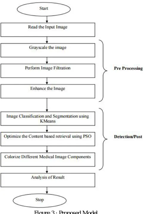
member estimation and local member estimation. If some value or the intensity value satisfy the local membership then it shows the existence of pixel or the component in particular cluster is valid. If the global membership satisfy, then the identification of the particular cluster will be done in which it actually satisfy the member. Based on this analysis, the component switching between the clusters will be done. At the final stage, the segmented area will be defined under some color model so that the colorization of different components over the image are performed. The recursive illustration of the work is given here underneath.
Proposed Model Workflow Diagram:
Figure 3 : Proposed Model
In the scheme the below steps explores the implementation along-with pseudo code:
1.Filter the Brain Image in terms of brightness, contrast and image size. 2. Define the Number of clusters over the Image called N clusters 3. Implement Min Max analysis over image to identify frequency range. 4. Identify Intensity Variation=Range/N
[The intensity of pixels that will change between cluster pixels] 5. For i=1 to N
6. Obtain Center for Cluster(i) called Center(i) and the relative cluster variation }
7. Set AvgChange=MinThreshold 8. While (AvgChange<> ActualMod)
[Repeat The cluster formation till the variation over the cluster pixel is obtained] {
9. Obtain the Segment Distance between pixel intensity and the cluster center 10. Update Variation min with minimum distance change using PSO 11. Include the pixels in different cluster based on intensity change 12. Take the Mean values to update the cluster centroids
} 42
13. Return ClusterList;
However the pseudo code s as under for ready reference and perusal :-
Step 1: Loading a gray scaled image Content Based Retrieval (Image)
/* Here Image is the actual medical image on which the content retrieval is performed*/ {
Image=To Gray(Image)
/*Check the image format and convert it to 8 bit grayscale image*/ Image=Adjust(Image)
/*preprocess the Image and perform the adjustment over the brightness and contrast*/
Step 2: Convert the image from RGB colour space to L*a*b* colour space
Unlike the RGB colour model, L*a*b* colour is designed to approximate human vision. There is a complicated transformation between RGB and L*a*b*.
(L*, a*, b*) = T(R, G, B). (R, G, B) = T’(L*, a*, b*).
Perform the Intensity Range Analysis called MinInt and MaxInt over the image.
Step 3: Undertake clustering analysis in the (a*, b*) colour space with the K-means algorithm In the L*a*b* colour space, each pixel has a properties or feature vector: (L*, a*, b*).
Like feature selection, L* feature is discarded. As a result, each pixel has a feature vector (a*, b*).
Applying the K-means algorithm to the image in the a*b* feature space where K = 3 (by applying the domain knowledge.
Perform the intensity based clustering over the Image and divide the area in small area segment under distance based analysis.
Step 4: Label every pixel in the image using the results from K-means Clustering (indicated by three different grey levels)
Step 5. Perform the convolution filter to highlight the edge points so that region classification will be performed
Step 6. Perform Region classification over the image.
Step 7. Obtain the clusters and take it as input set for PSO.
Step 8. Perform the cluster analysis in terms of frequency and variation analysis called velocity.
Step 9. Obtain the Local Best and Global Best analysis over each segment. For i=1 to Length(Image)
/*Process all area components over the Image using PSO*/ {
If (Dist1<Dist2) {
Identify the appropriate Cluster for Feature(i) and switch the clustering Update the cluster information
Update the Local Best and Global Best Parameters }
}
Perform the Colorization on obtained classification contents over the image }
III. RESULTSANDFINDINGS
Here figure 4 is showing the input image. The figure is showing the brain image. This applicable for brain, lung or any other medical image to perform the area image space classification and colorization. The input is here taken in jpg image form.
Figure 4 original gray scale medical image
Here figure 5 is showing the final output image after the colorization process. The colorization is here applied to perform the region separation so that the effective identification of component areas over the image will be done. Different components are here presented in different colors.
Classification Analysis:- Mean Value = 0.5000 Standard Deviation = 0.2932 Entropy Value = 5.9478
IV. CONCLUSION
In this present scheme, an effective Medical Image Space Classification for Disease Analysis using PSO Optimized K-Means Clustering approach is suggested by using hybrid three stage model. The presented model is defined with the integration of improved K=means algorithm followed by PSO based filtration approach. The presented work is defined on either medical DICOM or jpg images. These images can be brain images or the lung images. At the initial stage of work, Improved Kmeans algorithm is defined. This algorithm is based on the intensity based analysis followed by edge based analysis. This analysis is about to perform the region based segmentation over the image. At second stage, PSO is applied to perform the region filtering and switching of components. At the final stage, the color model is applied to perform the colorization. The result analysis is performed under different parameters such as frequency analysis, standard deviation analysis etc. The obtained results shows the effectiveness of the work.
ACKNOWLEDGMENT
The preferred spelling of the word “acknowledgment” in American English is without an “e” after the “g.” Use the singular heading even if you have many acknowledgments.
REFERENCES
[1] Ying Liu, Dengsheng Zhang, Guojun Lu,Wei-Ying Ma,“A survey of content based image retrieval with high level semantics ”,The Journal of the pattern
recognition society, vol. 40,pp. 262-282, 2007.
[2] Henning Müller, Nicolas Michoux, David Bandon, Antoine Geissbuhler, “A review of content-based image retrieval systems in medical applications–—
clinical benefits and future directions” International Journal of Medical Informatics, pp. 1-23, 2004.
[3] B.Ramamurthy, K.R.Chandran, “CBMIR: Shape-Based Image Retrieval Using Canny Edge Detection And K-Means Clustering Algorithms For Medical
Images”, International Journal of Engineering Science and Technology (IJEST), Vol. 3 No. 3, March 2011.
[4] V.S.V.S. Murthy, E.Vamsidhar, J.N.V.R. Swarup Kumar, P.Sankara Rao (2010) , “Content Based Image Retrieval using Hierarchical and K-Means Clustering
Techniques”, International Journal of Engineering Science and Technology, Vol. 2(3), pp. 209-212, 2010.
[5] Prof. Dr. K. Sakthivel, Dr. T.Ravichandran , Prof. Dr. C. Kavitha, “Performance Enhancement In Image Retrieval Using Modified K-Means Clustering
Algorithm” , Journal of Mathematics and Technology, ISSN: 2078-0257, February, 2010.
[6] Zhen Ma, João Manuel R. S. Tavares, R. M. Natal Jorgev, “A Review On The Current Segmentation Algorithms For Medical Images”. [7] Riries Rulaningtyas and Khusnul Ain, “Edge Detection For Brain Tumor Pattern Recognition”.
[8] Ehab F. Badran, Esraa Galal Mahmoud, and Nadder Hamdy, “An Algorithm for Detecting Brain Tumors in MRI Images”, 978-1-4244-7042-6/10 ©2010 IEEE,
pp. 368-373.
[9] N. Nandha Gopal , Dr. M. Karnan, “Diagnose Brain Tumor Through MRI Using Image Processing Clustering Algorithms Such As Fuzzy C Means Along With
Intelligent Optimization Techniques”, 978-1-4244-5967-4/10 ©2010 IEEE. 60
[10] Yanqing Xue, Shuicai Wu, Hongjian Gao, “Three Dimensional Visualization and Analysis of Brain Tumor”, 4th International Conference on Biomedical
Engineering and Informatics (BMEI), 2011.
[11] T.Logeswari, M.KARNAN, “An Enhanced Implementation of Brain Tumor Detection Using Segmentation Based on Soft Computing”, International
Conference on Signal Acquisition and Processing IEEE, pp. 243-247.
[12] Rashmi Saini, M Dutta, R Kumar, “A Comparative Study of Several Image segmentation Techniques”, Journal of Information and Operations Management,
[13] Aggarwal Preeti, Sardana H.K., Vig R(2012). Journal of Advances in Information Technology, Volume 3, Number 4, ISSN 1798-2340. [14] A.R. Fallahi, M. Pooyan and H. Khotanlou, “A New Approach For Classification of Human Brain CT Images Based on Morphological Operations”, Journal of
Biomedical Science and Engineering, vol. 3, pp. 78-82, 2010.
[15] Zhen Ma, Renato Natal Jorge, “Novel Approach to Segment the Inner and Outer Boundaries of the Bladder Wall in T2-Weighted Magnetic Resonance Images”,
Annals of Biomedical Engineering, Volume 39, Issue 8, pp 2287-2297, 2009.
[16] Prof. Dinesh D. Patil, Ms. Sonal G. Deore, “Medical Image Segmentation: A Review”, International Journal of Computer Science and Mobile Computing,
January, pp.22-27, 2013.
[17] Zhao Yu-qian, Gui Wei-hua, Chen Zhen-chen, Tang Jing-tian, Li Ling-yun, “Medical Images Edge Detection Based on Mathematical Morphology”,
Proceedings of the 2005 IEEE Engineering in Medicine and Biology 27th Annual Conference Shanghai IEEE, China, September 1-4, pp. 6492-6495, 2005.
[18] Anam Mustaqeem, Ali Javed, Tehseen Fatima, “An Efficient Brain Tumor Detection Algorithm Using Watershed & Thresholding Based Segmentation”, I.J.
Image, Graphics and Signal Processing, pp. 34-39, 2012.
[19] Shraddha Tripathi, Krishna Kumar, B.K.Singh, R.P.Singh, “Image Segmentation: A Review”, International Journal of Computer Science and Management
Research, November, pp. 838-843, 2012. 61
[20] R.Helen, Dr.Kamaraj, Dr.K.Selvi, V.Raman, “Segmentation of Pulmonary Parenchyma in CT lung Images based on 2D Otsu optimized by PSO”, IEEE, pp.
536-541, 2011.
[21] S.Derivaux, G.Forestier, C.Wemmert, S.Lefever, “Supervised Image SegmentationUsing watershed Transform, Fuzzy Classification, and Evolutionary
Computation”, Elsevier, May 12, pp. 1-25, 2010.
[22] Hanan Saleh S. Ahmed and Md Jan Nordin, “Improving Diagnostic Viewing of Medical Images using Enhancement Algorithms”, Journal of Computer Science,
pp.1831-1838, 2011.
[23] Amanpreet Kaur and M.D. Singh, “An Overview of PSO- Based Approaches in Image Segmentation”, International Journal of Engineering and Technology
(IJET), Vol. 2 No. 8, August, 2012.
[24] Selwyn O. Igwe and Adel S. Elmaghraby, “Medical Image Storage System for Content- Based Retrieval”, IEEE, pp. 520-528, 2011.
[25] Zhao Fang, Ma Yulei, Zhang Junpeng, “Medical Image Processing Based on Mathematical Morphology”, The 2nd International Conference on Computer
Application and System Modeling, pp.948-950, 2012.
[26] H. Lilian Tang, Rudolf Hanka, Member, IEEE, and Horace H. S. Ip, Member, IEEE “Histological Image Retrieval Based on Semantic Content Analysis” IEEE
Transactions On Information Technology In Biomedicine, Vol. 7, No. 1, pp. 26- 36, March 2003.
[27] Ramamurthy, B. and K.R. Chandran, “Content Based Medical Image Retrieval with Texture Content Using Gray Level Co-occurrence Matrix and K-Means
Clustering Algorithms”, Journal of Computer Science 8 (7): 1070-1076, Science Publications, pp. 1070-1076, 2012.
[28] Mamta Bharadwaj, Ankita Walia, Hemant Tulsani, “Comparative Research on Fuzzy C-Means and K-Means Clustering Segmentation”, International Journal of
Computer Applications & Information Technology, aug-sep, pp. 44-47, 2013. 62
[29] Meeshika Arora, “A Survey On Content Retrieval Schemes For Medical Images”, International Journal of Computer Sceince and Mobile Computing, Vol. 3, Issue.
3, March, pp. 431 – 437, 2014.
[30] Krivanek and Sonka, 2009, “New Approaches for Representation and Segmentation of Organs in CTand MR Scans” Doctoral Theses at Norwegian
University of Science andTechnology.
[31] Kumari Nirulata, 2009, “Study and Development of Some Novel Fuzzy Image Segmentation Techniques”, NIT.
[32] Mohamed Saleh Abuazoum, 2012, “Efficient Analysis of Medical Image Denoising for MRI and Ultrasound Images”, Universiti Tun Hussein Onn Malaysia.