Scholarship at UWindsor
Scholarship at UWindsor
Electronic Theses and Dissertations Theses, Dissertations, and Major Papers
2012
Expression of miRNA in different regions of the colon: An
Expression of miRNA in different regions of the colon: An
investigation in Zucker(fa/fa) rats
investigation in Zucker(fa/fa) rats
Vaishali BasuUniversity of Windsor
Follow this and additional works at: https://scholar.uwindsor.ca/etd
Recommended Citation Recommended Citation
Basu, Vaishali, "Expression of miRNA in different regions of the colon: An investigation in Zucker(fa/fa) rats" (2012). Electronic Theses and Dissertations. 63.
https://scholar.uwindsor.ca/etd/63
This online database contains the full-text of PhD dissertations and Masters’ theses of University of Windsor students from 1954 forward. These documents are made available for personal study and research purposes only, in accordance with the Canadian Copyright Act and the Creative Commons license—CC BY-NC-ND (Attribution, Non-Commercial, No Derivative Works). Under this license, works must always be attributed to the copyright holder (original author), cannot be used for any commercial purposes, and may not be altered. Any other use would require the permission of the copyright holder. Students may inquire about withdrawing their dissertation and/or thesis from this database. For additional inquiries, please contact the repository administrator via email
by
Vaishali Basu
A Thesis
Submitted to the Faculty of Graduate Studies through Biological Sciences
in Partial Fulfillment of the Requirements for the Degree of Master of Science at the
University of Windsor
Windsor, Ontario, Canada
2011
rats
by
Vaishali Basu
APPROVED BY:
______________________________________________ Dr. Siyaram Pandey
Department of Chemistry and Biochemistry
______________________________________________ Dr. Melania Cristescu
Department of Biological Sciences
______________________________________________ Dr. Ranjana Bird, Advisor
Department of Biological Sciences
______________________________________________ Dr. John Hudson, Chair of Defense
Department of Biological Sciences
DECLARATION OF ORIGINALITY
I hereby certify that I am the sole author of this thesis and that no part of this thesis has
been published or submitted for publication.
I certify that, to the best of my knowledge, my thesis does not infringe upon anyone’s
copyright nor violate any proprietary rights and that any ideas, techniques, quotations, or
any other material from the work of other people included in my thesis, published or
otherwise, are fully acknowledged in accordance with the standard referencing practices.
Furthermore, to the extent that I have included copyrighted material that surpasses the
bounds of fair dealing within the meaning of the Canada Copyright Act, I certify that I
have obtained a written permission from the copyright owner(s) to include such
material(s) in my thesis and have included copies of such copyright clearances to my
appendix.
I declare that this is a true copy of my thesis, including any final revisions, as approved
by my thesis committee and the Graduate Studies office, and that this thesis has not been
submitted for a higher degree to any other University or Institution.
ABSTRACT
The role of obesity in regulation of microRNA (miRNA) expression in distal and
proximal colon was assessed: 1) isolation and quantification methods for miRNAs were
established in rat liver tissue; 2) miRNA expression patterns were compared using
miRNA PCR arrays in lean proximal and distal colonic tissue; and 3) the influence of
obesity on miRNA expression in these colonic regions was investigated by screening
cancer miRNA coding genes in colonic mucosal samples from Zucker obese and lean
rats. Up-regulation of 20 miRNAs was observed in obese liver tissue. Colonic mucosal
miRNA expression patterns and abundance differed for distal and proximal colonic
regions in both lean and obese tissue. Obesity exerted a profound region-specific effect
on miRNA expression patterns and levels indicating biologically distinct tissue in distal
and proximal colon. Obesity markedly affects miRNA gene regulation, and miRNA is a
key molecular player in the genesis of colon cancer.
DEDICATION
Dedicated to my parents and grandparents
ACKNOWLEDGEMENTS
Sincere thanks, to my advisor, Dr. Ranjana P. Bird, for always motivating me to be a
critical thinker, and constantly question the direction of my research. Your dedication as
a teacher and a scientist is truly inspiring, and I gratefully appreciate the confidence,
guidance, and intellectual freedom you have supported me with throughout this journey.
I would also like to gratefully acknowledge my committee members, Dr. Siyaram Pandey
and Dr. Melania Cristescu for all their valuable suggestions, time, and efforts to guide me
through this dissertation.
A special thanks to various members of the Department of Biology for all their
assistance, whether by technical expertise or use of their facilities and equipments. An
analysis of inflammatory cytokines and biomarkers by Dr. Prem Kumarathasan at the
Ottawa Health Research Institute has provided significant insight towards this research,
and is much appreciated.
I would like to thank all my friends at Biology who helped me in some way or the other
either regarding my work or otherwise. Ashok K. Manickraj, Saqib Sachani, Biju,
Vasavan- thank you all so much for everything. I would like to thank Mohammad
Dezfulian and Espanta Jalili for helping me with the real time PCR machine. I also wish
to take this opportunity to thank Bob Hodge for helping me set up the lab.
Thanks to all the girls in wake house, my friends in India and abroad. Your good wishes
are always a source of inspiration.
In the end, I would like to thank my parents, sister and other family members. Keep
supporting me like you always do!
TABLE OF CONTENTS
Declaration of originality ……….. iii
Abstract ……….……… iv
Dedication ………..……… v
Acknowledgements ………...……… vi
List of Tables ……….. ..ix
List of Figures ………... x
CHAPTER I. INTRODUCTION Overview ……….. 1
Hypothesis ……… 2
Specific Aims of Research ………... 2
II. REVIEW OF LITERATURE Colon ……… 4
Right and Left Side of Colon...5
Colorectal Cancer ………. 7
Obesity Associated Inflammation and Colon Cancer ……….. 10
Physiological Parameters Linking Obesity and Cancer ………... 10
Metabolic Syndrome X ……… 12
Molecular Mediators Common to Inflammation and Cancer ………….. 12
Nuclear Factor Kappa B ………... 13
Reactive Oxygen and Nitrogen Species ………... 14
Chemokines and Cytokines ……….. 14
Chemokines and Adhesion Molecules ………. 16
Animal Models ………. 17
Zucker Obese Animal Model...17
microRNA- Brief Introduction...18
Gene Structure and miRNA gene transcription...19
Location in the Genome...19
MiRNA Biogenesis...19
Nuclear Processing by Drosha...20
Cytoplasmic Export by Nuclear Exportin-5...20
Cytoplasmin Processing by Dicer...20
Mechanisms of miRNA mediated Gene Regulation...23
Mechanism of transcriptional Repression...23
Mechanism of mRNA degradation/sequestration...23
MiRNA Signatures in Cancer ………..………....25
Oncogenesis and Tumor Suppression- Dual Role of microRNA...25
MicroRNA in Colorectal Cancer...27
MicroRNA Regulating EGFR pathway Signaling (KRAS and
Phosphatidylinositol 3 Kinase Pathways )...28
MiRNA Regulating p53 Pathway...28
MiRNA in Extra Cellular Matrix Breakdown and Epithelial Mesenchymal Transition...29
MiRNA Regulating Other Signaling Pathways in CRC...29
MiRNAs as Mediators of Inflammation In Colon Cancer...31
Distal and Proximal Colon Give Rise to Different Colonic Tumors...31
Biological Heterogeneity in Colonic Tumors Depending on their Spatial Origin...35
Behavioral Heterogeneity in Colonic Tumor...35
Molecular Heterogeneity Evident in Profiling Studies in Clinical Systems...36
MiRNA profiling in Distal and Proximal Colonic Studies : Evidences from Clinical Studies...37
III. DESIGN AND METHODOLOGY Animals ………...39
Blood Analysis ………....39
Colon Preparation ………...40
Total RNA isolation...40
MiRNA Isolation ………..40
Real Time Quanitification of miRNA ……….42
MiRNA Rat Cancer PCR Array ………..42
Statistical Analysis ………..44
Target Prediction by Bioinformatics Analysis ………44
IV. ANALYSIS OF RESULTS Body Weight, Kidney Weight, Liver Weight ………..45
Blood Parameters ……….45
Differential Expression of miRNA in Zk-Ob and Zk-Ln Liver ………..46
Differential Expression of miRNA in Distal and Proximal Colon of Zk-Ln (control) ………...54
Differential Expression of miRNA in Distal and Proximal Region of Zk-Ob Rats Under Conditions of Chronic Inflammation …………..58
Differential Expression of miRNA in Zk-Ob and Zk-Ln rat colon …...63
Discussion ………..73
V.CONCLUSIONS AND RECOMMENDATIONS Summary and Future Research ……….84
APPENDICES ………..85
REFERENCES ………..91
VITA AUCTORIS ………....104
LIST OF TABLES
Table 1.1: Differences in right sided (proximal) and left sided (distal) colon ………...33
Table 4.1: Significantly up-regulated miRNAs in Zk-Ob liver compared to Zk-Ln liver
and their function/target ……….……….. 49
Table 4.2: Significantly up-regulated miRNAs in Ln proximal colon compared to
Zk-Ln distal colon and their function/targets ……….………..………. 56
Table4.3:Significantly down-regulated miRNAs in Zk-Ob proximal colon compared to
Zk-Ob distal colonic mucosa ……….………...60
Table 4.4:Significantly down-regulated miRNAs in Zk-Ob proximal colon compared to
Zk-Ln proximal colon and their function/targets ………... 66
Table 4.5:Significantly up-regulated miRNAs in Zk-Ob distal colon compared to Zk-Ob
distal colon ………....………….………... 71
Table 4.6: Summary of miRNAs showing significant differential regulation in Zk-Ob
versus Zk-Ln and proximal versus distal colonic mucosa ………...……79
LIST OF FIGURES
Figure 1.1: Structure of colon ………... 5
Figure 1.2: Colon anatomy ……… 6
Figure 1.3: Differentiation of colonic epithelium ……… 7
Figure 1.4: Fearon–Vogelstein model of colon carcinogenesis ………... 9
Figure 1.5: Schematic diagram showing pathways linking obesity to cancer …….. 16
Figure 1.6: Biogenesis of miRNA...22
Figure 1.7: Mechanisms of miRNA mediated repression of gene expression ……...24
Figure1.8:Aberrant miRNA expression affecting key pathways in colon carcinogenesis ……….. 30
Figure 4.1: Differential expression of miRNAs in liver tissue of Zk-Ob versus Zk-Ln ………... 47
Figure 4.2: Validation of selected miRNAs in independent sample set ……… 48
Figure 4.3:Differential expression of miRNAs in Zk-Ln proximal colonic mucosa versus Zk-Ln distal colonic mucosa ……… 55
Figure 4.4: Differential expression of miRNAs in Zk-Ob proximal colonic mucosa versus Zk-Ob distal colonic mucosa ………... 59
Figure 4.5: Differential expression of miRNAs in Zk-Ob proximal colonic mucosa versus Zk-Ln proximal colonic mucosa ……….. 65
Figure 4.6: Differential expression of miRNAs in Zk-Ob distal colonic mucosa
versus Zk-Ln distal colonic mucosa ……… 70
1
CHAPTER1
INTRODUCTION
Overview
Development of colon cancer is a complex multistep process involving sequential
clonal selection and propagation of transformed cells. It is recognized that there are
multiple players which orchestrate the important events in the life of a transformed
cells. These players include host’s physiological state and interaction of the
transformed cells with the normal surrounding cells of the host organ as well other
organ systems. The complex orchestrated events allow the transformed cells to either
complete their life cycle and emerge as tumor or lead to premature death.
It is recognized now that the term “colon cancer” is normally used to represent
multiple cancer sub-types based on the mutational spectra of the cancers and their
morphology. Ample evidence exist to support the concept that colon is not a single
organ and that proximal (right sided colon) and distal colon (left sided colon) are
embryologically and physiologically different. Subsequently, the tumors appearing in
these regions are also biologically different. A majority of these evidences are from
epidemiological and clinical studies. A limited exploration has been conducted in
animal models.
Our ability to dissect the process of colon carcinogenesis at the molecular level has
added a number of new molecular players, miRNA being prominent among them.
miRNAs or miRNAs are known as the master regulators of genes involved in normal
functioning of the cell as well as pathogenesis of cancer. Hence the study of their
functioning and regulation in normal versus tumor cell is important for identification
of new molecular targets in cancer preventive therapies.
Obesity is linked to heightened risk of colon cancer. In our laboratory we have used
the Zucker obese (Zk-Ob) rats as a model to study the link between obesity,
2
distal colonic regions however, a number of tumors also appear in the proximal
colonic regions. Now it is known that several miRNAs are associated with
inflammation and associated diseases like colitis, rheumatoid arthritis,
atherosclerosis, allergic airway inflammation. In our laboratory, we are interested to
develop animal model of carcinogenesis focusing on the biology of the tumors. The
purpose of this research was to establish the method of miRNA isolation and to
quantify their expression levels in rat colon under obese conditions. We used the
Zucker obese (Fa/fa) and lean (fa/fa) rats as model system.
Hypothesis
The hypothesis which provides the basis of this dissertation is that obese state will
influence the composition and expression levels of miRNAs in the colons of Zucker rats.
During this investigation it was of interest to further determine if obesity known to
heighten the risk of colon cancer will influence miRNA expression in distal versus
proximal colon. This hypothesis is based on the fact that the clinical tumor samples from
proximal colon exhibit different mutational spectra than those from distal colon. We have
recently published that in Zucker obese rats, the molecular feature of the tumors
appearing in proximal colon also differ from those that appear in the distal regions. We
speculated that the ability of proximal and distal colonic regions to respond to DNA
damage and repairs as well as sustain the growth of transformed cells with specific
mutations depends on the biology of the host tissue.
Specific aims of research
Aim 1: To establish the method of isolation and quantification of miRNAs in rat
tissue (liver).
Aim 2: To determine if obesity influences miRNA expression level and pattern in
3
To meet the specific aim 1 we used liver tissue from Zucker obese and lean rats. . To
meet the aims 2 we assessed the expression levels of miRNA in lean rats and compared
the expression pattern in the distal and proximal colonic regions. This study was followed
by the assessment of miRNAs in obese rat colonic tissues by the regions. To put this
dissertation in proper perspective a background information on colonic structure and
anatomical differences between the distal and proximal colonic regions, development of
colon cancer, link between obesity and colon cancer and role of miRNAs structure and
function are provided. For brevity the key information is provided with specific
4
CHAPTER 2
REVIEW OF LITERATURE
Colon
The large intestine or the colon is a muscular tubular organ about 1.5 meters in
length which represents the terminal part of the gastro-intestinal tract. The colon
consists of the caecum (with an attached appendix), ascending, transverse,
descending and sigmoid components ending with the rectum. Primary functions of
the colon involve processing of waste material, reabsorption of water and nutrients,
and mucous production.(Arthur F. Dalley 1999).
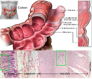
Histologically, the colon has four distinct layers: innermost mucosa, sub mucosa,
muscularis externa and outermost serosa. (Arthur F. Dalley 1999) (Figure1.1). The
innermost mucosal layer consists of invaginations known as crypts lined with colonic
epithelium. Unlike the small intestine, colonic crypts have no villi for nutrient
absorption. Crypts are supported by the lamina propria consisting of connective
tissue, blood vessels, and immune cells including lymphocytes and macrophages.
(Arthur F. Dalley 1999) The mucosal layer is the most common origin for colon
5
Figure 1.1 Structure of colon. The colon or the large intestine is a hollow, muscular
and tubular organ with four layers: the innermost mucosa, sub mucosa, muscularis
externa and outermost serosa. The innermost mucosal epithelium has invaginations
called ‘colonic crypts’ which are the site of origin of colon cancer. (adapted and
modified from Yi-Ben Chen, updated 2010)
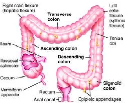
Right and left side of colon
Embryologically, the colon develops partly from the midgut (ascending colon to
proximal transverse colon) and partly from the hind gut (distal transverse colon to
sigmoid colon). (Figure1.2) The ascending colon which lies vertically on the right
side of the splenic flexure, is also referred to as the ‘proximal’ colon. The transverse
6
becomes the descending (left) colon, which lies vertically in the most lateral left part
of the abdominal cavity.
Figure 1.2 Colon anatomy. Colon has been divided into three parts depending on its
location with respect to splenic flexure. On right side of the splenic flexure is the
ascending colon also known as ‘proximal’ colon and on the left side of the splenic
flexure is the descending colon also known as the ‘distal’ colon. Transverse colon
runs horizontal.
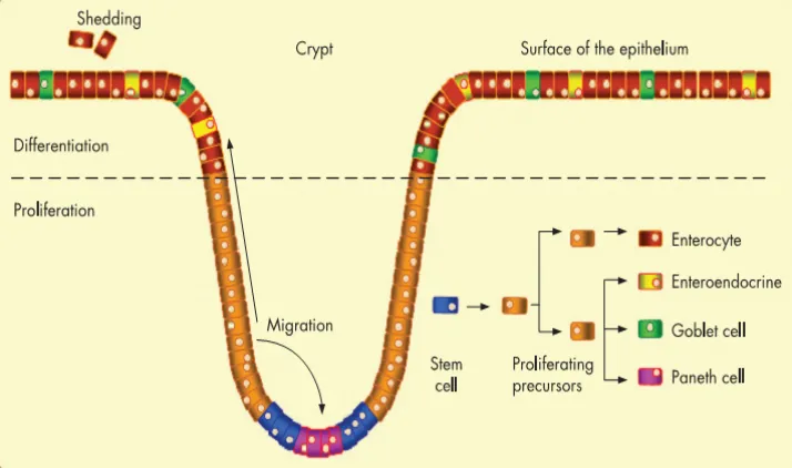
Colonic epithelium is a highly diverse and dynamic system involving constant cell
renewal. (Ding, Ko et al. 1998). Continuously proliferating stem cells situated at the
base are responsible for replenishing the entire epithelium every 3-8 days.(Cotran
R.S. 1999) This cell regeneration occurs within the basal two-third portion of the
crypt. As the cells migrate upwards they differentiate into specialized cell types
including absorptive, mucous secreting, endocrine, and anti-bacterial protein
secreting Paneth cells. (Radtke and Clevers 2005; Schneikert and Behrens 2007)
7
Figure 1.3 Differentiation of colonic epithelium. The colonic epithelium has
invaginations called crypts which have a population of stem cells at the base that give rise
to actively dividing precursors. These precursor cells migrate to the top of the colon and
and differentiate as enterocytes (absorptive cells), entero endocrine (hormone secreting
cells), goblet cells (mucus secreting cells) and paneth cells (antimicrobial toxin secreting
cells) ( adapted and modified from Jean Schneikert, Jurgen Behrens, Gut 2007)
Colorectal Cancer
Colorectal cancer (CRC) is the 3rd most common malignancy and 4th most common
cause of cancer related mortality worldwide. (Tenesa and Dunlop 2009) It happens to
be the 2nd leading causes of cancer related deaths in the United States and other
developed countries, despite considerable advances in diagnosis and treatment
techniques.(Jemal, Murray et al. 2005; Jemal, Siegel et al. 2009). It alone accounts
for 11% of worldwide cancer cases, preceded only by prostate, lung and breast
cancer. In addition, it is estimated that 50% of the population will develop an
8
Approximately, 20% of CRC cases have a familial basis, (Rustgi 2007) where some
are associated with well-defined syndromes, such as hereditary non polyposis
colorectal cancer (HNPCC) and familial adenomatous polyposis (FAP). However,
majority of the CRC cases have been linked to environmental factors and food borne
mutagens, specifically intestinal microflora including pathogens and chronic
intestinal inflammation, which predisposes tumor formation.
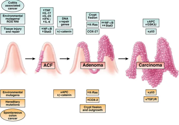
CRC is multistep, multi factorial disease that involves the clonal selection and
clonal propagation of initiated colonic epithelial cells, which progress from normal
to precancerous to malignant states over a span of 5-40 years.(Harris 1991; Roncucci
L. 1991.; Lei Cheng 2003 December) . The sequential events of colon carcinogenesis
concerning genetic and phenotypical changes are very well characterized by the
adenoma-carcinoma model first described by Fearon and Vogelstein (1990).
According to this model, CRC is caused by accumulation of mutations in the
classical oncogenes and tumor suppressors genes, which correspond to a sequential
transition of single pre-neoplastic cell to aberrant crypt foci (ACF) and then to
adenoma and metastatic carcinoma. (Figure 1.4) The earliest molecular alteration in
this pathway is mutation in the adenomatous polyposis coli (APC) tumor suppressor
gene which is a component of the Wnt signaling pathway. This is followed by
mutation in the KRAS and TP53, in the larger adenomas and invasive carcinoma.
(Fearon and Vogelstein 1990) This pathway is referred to as the ‘APC pathway’ or
the ‘chromosomal instability (CIN) pathway’ because the colorectal tumors arising
by this pathway are characterized by gross chromosomes abnormalities including
deletions, insertions, and loss of heterozygosity. Another molecular pathway
proposed is the DNA mismatch repair pathway.(Aaltonen, Peltomaki et al. 1993) The
key element of this pathway is mutation in the DNA repair enzymes e.g MLH1,
MSH2, which results in accumulation of mutations in the microsatellite regions of
the genome. (H Iino 2000 July) This results in microsatellite instability in the tumors
arising from this pathway and hence it is also called microsatellite instability (MSI)
9
Figure 1.4 Fearon–Vogelstein model of colon carcinogenesis. The sequential events of
colon carcinogenesis concerning genetic and phenotypical changes are very well
characterized by the adenoma-carcinoma model first described by Fearon and Vogelstein
(1990)
Evidences indicating that adenomas might not be the only CRC precursor began to
emerge in 1990. Longacre and Fenoglio - Preiser studied a group of polyps that
showed mixed histology of both hyperplastic and adenomatous polyps.(Longacre TA
1990) These lesions had glands with serrated appearance and showed nuclear
features of adenomas, hence they were called serrated adenomas. The adenoma
carcinoma pathway for serrated polyps is different from the traditional CIN or MSI
pathway. For e.g- APC and TP53 mutations are rare, whereas alterations in
microsatellite sequence and hypermethylation of CpG islands are more common.
There are two overlapping pathways for development of serrated lesions. The first
10
level of MSI and CpG island methylation and BRAF mutation.(Spring, Zhao et al.
2006) The second one is more common in left sided colon (distal) and shows
microsatellite stability (MSS) or low grade of MSI (MSI-L) and also show mutations in
KRAS. (Jass, Whitehall et al. 2002)
Obesity associated inflammation and colon cancer
Obesity is a chronic inflammation state and a rising epidemic in North America, with
both environmental and genetic etiological factors.(Formiguera and Cantón 2004)
The connection between obesity and risk of colon cancer is well established and has
a great deal of supporting evidence from genetic, pharmacological and
epidemiological data. (Rapp, Schroeder et al. 2005). While the biochemical and
molecular links between the two physiological disorders are still under review, the
interconnected roles of insulin resistance , adipose tissue and inflammation are well
documented in literature. (Figure 1.5) Key mediators of inflammation-induced cancer
include nuclear factor kappa B (NFκB), reactive oxygen and nitrogen species
(RONS), inflammatory cytokines, chemokines, adhesion molecules and specific
miRNAs. Chronic inflammation predisposes cells for oncogenic transformation by
inducing genomic instability, increasing cell proliferation and angiogenesis, altering
the genomic epigenetic state.
Physiological parameters linking obesity and cancer
Obesity or chronic inflammation state is often encountered with high levels of
glucose, triglycerides, cholesterol in the blood plasma due to inactivity of the insulin
hormone or its receptor. Insulin resistance implies a cells inability to respond to
normal levels of insulin, thus requiring abnormally high amounts of insulin hormone
for glucose metabolism. This condition further leads to irregular triglycerides, fatty
acids and glucose levels, all metabolic aberrations that also correlate with an
increased risk of colorectal cancer.(Gunter and Leitzmann 2006). There are
convincing evidences that suggest the connection between obesity and colon cancer
risk in three possible ways. (reviewed by Gunter and Leitzmann 2006) The first
11
growth and proliferation. Hence, the exposure of high levels of insulin to parts of the
body that are not accustomed to it e.g- colon, can interfere with the normal cell
signaling and promote cell proliferation. (Gunter and Leitzmann 2006) This has been
experimentally proved by Kiunga et al 2004, who reported the presence of high
levels of insulin and insulin receptor in colon tumor cells compared to normal
mucosa. Secondly, an insulin resistant state also promotes high levels of circulating
insulin-like growth factor (IGF), which can also promote colonocyte division and
block apoptosis by receptor binding. (Giovannucci 2001) Increased IGF-1-receptor
(IGF-IR) also stimulates cell division in intestinal epithelial cells (Ma J. 1999) and
several epidemiological studies have shown positive correlation between IGF and IGF-IR
levels and colon cancer risk.(Komninou, Ayonote et al. 2003) Finally, the high
circulating plasma glucose and lipids associated with insulin resistance serve as a
reservoir for reactive oxygen species (ROS) generation, known contributors to
carcinogenesis. High visceral abdominal fat found in obese individuals is especially a
critical source for ROS. (Furukawa, Fujita et al. 2004; Frezza, Wachtel et al. 2006)
Along with oxidative stress, increased amount of fat tissue promotes a
pro-inflammatory environment, also strongly correlates with increased cancer risk.
Cytokines/adipokines such as tumor necrosis factor-α (TNF-α) are readily secreted
by adipocytes and further enhance inflammation, insulin resistance, impaired glucose
metabolism and ROS production. (Furukawa, Fujita et al. 2004; Sonnenberg G.E.
2004.)
Leptin, which is also known as the hormone of satiety, has been implicated in obesity
associated colon cancer. Though its role is not very clear, it has been shown to
accelerate HT-29 cell proliferation and promote ACF formation in vivo.(Liu Z 2001.)
Interestingly, F344 rats administered with continuous high dose of leptin have shown
reduction in the number of ACF compared to controls. (Aparicio, Guilmeau et al.
2004) Leptin secreted from adipose tissue has also been shown to enhance insulin
12
Metabolic Syndrome X
Many of the physiological abnormalities associated with obesity or insulin resistance
are collectively referred to as Metabolic syndrome, or syndrome X. It is a term given
by Reavan in 1988, to the physiological disorders associated with obesity,
specifically abdominal/visceral obesity. Although there are contradicting views
regarding the inclusion of insulin resistance and dyslipidaemia (including
hypercholesterolemia and hypertriglycerolemia) in this syndrome, hyperglycemia
and hypotension have been unanimously included in this category.(Shaw, Hall et al.
2005; Sorrentino 2005). These symptoms or abnormalities have been associated with
diabetes, atherosclerosis and cancer.
Oncogenic mechanisms associated with inflammation
Prolonged inflammation can induce DNA mutations and contribute to genomic
instability in more than one ways. (Perwez Hussain and Harris 2007; Colotta,
Allavena et al. 2009) Free radicals (RONS), produced during inflammation can
reduce the expression and enzymatic activity of the critical DNA repair enzymes eg.
mut S homolog 2 and 6. It can further cause increased expression of DNA
methyltransferases that can cause global hypermethylation of the genome. The
downstream effect of this includes silencing of DNA mismatch repair enzymes (eg-
hMLH1 and hSMH2) and several tumor suppressor genes (eg- APC, CDKN 2,
BRCA1,Rb and MDM2) by promoter methylation. (Fleisher, Esteller et al. 2000; Das
and Singal 2004)
Molecular mediators common to inflammation and cancer
Inflammation can induce cancer by both extrinsic and intrinsic pathways. In extrinsic
pathway, chronic inflammation itself is the driving force, whereas in intrinsic
pathway, the genetic alteration of oncogenes and tumor suppressors as well as
various inflammatory mediators play an important role in carcinogenesis. The
13
NFκB. For eg- a dominant mutation in Ras proto-oncogene, can induce inflammatory
response through induction of pro-inflammatory cytokines interleukins (ILs) IL1,
IL6 and IL11 and the chemokine IL8, which aid in Ras mediated oncogenesis. (Bos
1989) Ras also induces IL-8 that mediates tumorigenesis and angiogenesis.
(Sparmann and Bar-Sagi 2004) Genetic mutations stimulate the tumor cells to
produce inflammatory cytokines and free radicals, which creates a feedback loop
where the tumor cells recruit inflammatory cells that can produce additional
cytokines and free radicals to the site of tumor that can aid tumorigenesis. (Figure
1.5)
Some of the key players are described in detailed in the following sections:
Nuclear Factor Kappa B
Nuclear factor kappa B or NFκB as it is commonly called is a transcription factor
and key mediator of inflammation induced carcinogenesis. (Shen and Tergaonkar
2009) Under normal condition, NFκB is bound to its negative regulator also called
the inhibitor of kappa B (IκB) and stays inactive in the cytoplasm. Following an
inflammation stimulus, IκB kinase phosphorylates IκB and targets it for proteosomal
degradation. Activated NFκB translocates to the nucleus where it drives the
transcription of its target genes, many of which are inflammation related
eg-cytokines and chemokines, nitric oxide synthase (NOS) , cyclooxygenase (Cox-2)
and tumor necrosis factor (TNF)α.(Naugler and Karin 2008)
NFκB creates a protumorigenic environment by turning on the expression of
following set of genes:
1.) Cell cycle related genes eg c-Jun-N-terminal kinase that increase cell
proliferation
2.) Vascular endothelial growth factor (VEGF) and angiopoetin that stimulate
angiogenesis
3.) Bcl2, Bcl-xL, and cFLIP that makes cell resistant to apoptosis and necrosis.
14
Inflammatory bowel disease (IBD) which is associated with colitis associated cancer
(CAC), show persistant activation of NFκB in the myeloid and epithelial cells of
colonic mucosa. (Rogler and Andus 1998; Chung 2000)
Reactive oxygen and nitrogen species (RONS)
These are highly reactive free radicals produced by inflammatory cells, collectively
known as the reactive oxygen and nitrogen species (RONS). They have the ability to
induce DNA strand breaks, mutations and aberrant cross linking, causing genomic
instability. Chronic inflammation results in elevated levels of RONS that causes
oxidative and nitrosative stress that contributes to tumorigenesis. (Hussain, Trivers et
al. 2004; Hofseth 2008) In colitis associated cancer (CAC), chronic inflammation
causes oxidative damage to DNA, leading to the p53 mutations observed in tumor
cells.(Choi, Yoon et al. 2002; Kraus and Arber 2009). ROS can also directly oxidize
and inactivate mismatch repair enzymes at protein level. (Choi, Yoon et al. 2002;
Hussain, Hofseth et al. 2003; Kraus and Arber 2009). Nitrogen oxide (NO) is a
reactive free radical produced in large amounts by the inducible nitric oxide synthase
2 (NOS2) under conditions of chronic inflammation. In its pro-tumorigenic role, NO
induces DNA strand breaks, promotes angiogenesis by induction of VEGF and
increases cell proliferation and invasion. (Hussain, Trivers et al. 2004). In its
anti-tumorigenic role, NO can cause cytotoxic cell death of malignant cells and modulate
the immune system to eradicate the cancerous cells. There is a negative loop between
NO and tumor suppressor protein 53 (TP53), where NO causes the stabilization and
accumulation of TP53 which induces apoptosis, cell cycle arrest in the malignant
cells and represses levels of NO. (Forrester, Ambs et al. 1996; Ambs, Ogunfusika et
al. 1998).
Cytokines and chemokines
Cytokines are signaling molecules that are key mediators of inflammation or an
15
that leads to cell proliferation, apoptosis, angiogenesis and cellular senescence. They
broadly fall under two categories 1.) Pro-tumorigenic and 2.) Anti-tumorigenic. The
pro-tumorigenic cytokines include IL1, IL6, IL15, IL17, IL23 and tumor necrosis
factor α (TNFα) , whereas the anti-tumorigenic cytokines include IL4, IL10, IL13,
transforming growth factor (TGFβ) and interferon (IFN α). In case of CRC and CAC,
the role of IL-6, IL-1 and TNF-α in tumorigenesis has been well studied. (Tang,
Katuri et al. 2005; Popivanova, Kitamura et al. 2008; Wang, Liu et al. 2009). IL-6 is
a potent stimulator of colon cancer cell proliferation and tumor growth. (Becker,
Fantini et al. 2005) It has been implicated in pro-tumorigenic activity for many
cancers and has been found to be required for CAC in mouse models. (Grivennikov,
Karin et al. 2009) TNFs are produced during early inflammation; they trigger
production of other cytokines, chemokines and endothelial adhesion
molecules.(Balkwill 2009; Li, Vincent et al. 2009).TNF-α expression increases
during colon carcinogenesis which further confirms its role in inflammation,
angiogenesis and tumor promotion. (Popivanova, Kitamura et al. 2008). One of the
key functions of TNF-α is to activate the pro-inflammatory transcription factor
NFκB. The anti inflammatory cytokines like IL-10 and TGF-β , have a general role
in tumor suppression. IL-10 exerts its anti-tumorigenic effect by down-regulating
NFκB, which results in low levels of pro-inflammatory cytokines eg- IL-6, IL-12 and
TNF-α. (Zhang 2008 December) TGF-β is another anti-inflammatory cytokine that
suppresses tumor growth by inhibiting proliferation, promoting apoptosis,
stimulating the release of anti-inflammatory cytokines and suppressing the
expression of pro-tumorigenic cytokines.(Yang and Moses 2008) Mutation in TGF-β
pathway within epithelial cells predisposes to or facilitates colonic tumor
development and growth.
Chemokines are a class of cytokines that recruit leukocytes at the site of
inflammation. They are released by the inflammatory cells after the initial
stimulation by the cytokines. Tumors generally show high levels of chemokines that
recruit leukocytes to the site of tumor, creating a inflammatory and
16
Chemokines and adhesion molecules
Early feature of inflammation is the release of chemokines like Monocyte
chemoattractant protein (MCP). These factors increase the expression of interstitial
and vascular cellular adhesion molecules like Interstitial Cellular Adhesion
Molecules (ICAM) and E-selectin that attract monocytes and immune cells.
Chemokines like MCP also induce the proliferation and pro-inflammatory gene
activation producing cytokines like IL-1α, IL-6, IL-18 etc. Other factors that
stimulate gene expression of pro-inflammatory cytokines in obese state are RONS,
oxidized lipids, free fatty acids.
Figure 1.5 Schematic diagram showing pathways linking obesity to cancer.
Insulin resistance and insulin like growth factor (IGF) are one of the several links
between obesity and cancer. High levels of circulating insulin up-regulates growth
hormone (GH) that induces hepatic production of IGF-1. IGF-1can inhibit apoptosis
17
including the phosphatidylinsitol3-kinase (PI3-K)-AKT system and the
Ras/Raf/mitogen activated protein kinase (MAPK) systems, respectively. Leptin,
also secreted by adipocytes has mitogenic effects and anti apoptotic effects through
MAPK and PI-3-Kinase pathways. Adiponectin exerts anti-carcinogenic effect
through AMP activated protein kinase (AMPK) through receptors AdipoR1 and
AdipoR2. Adipopectin regulates growth arrest and apoptosis through p53 and p21.
IL-6 and TNF-α are the two major inflammatory cytokines that are elevated in
obesity. They exert pro-tumorigenic effect by increasing cell survival and cell
proliferation through activation of NF-κB
Animal Model
Preclinical or animal models provide a system to study the biological process of a
disease in a physiological state relevant to human beings. They provide valuable
preliminary data upon which human clinical trials can be based. However, they
should always be evaluated with regards to how relevant they are to human
conditions, and how predictive they can be for the disease process. (Green and
Hudson 2005)
With regards to colon carcinogenesis, molecular and pathological similarities to the
human condition should be observed in order to have an effective model.(Reddy
2004) Currently the APC min+/-mouse and azoxymethane rat model are the two main
animal models used to study the effect of dietary supplements on colon cancer.
Zucker obese rats, traditionally used to study obesity and related metabolic disorders,
offer a novel way of studying the progression of colon cancer in an altered
physiological state.
Zucker-Obese Animal Model
Zucker obese (Zk-Ob) rats are an excellent model of human obesity, and provide an
ideal opportunity to study colon carcinogenesis in an altered physiological state. The
Zucker or ‘fatty’ rat was developed in the laboratory of Zucker and Zucker in 1961,
18
normal, while fa is the fatty mutation). Zk-Ob rats inherit obesity as an autosomal
Mendelian recessive trait, fa/fa homozygous for nonfunctional leptin receptors, in
comparison to their lean (Fa/fa or Fa/Fa) counterparts.(A. 1977.) Leptin is a peptide
hormone produced by adipocytes that regulates body weight and fat metabolism by
sending signals to the hypothalamus to suppress apetite.(Arthur F. Dalley 1999) In
Zk-Ob rats, obesity is associated with metabolic dysfunction of fat metabolism
characterized by latescence and increased adipose tissue formation. (Zucker T.F.
1962. ) Genetic association of Fa gene to obesity is confirmed as Zk-Ob rats on low
fat or energy restricted diet also exhibit these symptoms. Interestingly, female
Zucker rats are sterile, which has implications for breeding programs.
Average weights of these animals at 40 weeks is 800g and 625g for Zk-Ob, and
480g and 295g for Zucker lean (Zk-Ln), males and females respectively.(Zucker T.F.
1962. ) Zk-Ob rats exhibit hyperphagia, hypertriglyceridemia, hypercholesterolemia,
hyperinsulinaemia and mild hyperglycemia at about six weeks of age. Pathological
findings at death often include hydronephrosis and polycystic kidneys, as well as
fatty livers.(Zucker T.F. 1962. ) It has been shown that Zk-Ob rats are more sensitive
to chemically induced colon carcinogenesis in comparison to their lean
counterparts.(Raju and Bird 2003) Since there is little difference in ACF number
between Fa/fa and Fa/Fa animals, the recessive gene linked to leptin receptor
deficiency is not solely responsible to higher susceptibility of colon cancer.(Weber,
Stein et al. 2000) Use of this animal model helps understanding carcinogenesis in
relation to obesity and associated metabolic disorders, providing valuable
information for treatment and prevention strategies.
Micro-RNA
Brief introduction
Micro RNAs (mi RNAs) are 19-22 nucleotide long single stranded non protein
coding RNAs that act as guide molecules in post transcriptional gene silencing by
19
repression.(CULLEN 2003; Bartel 2004; VICTOR AMBROS 2008). With >200
members per species in higher eukaryotes, miRNAs form one of the largest gene
families accounting for approx 3% of the genome and targeting 30% of coding
regions of the genome (Lewis, Shih et al. 2003; Bartel 2004). Since their discovery
back in 1993 by Victor Ambros and colleagues, who identified the first miRNA
targeting lin-4 gene in C.elegans, miRNAs have come a long way in being
recognized as a major player in post transcriptional gene regulation. Even though we
are far from unraveling the complex role of miRNAs in gene regulation, it is evident
that they have important role in almost all biological process known, including
developmental timing, growth control, differentiation and apoptosis. Accordingly,
altered miRNA expression can have serious implication on the regular metabolism of
the cell and be a potential cause for diseases like cancer.
Gene Structure and miRNA gene transcription
Location in the genome
Early annotation to find out the genomic position of miRNAs indicated that most
them are located in the intergenic regions , although a sizeable minority are found in
the intronic region of known genes in sense or anti sense orientation.(Lau, Lim et al.
2001; Kim 2005) This implied that most of the miRNAs are transcribed as
autonomous replication unit with their own promoter. Approximately 50% of the
miRNAs are found in close proximity to known genes, (Lagos-Quintana, Rauhut et
al. 2001; Lim, Lau et al. 2005) which allows one to speculate that they are
transcribed from single polycistronic transcription unit. So the conclusion drawn was
that they can be transcribed as discrete as independent transcription unit or in a
cluster by polycistronic transcription unit.
MiRNA biogenesis
miRNAs are transcribed as long primary transcripts by RNA polymerase II;
subsequently trimmed into short hairpin intermediates called primary miRNAs and
20
of the enzymes responsible for first and second miRNA processing is
compartmentalized into the nucleus and cytoplasm and is tightly regulated. (Figure
1.6)
Nuclear processing by Drosha
RNase III endonuclease Drosha is a 160 kDa protein that is highly conserved in
higher eukaryotes. (Filippov, Solovyev et al. 2000; Wu, Xu et al. 2000; Kristine R
Fortin 2002) It contains two random RNAIII domains (RIIID) and a double stranded
RNA binding domain (dsRBD) that are critical for its catalytic activity. Transcription
of miRNA genes by RNA pol II yields primary transcripts (pri-miRNA) that are
usually several kilobases long and contain a local hairpin structure. The pri-miRNA
is first cleaved by RNase III endonuclease Drosha at sites near the base of stem loop,
that releases ~60-70 nucleotide precursor miRNA. (Lee, Ahn et al. 2003) Drosha
cleaves the pri-miRNA duplex with a staggered cut typical of RNase III
endonucleases, leaving 2 nucleotide overhang at the 3’ end.(Lee, Ahn et al. 2003)
The specificity with which Drosha recognizes and cleaves pri-miRNA has been
under investigation. It was proposed on the basis of evidences from mutational
studies that the tertiary structure of the pri miRNA is the factor that decides the
specificity of Drosha. (CULLEN 2003; Lee, Ahn et al. 2003; Zeng, Yi et al. 2005)
Nuclear export by exportin-5
Following nuclear processing by Drosha, pre-miRNA is translocated to the
cytoplasm through the nuclear pore complex embedded in the nuclear
membrane.(Nakielny and Dreyfuss 1999) Export of the pre-miRNA is mediated by
one of the nuclear transport receptors, exportin-5. (Rui Yi 2003; Lund, Güttinger et
al. 2004) . Owing to the compartmentalization of the two processing events, the
nuclear transport of pre-miRNA is a crucial step in miRNA biogenesis. (Kim 2004;
Murchison and Hannon 2004)
21
On reaching the cytoplasm, pre-miRNA is cleaved by cytoplasmic RNase III
endonuclease Dicer, into ~ 22 nucleotide miRNA duplex. (Bernstein, Caudy et al.
2001; Grishok, Pasquinelli et al. 2001; Hutvágner, McLachlan et al. 2001; Ketting
2001; Knight and Bass 2001). Dicer is a highly conserved protein, found in all
eukaryotes including Saccharomyces pombe, plants and animals. Apart from the two
RIIID domains and dsRBD domain, Dicer contains long N terminal segment
containing ‘dead box RNA helicase’ domain and PAZ domain.(Zhang, Kolb et al.
2004) PAZ domain belongs to a group of highly conserved proteins called Argonaute
preteins. Structural and biochemical studies of AGOI and AGOII in D.melanogaster
revealed that PAZ domain binds to the 3’ protruding end of the small RNAs.(Song,
Liu et al. 2003; Yan, Yan et al. 2003; Lingel, Simon et al. 2004) . Usually one strand
of this short lived duplex disappears, whereas the other strand remains as mature
miRNA. Mature miRNAs are incorporated into effector complexes that are known as
microRNP (miRNA containing ribonucleoprotein complex), migronaute’ or
microRISC (miRNA containing RNA inducing silencing complex). The strand that
gets loaded in the microRISC complex is thought to be the one that has unstable base
pairing at the 5’ end. (Khvorova, Reynolds et al. 2003; Schwarz, Hutvágner et al.
Figure 1.6 Biogenesis of
independent promoters as monocistronic whereas most of the
and found in clusters with other genes.
miRNAs are transcribed by RNA polymeraseII as several kilobases long pri
Pri- miRNA is processed in the nucleus by RNAIII endonuclease (Drosha) which
cleaves it into 60-70nt
3’end. This pre-miRNA
exportin-5. In the cytoplasm, pre
endonuclease (Dicer), which generates the mature
resulting duplex is loaded onto
Mature ss miRNA guides RISC to the target mRNA. After that there are several
mechanisms to downregulate mRNA/protein expression depending on the de
sequence complimentarity.
2010)
22
Biogenesis of miRNA. Intergenic miRNAs are transcribed from
independent promoters as monocistronic whereas most of the miRNA
in clusters with other genes. They are transcribed as polycistronic units.
miRNAs are transcribed by RNA polymeraseII as several kilobases long pri
is processed in the nucleus by RNAIII endonuclease (Drosha) which
long pre-miRNA with a 2 nucleotide long overhang at the
miRNA is transported to the cytoplasm by nuclear transporter
5. In the cytoplasm, pre-miRNA is cleaved by another RNAIII
(Dicer), which generates the mature miRNA. One of the strands of the
resulting duplex is loaded onto miRNA inducing silencing complex called miRISC.
guides RISC to the target mRNA. After that there are several
mechanisms to downregulate mRNA/protein expression depending on the de
sequence complimentarity. (adapted and modified from Christian Marin
s are transcribed from
miRNAs are intragenic
They are transcribed as polycistronic units.
miRNAs are transcribed by RNA polymeraseII as several kilobases long pri-miRNA.
is processed in the nucleus by RNAIII endonuclease (Drosha) which
with a 2 nucleotide long overhang at the
is transported to the cytoplasm by nuclear transporter
is cleaved by another RNAIII
. One of the strands of the
inducing silencing complex called miRISC.
guides RISC to the target mRNA. After that there are several
mechanisms to downregulate mRNA/protein expression depending on the degree of
23
Mechanism of miRNA mediated gene regulation
miRNA can direct RISC to downregulate gene expression by either of the two
post-transcriptional mechanisms:
1.) Translational repression
2.) mRNA degradation/sequestration
Mechanism of translational repression
Once incorporated in the RISC complex, miRNA directs translational repression or
target cleavage depending on degree of sequence complimentarity with the
target.(Hutvágner, McLachlan et al. 2001). In animals, there are several evidences
suggesting that miRNAs recognize their target mRNA by limited base pairing
between the 2-8 nucleotide seed region at the 5’end of the miRNA and the 3’ UTR of
target mRNA.(Lewis, Shih et al. 2003; Farh, Grimson et al. 2005; Stark, Brennecke
et al. 2005) The interaction between microRNPs and mRNA can have several
consequences which can be direct or indirect. Direct effects include inhibition of
translational initiation by preventing ribosome association with the internal ribosome
entry site (IRES) on the target mRNA or inhibition post translation. Post
translational repression includes premature ribosome drop off, stalled or slowed
down elongation, and/or co-translational protein degradation; the repressed mRNA
seems to be present in polyribosomes.(Nilsen 2007) (Figure 1.7)
Mechanism of mRNA degradation/sequestration
Perfect sequence complimentarity between the seed region of miRNA and the 3’UTR
of target sequence leads to the destabilization and subsequent degradation of the
transcript. miRNAs achieve this by deadenylation and decapping of the target
sequence, which results in degradation and increased turnover of the transcript. The
site of miRNA directed target sequestration are thought to be the processing bodies
(P bodies) also known as the GW bodies. The P bodies, which were originally
described in budding yeasts, are known as the cytoplasmic foci rich in decapping ,
24
(THEOPHANY EYSTATHIOY 2003 October; Liu, Rivas et al. 2005; Eulalio,
Behm-Ansmant et al. 2007) miRNA bound to the Argonaute protein in the RISC
complex recognizes its target by complimentarity base pairing. The Argonaute
protein interacts with the GW182 protein; miRNA-mRNA and argonaute protein
complex is delivered to P bodies. In the P bodies, the mRNA is decapped and
deadenylated, subsequently degraded or held in stasis i.e spatially isolated from the
translational machinery since P bodies do not have ribosomes. (THEOPHANY
EYSTATHIOY 2003 October; Liu, Valencia-Sanchez et al. 2005) In stasis, the P
bodies are thought to act as repository for untranslated mRNAs and this has been
experimentally proved in budding yeast as well as mammalian cells.(Brengues,
Teixeira et al. 2005; Bhattacharyya, Habermacher et al. 2006)
Figure 1.7 Mechanisms of miRNA mediated repression of gene expression.
Mature single stranded miRNA loaded in a RISC complex targets and represses the
translation of mRNA in more than one way, either directly or indirectly. In direct
interaction, miRNA inhibits the initiation of translation by blocking the entry of
ribosomes into the internal ribosomal entry site(IRES). Post translational initiation,
miRNA can cause premature ribosome drop off , slowed or stalled elongation or
P-25
bodies where they might undergo deadenylation and decapping by the enzymes
present in the P-bodies and hence degraded or they might simply be sequestered from
the translational machinery of the cell. In the later case, which is also called ‘stasis’
the P-bodies act as repository for all those mRNA that indirectly targeted by miRNA.
(adapted and modified from Timothy Nilsen, 2007)
MiRNA signatures in cancer
Cancer is a complex genetic disease that involves aberrant expression pattern of
coding as well as non-coding genes. Earlier it was thought that only the protein
coding genes and their altered expression are involved in the pathogenesis of cancer
but with the discovery of miRNAs, the understanding of cancer mechanisms and
genomic complexity of cancer cell has completely changed. miRNAs have been
proposed to contribute to oncogenesis either as tumor suppressors (eg- miR-15a and
miR-16-1) or oncogenes ( eg- miR-155 or members og the miR-17-92 cluster). The
genomic abnormalties influencing miRNA expression in cancer are the same as for
the protein coding genes eg- chromosomal rearrangement, genomic amplification,
deletion and polymorphisms in the target site of the mRNA.(Abelson, Kwan et al.
2005; He, Jazdzewski et al. 2005)
Oncogenesis and tumor suppression - dual role of miRNAs
Majority of the miRNAs are located in regions of genomic instability that undergo
amplification or deletion which is why they have a huge role to play in the regulation
of all major cancer pathways. miRNAs lying in regions that undergo genomic
amplification act as oncogenes eg-miRNA 17-92 cluster; whereas the chromosomal
regions that undergo deletion in cancer, act as tumor suppressors eg- miRNAs
15s-miR-16-1 cluster. O’Donnels in his work on cell model system showed that, human
B-cell line P493-6, which overexpresses c-MYC, shows tumor suppressor activity
through miRNA 17-92 cluster, which targets E2F1 and hence inhibits c-MYC
regulated cell proliferation.(O'Donnell, Wentzel et al. 2005) The same cluster of
miRNA when undergo amplification in the genome, act as oncogene by cooperating
26
by concluded that miRNAs can participate in distinct cancer pathways with either the
role of oncogene or tumor suppressor depending on the cell type and expression
pattern. Apart from their dual role, miRNAs can also act by targeting more than one
mRNAs at the same time and have varied response .For eg-miRNA 15a and
miRNA-16-1 target the anti-apoptotic Bcl-2 in leukemia cells , however in 293 fetal kidney
cells, where they are expressed, they don’t show any such response.(Calin, Liu et al.
2004). The loss of miRNA 15a and miRNA 16-1, leads to the over-expression of
Bcl-2 and consequently B-cell malignancies. In a different tissue, the
over-expression of miRNA 15a and miRNA 16-1 causes the loss of activity of a tumor
suppressor that is critical to check cell proliferation and promote apoptosis in
malignant cells. There are different mechanisms that can lead to aberrant miRNA
expression in a cancerous cell-
1.) Location of miRNA in cancer associated genomic regions
More than 50% of known miRNAs are found in genomic regions that are prone to
alteration in cancer cells.(Calin, Sevignani et al. 2004). Such regions include
minimal regions of LOH (loss of heterozygosity), which are thought to harbor tumor
suppressors genes, minimal regions of amplification, which mostly harbor
oncogenes, common breakpoint region in or near possible oncogenes or tumor
suppressor genes and fragile sites.
2.) Epigenetic regulation of miRNA expression
DNA hypomethylation, CpG island hypermethylation and histone modification
losses represent epigenetic markers of malignant transformation and have been
known to be one of the mechanisms involved in miRNA abnormal expression in
cancer.((Fraga and Esteller 2005)
3.) Abnormalities in miRNA processing genes and proteins
Alterations in the proteins machinery that is involved in miRNA biogenesis, has
27
Dicer mutation, could explain the downregulation of miRNA observed in primary
tumors.(J. Michael Thomson1 2006)
MiRNAs in colorectal cancer
Dysregulation of miRNA targeting tumor suppressors and oncogenes, have been
reported in carcinogenesis of colorectal cancer. Two approaches are applied today to
investigate the connection between miRNAs and CRC: functional and profiling
studies. Many proteins involved in key signaling pathways of CRC, such as members
of the Wnt/β-catenin and phosphatidylinositol-3-kinase (PI-3-K) pathways, KRAS,
p53, extracellular matrix regulators as well as epithelial-mesenchymal transition
(EMT) transcription factors (Fearon and Vogelstein 1990) are altered by miRNA in
CRC (Figure 1.8). Study of these miRNAs crucial for better understanding CRC
pathogenesis with an aim to eventually identify novel therapeutic targets. Expression
profiling of miRNAs have shown to have same potential for identification of
biomarkers as profiling of their mRNA or protein counterparts. Together these
studies help to determine the clinical prognosis of the disease along with
identification of therapeutic targets.
Few pathways that are prominent in CRC and provide evidence of involvement of
miRNAs, are detailed below-
MiRNA altering regulation of Wnt/β-catenin pathway in CRC
The Wnt/β-catenin pathway plays a central role in an early colorectal tumor
development. Inactivation of the adenomatous polyposis coli (APC) gene is a major
initiating event in colorectal carcinogenesis occurring in more than 60% of colorectal
adenomas and carcinoma. (Fearon and Vogelstein 1990). According to a recent study
by Nagel et al.(Nagel, le Sage et al. 2008), miRNAs represent a novel mechanism for
APC regulation in CRC. miR-135a and miR-135b decrease translation of the APC
transcript in vitro. miR-135a and miR-135b were also found to be up-regulated in
28
(Nagel, le Sage et al. 2008). These observations suggest that alteration in the
miR-135 family can be one of the early events in CRC's molecular pathogenesis.
MiRNA regulating EGFR signaling (KRAS and phosphatidylinositol-3-kinase
pathways)
The epidermal growth factor receptor (EGFR) pathway contribute to promotion and
progression of several solid tumors including colonic tumor. Stimulation of the
EGFR and KRAS signaling lead to the activation of numerous signal transduction
molecules initiating a cascade of downstream effectors that mediate tumor growth,
survival, angiogenesis and metastasis. (Ciardiello and Tortora 2008) KRAS
oncogene has been reported to be a direct target of the let-7 miRNA
family.(Johnson, Grosshans et al. 2005). Another miRNA associated with KRAS
regulation in CRC is miR-143.(Chen, Guo et al. 2009). Inhibition of KRAS
expression by miR-143 blocked constitutive phosphorylation of MAPK.(Chen, Guo
et al. 2009). Another central signaling pathway downstream from EGFR and
important in CRC development is the phosphatidylinositol-3-kinase (PI-3K)
pathway. The p85β regulatory subunit involved in stabilizing and propagating the
PI-3K signal was mechanistically proven to be a direct target of miR-126. Furthermore,
this p85β reduction mediated by miR-126 was accompanied by a substantial
reduction in phosphorylated AKT levels in the cancer cells, suggesting an
impairment in 3K signaling. Another important regulatory component of the
PI-3K pathway, the tumor suppressor gene PTEN, is strongly repressed by
miR-21.(Meng F 2007 Aug) miR-21 is the miRNA most frequently up-regulated in
CRC.(Slaby O 2007; Schetter, Leung et al. 2008; Krichevsky and Gabriely 2009) It
appears that suppression of PTEN controlled by miR-21 is associated with
augmentation of PI-3K signaling and progression of CRC.
MiRNAs regulating p53 pathway
Tumor suppressor gene p53 is mutated in about 50-75% of all CRCs and many other
human tumors. (Hussain, Amstad et al. 2000) It has been speculated that
29
gene expression at the post-transcriptional level. Recently, several groups have
unraveled important aspects of connection between p53 and the miRNA
network.(Hermeking 2007). The miRNA 34a-c family was found to be direct
transcriptional targets of p53 and their targets regulate cell-cycle progression ,
cellular proliferation, apoptosis, DNA repair and angiogenesis. (Chang, Wentzel et
al. 2007). Among the downregulated targets of the miR-34 family were
well-characterized p53 targets like CDK4/6, cyclin E2, E2F5, BIRC3 and
Bcl-2.Epigenetic silencing was found to be the mechanism of downregulation of miRNA
34a-c in several CRC tumors. (Lodygin, Tarasov et al. 2008) Hence, miRNA 34 a-c
was identified as a tumor suppressor that is downregulated by epigenetic silencing in
CRC, whereas in a normal cell it is associated with the p53 network.
MiRNAs in extracellular matrix breakdown and epithelial-mesenchymal transition
Epithelial mesenchymal transition (EMT) is the conversion of an epithelial cell into a
mesenchymal cell. ECM remodeling is one of the processes in tumor growth,
survival, invasiveness, and metastasizing. The key enzymes involved in ECM
breakdown are the urokinase plasminogen activator (uPA) cascade and the matrix
metalloproteinases (MMPs). (Takayama, Miyanishi et al. 2006) Morphologically,
EMT is characterized by a decrease of E-cadherin, loss of cell adhesion, and
increased cell motility leading to promotion of metastatic behavior of cancer cells
(including CRC ). (Asangani, Rasheed et al. 2007). The functional links to EMT
comes from members of the 200 family (200a, 200b, 200c,
miR-141 and miR-429).
MiRNAs regulating other signaling pathways in CRC
miR-145 has been proposed as a tumor suppressor and is known to target the 3' UTR
of insulin receptor substrate-1 (IRS-1) which dramatically inhibits the growth of
colon cancer cells. (La Rocca, Badin et al. 2009) More recently, IGF-IR was proven
to be another direct target of miR-145. (Gregersen, Jacobsen et al. 2010)
Cyclooxyygenase-2 (COX-2) strongly contributes to the growth and invasiveness in
30
repression, hence playing the role of a tumor suppressor miRNA. (Strillacci, Griffoni
et al. 2009)
Figure 1.8 Aberrant miRNA expression affecting key pathways in colon
carcinogenesis. Several miRNAs are involved in various stages of colon
carcinogenesis. Inactivation of the APC gene of the Wnt/β catenin signaling pathway
by miRNA 135 which is up-regulated in most CRC cases, leads to the stabilization
and constitutive activation of β-catenin. This leads to transcription of genes
promoting cell proliferation. Similarly, down-regulation of miRNA 145, which
directly targets c-MYC increases cell proliferation. miRNA 143 and miRNA 126,
which are down-regulated in CRC target KRAS and p85β subunit of P-I-3 kinase
AKT pathway which leads to increased cell proliferation and survival. miRNA
21,up-regulated in majority of CRC cases directly targets tumor suppressor PTEN.
miRNA 34 a-c are p53 regulated miRNAs that target several of downstream p53
targets eg- CDK4/6, cyclin E2, E2F5, BIRC3 and Bcl-2. miRNA21 also targets