ISSN(Online): 2320-9801 ISSN (Print) : 2320-9798
I
nternational
J
ournal of
I
nnovative
R
esearch in
C
omputer
and
C
ommunication
E
ngineering
(An ISO 3297: 2007 Certified Organization)
Vol. 4, Issue 10, October 2016
High Performance on Edge Detection Using
Modified SOBEL Filter Method
Dr. S. Gopinathan1, S. Gayathri2, G. Lalitha2
Associate Professor, Dept. of Computer Science, University of Madras, Chennai, India1
Research Scholar, Dept. of Computer Science, University of Madras, Chennai, India2
ABSTRACT:
Edge detection is basically a method of segmenting an image into regions of discontinuity. Edge detection plays an important role in digital image processing. In our study on comparing the LAPLACIAN, SOBEL Edge Detection method, it is inferred that modified SOBEL Edge Detection Method performs better than LAPLACIAN Method and existing SOBEL detection method, in terms of detecting Missing Edges. A new modified set of kernel is designed for the proposed method. From the result it is deduced that proposed edge detector, detects the Edges better than existing Edge Detectors. We calculated Noise Ratios for Proposed and Existing Methods. The Results shown that proposed Method (Modified SOBEL) is better in detecting Noise Ratio when compared to Existing Methods.KEYWORDS: SOBEL, LAPLACIAN, Gradient, Kernel, Statistical parameter, visualization.
I. INTRODUCTION
Image processing allows converting an image in to digital form and performing some operations, in order to get an enhanced image or to extract some useful information from an input image. Segmentation partitions an image into distinct regions containing each pixel with similar attributes. Segmentation based on Discontinuity of pixels like line, Point, Edge. An Edge is a Preset of Linked pixels that forms a boundary between two disjoints regions. Edges characterize object boundaries and are useful features for segmentation, registration and object identification in scenes. Edge detection is one of the most fundamental operations on image processing and computer vision. The use of edge enhancement operator is to highlight the local edge of an image, then define the pixel "edge strength" and set the threshold to extract the edge point set. Because of the noise and the blur present in the image, the edge detected may not be continuous. So, edge detection includes two contents. First is using edge operator to extract the edge point set. Second is removing some of the edge points from the edge [5].
II. RELATED WORK
K. J. Anil Discusses the basic idea of image processing, their types. Also discussed about the edge detection and working methodology.[1] C. Gonzalez, (2002) disscuses about topics on Image segmentation, edge detection concepts. etc..,[2] Amit Chaudhary, et al (2013) discussed about Edge Detection techniques like SOBEL, LAPLACIAN methods and their working. [3]G. Wenshuo, et al (2010) focuses on SOBEL method with its working of mask with its gradient calculation in horizontal,vertical direction[4]. Sonam Saluja, et al (2013) presented a theoretical study of edge based image segmentation methods which provide insight into most widely used edge detection techniques of Gradient-based and LAPLACIAN based Edge Detection. [5]C.Umamaheswari, et al (2015) attempt is made to study the performance of most commonly used edge detection techniques for edge based image segmentation. The comparisons of these techniques are carried out with a tiles image as an experiment by using MATLAB software. By visual inspection it is clear that gradient based classical operators like Robert, Prewitt were used for edge detection did not give sharp edges and they were highly sensitive to noise image. LAPLACIAN based LoG operators also suffers from two limitations, the probability of detecting false edges is high and at the curved edges, the localization error may be severe .[6]
ISSN(Online): 2320-9801 ISSN (Print) : 2320-9798
I
nternational
J
ournal of
I
nnovative
R
esearch in
C
omputer
and
C
ommunication
E
ngineering
(An ISO 3297: 2007 Certified Organization)
Vol. 4, Issue 10, October 2016
Maximization (EM) algorithm, OSTU Algorithm and Genetic Algorithm are studied. MATLAB 7.9 was used for experimentation image. Expectation-Maximization algorithm and OTSU algorithm exhibited stable segmentation effect. [7]
Tzu-Heng Henry Lee, et al (2012) ROCdescribed the various edge detection algorithms and detector design methods. Since edge detection is the initial step in object recognition, it is necessary to know the differences between edge detection algorithms. In Marr-Hildreth, locality is not especially good. SOBEL method is preferred since it produces single pixel thick, continuous edges. [8]Li Bin, et al (2013) discusses on One-dimensional operators Roberts, SOBEL and Prewitt are able to handle treatment effect of images of more gray-scale gradient and noise. The SOBEL operator is more sensitive to the diagonal edge is than to the horizontal and vertical edges. On the contrary, Prewitt operator is more sensitive to horizontal and vertical edges.LOG often produces the edge of double pixels wide; therefore, LOG operator is rarely directly used for edge detection. It is mainly used to determine pixels to determine if the pixels of image are in the dark areas or bright area of the known edge.[9]
Rashmi, et al (2013), discussed various edge detection techniques as Prewitt, Robert, SOBEL, Marr Hildrith and Canny operators. On comparing them we can see that canny edge detector performs better than all other edge detectors on various aspects such as it is adaptive in nature, performs better for noisy image, gives sharp edges , low probability of detecting false edges etc. [10] B.Poornima, et al (2011) focuses mainly on the noise elimination and image segmentation using edge detection operators. The edge detection with the SOBEL, Prewitt, Robert, Canny, loG,Expectation-Maximization (EM) algorithm, OSTU and Genetic Algorithm are studied. A new edge detection technique is proposed which detects the sharp and accurate edges that are not possible with the existing techniques. The proposed method with different Threshold values for given input image is shown that ranges between 0 and 1 and it are observed that when the threshold value is 0.68 the sharp edges are recognised properly. [11] K. J. Pithadiya, et al (2011) Proposed idea on edge detection techniques Optimal & Template Based edge detections are compared to inspect the over and under fill liquid level of bottle in machine vision system. Compared to LOG, the Prewitt, the SOBEL operators based edge detectors give better performance. Very high variation is obtained in case of Roberts operator based edge detection. [12]
III. METHODOLOGY
The present work ‘Modified SOBEL method’ performs better than existing LAPLACIAN Method in terms of detecting Missing Edges. A new modified set of kernel is designed. From the result it is deduced that proposed edge detector, detects the edges better than existing Edge Detectors. We calculated Noise Ratios for Proposed and Existing Methods. The Results shown that proposed (Modified SOBEL) Method is best in Noise Ratio when compared to Existing Methodologies.
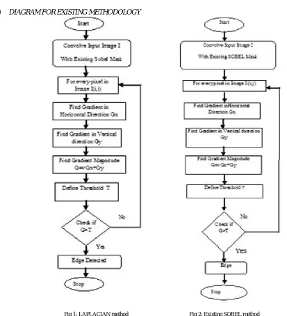
A. EXISTING METHODOLOGY
a) LAPLACIAN METHOD
The LAPLACIAN method is second derivative method, which searches for zero crossings in the second
derivative of the image to find edge.
Also called as Marr-Hildreth or Mexican hat operator edge detector. Edge is detected whenever second derivative
of image tends to zero and the first derivative is at a maximum [8-10].
b) SOBEL METHOD
The SOBEL method of edge detection for image segmentation finds edges using the SOBEL approximation to the
derivative.
ISSN(Online): 2320-9801 ISSN (Print) : 2320-9798
I
nternational
J
ournal of
I
nnovative
R
esearch in
C
omputer
and
C
ommunication
E
ngineering
(An ISO 3297: 2007 Certified Organization)
Vol. 4, Issue 10, October 2016
c) DIAGRAM FOR EXISTING METHODOLOGY
Fig 1: LAPLACIAN method Fig 2: Existing SOBEL method
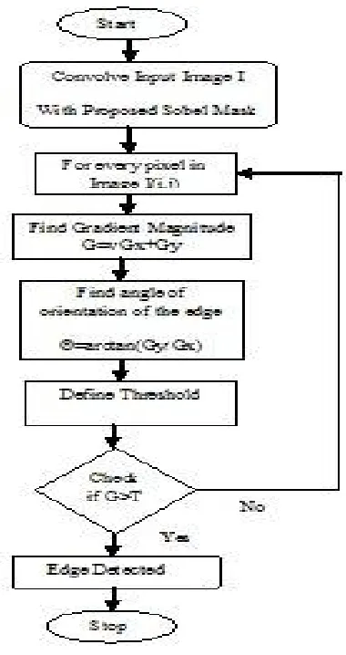
B. PROPOSED METHODOLOGY
In the proposed method (Modified SOBEL method) we design a new kernel to detect edges in given different kind of images like RGB, Gray, Medical, etc., compared to existing method Sobel, Laplacian methods we design a 3*3 convolution mask given in matrix GX,GY.[4]
GX= 2 1 0
1 0 -1
ISSN(Online): 2320-9801 ISSN (Print) : 2320-9798
I
nternational
J
ournal of
I
nnovative
R
esearch in
C
omputer
and
C
ommunication
E
ngineering
(An ISO 3297: 2007 Certified Organization)
Vol. 4, Issue 10, October 2016
GY=
The kernels can be applied separately to the input image, to produce separate measurements of the gradient
component in each orientation (Gx and Gy).
The modified SOBEL operator provide a simple approximation to the gradient magnitude:
GX=
Where Gx=(2(i-2,j)+(i-1,j)+(i-2,j-1))-((i,j-1)+(i-1,j-2)+2(i,j-2)
GY=
Where GY=((i-2,j-1)+2(i-2,j-2)+(i-1,j-2))-(2(i,j)+(i-1, j)+(i,j-1))
The gradient magnitude is given by:
G=√Gx+Gy
Typically, an approximate magnitude is computed using:
│G│=│GX│+│GY│ Maximum of Gradient is Edge.
The angle of orientation of the edge (relative to the pixel grid) giving rise to the spatial gradient is given by:
Ѳ=arctan(GY/GX)
0 -1 -2
1 0 -1
2 1 0
i-2,j i-1,j
i-2,j-1 i,j-1
i-1,j-2 i,j-2
i-1,j i ,j
i-2,j-1 i,j-1
ISSN(Online): 2320-9801 ISSN (Print) : 2320-9798
I
nternational
J
ournal of
I
nnovative
R
esearch in
C
omputer
and
C
ommunication
E
ngineering
(An ISO 3297: 2007 Certified Organization)
Vol. 4, Issue 10, October 2016
a) BLOCK DIAGRAM FOR PROPOSED METHOD
Fig 3: Modified SOBEL method
IV. PSEUDO CODE
Step 1: Read the input image I.
Step2: Convolve the proposed modified SOBEL mask to the input image I. Step 3: Find the gradient G and direction of edge by using the formula. Step 4: Set the threshold value T.
Step 5: Threshold the gradient image. Step 6: Maximum of Gradient is the output
V. STIMULATION RESULTS
ISSN(Online): 2320-9801 ISSN (Print) : 2320-9798
I
nternational
J
ournal of
I
nnovative
R
esearch in
C
omputer
and
C
ommunication
E
ngineering
(An ISO 3297: 2007 Certified Organization)
Vol. 4, Issue 10, October 2016
A. ESTIMATION OF STATISTICAL PARAMETERS
The parameters which are used in estimation of performance are Peak Signal to Noise Ratio (PSNR), Mean Square Error (MSE).
a) ESTIMATION OF PSNR
PSNR gives the ratio between possible power of a signal and the power of corrupting noise present in the image.
PSNR = 10 log 10 (MAXI / MSE)
Higher the PSNR gives more closure to clear edge detected image i.e. higher image quality. Here MAXI is maximum possible pixel value of the image.
b) ESTIMATION OF MSE
Mean Square Error (MSE) is given by MSE = ∑Ni=j=1 [ f(i, j) - F(i, j) ]2 / N2
Where, f is the original image F is the final image detected with some filter and N is the size of image.
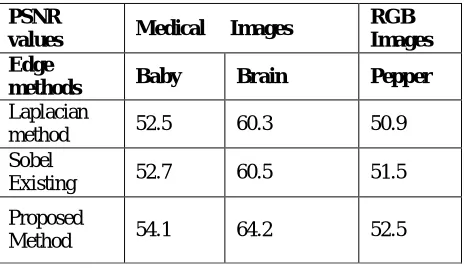
From the results it is deduced that for Existing Edge detection methods, missing and false edges persist. The newly proposed method (modified SOBEL method) comes with best PSNR and MSE values. Edges clearly detected are shown in the results.
PSNR
values Medical Images
RGB Images Edge
methods Baby Brain Pepper
Laplacian
method 52.5 60.3 50.9
Sobel
Existing 52.7 60.5 51.5
Proposed
Method 54.1 64.2 52.5
Table 1: PSNR values for various types of images in edge detection-automatic threshold method
MSE values Medical Images RGB
images
Edge
Methods Brain Baby Pepper
Laplacian
Method 0.06 0.37 0.4
Sobel
Existing 0.06 0.35 0.42
ISSN(Online): 2320-9801 ISSN (Print) : 2320-9798
I
nternational
J
ournal of
I
nnovative
R
esearch in
C
omputer
and
C
ommunication
E
ngineering
(An ISO 3297: 2007 Certified Organization)
Vol. 4, Issue 10, October 2016
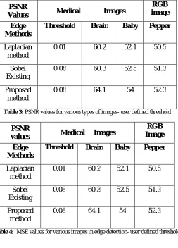
PSNR
Values Medical Images
RGB image
Edge Methods
Threshold Brain Baby Pepper
Laplacian method
0.01 60.2 52.1 50.5
Sobel Existing
0.08 60.3 52.5 51.3
Proposed method
0.08 64.1 54 52.3
Table 3: PSNR values for various types of images- user defined threshold
PSNR
values Medical Images
RGB Image
Edge Methods
Threshold Brain Baby Pepper
Laplacian method
0.01 60.2 52.1 50.5
Sobel Existing
0.08 60.3 52.5 51.3
Proposed method
0.08 64.1 54 52.3
Table 4: MSE values for various images in edge detection- user defined threshold
B. CHARTS
Figure 4: chart for PSNR values for various types of images in edge detection-automatic threshold
PSNR VALUES FOR VARIOUS IMAGES IN EDGE DETECTION 0 50 100 150 200 B a b y C a n c e r B ra in C h e s t P e p p e r B a b y 2 B u tt e rf ly B a rb a ra B a b o o n H o u s e
RGB IIMAGES GREY IMAGES
MEDICAL IMAGES
VARIOUS TYPES OF IMAGES
ISSN(Online): 2320-9801 ISSN (Print) : 2320-9798
I
nternational
J
ournal of
I
nnovative
R
esearch in
C
omputer
and
C
ommunication
E
ngineering
(An ISO 3297: 2007 Certified Organization)
Vol. 4, Issue 10, October 2016
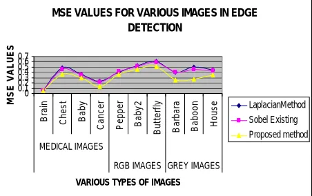
Figure 5: chart for MSE values for various images in edge detection-automatic threshold
Figure 6: chart for PSNR values for various images in edge detection-user defined threshold
Figure 7: chart for MSE values for various images in edge detection-user defined threshold
MSE VALUES FOR VARIOUS IMAGES IN EDGE DETECTION 0 0.1 0.2 0.3 0.4 0.5 0.6 0.7 B ra in C h e s t B a b y C a n c e r P e p p e r B a b y 2 B u tt e rf ly B a rb a ra B a b o o n H o u s e MEDICAL IMAGES
RGB IMAGES GREY IMAGES
VARIOUS TYPES OF IMAGES
M S E V A L U E S LaplacianMethod Sobel Existing Proposed method
MSE VALUES FOR VARIOUS IMAGES IN EDGE DETECTION
0 0.1 0.2 0.3 0.4 0.5 0.6 0.7 T h re s o ld B ra in C h e s t B a b y C a n c e r B a rb a ra B a b o o n H o u s e P e p p e r B a b y 2 B u tt e rf ly
GREY IMAGES RGB IMAGES
VARIOUS TYPES OF IMAGES
ISSN(Online): 2320-9801 ISSN (Print) : 2320-9798
I
nternational
J
ournal of
I
nnovative
R
esearch in
C
omputer
and
C
ommunication
E
ngineering
(An ISO 3297: 2007 Certified Organization)
Vol. 4, Issue 10, October 2016
C. PROPOSED METHOD OF EDGE DETECTION IN PICTORIAL REPRESENTATION
Figure 8: SOBEL METHOD BY USER DEFINED THRESHOLD T=0.08 ON RGB IMAGE –BABY
Figure 9: EDGE DETECTION ON RGB IMAGE- BUTTERFLY
Figure 10: PROPOSED EDGE DETECTION WITH THRESHOLD VALUE 0.08 ON RGB IMAGE-PEPPER
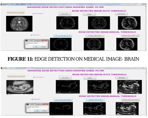
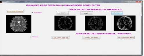
FIGURE 11: EDGE DETECTION ON MEDICAL IMAGE- BRAIN
ISSN(Online): 2320-9801 ISSN (Print) : 2320-9798
I
nternational
J
ournal of
I
nnovative
R
esearch in
C
omputer
and
C
ommunication
E
ngineering
(An ISO 3297: 2007 Certified Organization)
Vol. 4, Issue 10, October 2016
FIGURE 13: EDGE DETECTION ON MEDICAL IMAGE- CANCER
Figure 14: EDGE DETECTION ON MEDICAL IMAGE-CHEST CT
Figure 15: EDGE DETECTION ON GREY IMAGE –HOUSE
Figure 16: EDGE DETECTION ON GREY IMAGE- LENA
ISSN(Online): 2320-9801 ISSN (Print) : 2320-9798
I
nternational
J
ournal of
I
nnovative
R
esearch in
C
omputer
and
C
ommunication
E
ngineering
(An ISO 3297: 2007 Certified Organization)
Vol. 4, Issue 10, October 2016
Figure 18: EDGE DETECTION ON LOW RESOLUTION IMAGE –BARBARA
Figure 19: LAPLACIAN METHOD OF AUTOMATIC EDGE DEECTION ON MEDICAL IMAGE-BRAIN
Figure 20: SOBEL METHOD OF AUTOMATIC EDGE DEECTION ON MEDICAL IMAGE-BRAIN
Figure 21: PROPOSED METHOD OF AUTOMATIC EDGE DEECTION ON MEDICALIMAGE-BRAIN
VI. CONCLUSION AND FUTURE WORK
This proposed work presents the new modified SOBEL method which is capable of Detecting Edges keenly and tested on various types of images like medical, RGB, Gray images. This performs Edge detection with user defined threshold values and gives best results for specific threshold values. The earlier LAPLACIAN Edge Detection method falls under main drawback of Missing Edges and more False Edges. In this type of edge detection ,noises replicate twice more times as this performs second derivative of image. Existing Sobel method reduces the drawback of earlier one,but still false edges persist.
ISSN(Online): 2320-9801 ISSN (Print) : 2320-9798
I
nternational
J
ournal of
I
nnovative
R
esearch in
C
omputer
and
C
ommunication
E
ngineering
(An ISO 3297: 2007 Certified Organization)
Vol. 4, Issue 10, October 2016
From the results it is deduced that proposed method is best in Noise Ratio and Detects Edges clearly when compared to Existing Methods. In future the current technique called “Modified SOBEL Method” can be extended to work with Image segmentation with wavelet Thresholding methods for best results.
REFERENCES
[1]Anil .K.J, “Fundamentals of Digital Image Processing”, Prentice Hall Information and System Sciences Series, Editor Thomas Kailath, 1989.
[2] Rafael Gonzalez .C &Woods.R.E, “Digital Image Processing “,Pearson Education India, New Delhi, 2009. [3] Amit Chaudhary, Tarun Gulati, “Segmenting Digital Images Using Edge Detection”, Volume 2, Issue 5, May 2013. [4] Wenshuo.G, Lei.Y, Xiaoguang.Z and Huizhong.L, "An Improved Sobel Edge Detection", IEEE, Volume 3, Issue 2, 2010. [5] Sonam Saluja, Aradhana Kumari Singh, Sonu Agrawal “A Study of Edge-Detection Methods”Vol. 2, Issue 1, January 2013.
[6] Umamaheswari , Bhavani .R ,Thirunadana Sikamani.K “Segmentation of Tiles Image Using Various Edge Detection Techniques”,Volume: 04, Special Issue: March 2015.
[7]Ramadevi.Y, Sridevi.T, Poornima.B, Kalyani.B,“Segmenting and Object Recognition using various Edge Detection Techniques ”,Vol 2, No 6, December 2010.
[8] Tzu-Heng Henry Lee and Taipei, Taiwan ROC, “Edge Detection Analysis”, IJCSI International Journal of Computer Science Issues, Vol. 5, Issue 6, No 1, September 2012.
[9] Li Bin, Mehdi Samiei yeganeh Comparison for Image Edge Detection Algorithms” ISSN: 2278-0661 Volume 2, Issue 6 (July-Aug. 2012) [10] Rashmi, Mukesh Kumar, and Rohini Saxena, “Algorithm and Technique on various Edge Detection-A Survey “, Vol.4, No.3, June 2013 [11] Poornima .B, Ramadev.Y, Sridevi .T,” Threshold Based Edge Detection Algorithm “,IACSIT International Journal of Engineering and Technology, Vol.3, No.4, August 2011
[12] Pithadiya .K.J, Modi.C.K & Chauhan J.D, “Selecting the Most Favourable Edge Detection Technique for Liquid Level Inspection in Bottles”, Vol.3, 2011.
[13] Lakshmi S & Sankaranarayanan .V ,“A Study of edge detection techniques for segmentation computing approaches”, IJCA special issue on Computer Aided Soft Computing Techniques for Imaging and Biomedical Applications CASCT, 2010.
[14] Raman Maini, Sohal .J.S, “Performance Evaluation of Prewitt Edge Detector for Noisy Images” GVIP Journal, Volume 6, Issue 3, December 2006
[15] Bhadauria.H.S , Annapurna Singh, Anuj Kumar ”Comparison between Various Edge Detection Methods on Satellite Image “ISO 9001:2008 Certified Journal, Volume 3, Issue 6, June 2013
BIOGRAPHY
Dr.S.Gopinathan1
working as an Associate Professor in the Department of Computer Science, University Of Madras, Chennai, India. He has 16 years of teaching experience for post graduate in the field of Computer Science and Research. He has published number of papers. He has produced 9 M. Phil Scholars in the Computer Science; PhD Research Scholars are registered under him. He also has been serving as a panel member for various competitive examinations and universities. His interested area of research is Image Processing, Software Engineering.
S. Gayathri2 currently being as a Research Scholar in the Department of Computer
Science in University Of Madras, Chennai, India. She received BCA, MCA and M. Phil Degree in Department of Computer Science in University Of Madras, Chennai, India. She is interested in doing the research in the area of Image Processing.
G. Lalitha2 currently being as a Research Scholar in the Department of Computer