_____________________________________________________________________________________________________ ISSN: 2231-0614
SCIENCEDOMAIN international www.sciencedomain.org
The Results of Intravitreal Bevacizumab Injections
and Laser Photocoagulation for the Treatment of
Macular Edema Secondary to Retinal Vein Occlusion
Ihsan Yilmaz
1*, Ahmet Taylan Yazici
1, Ahmet Demirok
1and Omer Faruk Yilmaz
11
Beyoglu Eye Training and Research Hospital, Istanbul, Turkey.
Authors’ contributions
This work was carried out in collaboration between all authors. Author IY designed the study, performed the statistical analysis, wrote the protocol, and wrote the first draft of the manuscript. Authors ATY and AD managed the analyses of the study. Author OFY managed the literature searches. All authors read and approved the final manuscript.
Article Information
DOI: 10.9734/BJMMR/2015/13100
Editor(s):
(1) Ian Dooley, Limerick Regional Hospital, Republic of Ireland. (2) Jimmy T. Efird, Department of Public Health, Epidemiology and Outcomes Research East Carolina Heart Institute, Brody School of Medicine, Greenville, North Carolina, USA.
Reviewers:
(1) Anonymous, Mansoura University, Egypt. (2) Anonymous, Gaziantep University Medical School, Turkey. (3) Anonymous, Mie University, Japan. (4) Anonymous, Dicle University, Turkey. (5) Anonymous, USA. Complete Peer review History: http://www.sciencedomain.org/review-history.php?iid=696&id=12&aid=6422
Received 2nd August 2014 Accepted 20th September 2014 Published9th October 2014
ABSTRACT
Aims: To evaluate the efficacy and safety of intravitreal bevacizumab injections on macular edema secondary to retinal vein occlusion.
Study Design: Retrospective case series.
Place and Duration of Study: Department of Retina, Beyoglu Eye Training and Research Hospital, between July 2012 and July 2013.
Methodology: Forty two eyes (22 CRVO and 20 BRVO) were included in this study. Follow up time was a year for all patients. All patients were newly diagnosed and 3 intravitreal bevacizumab injections are used monthly and additional injection is used as needed. Scatter laser photocoagulation is used after macular edema disappears in all patients. Macular thickness was measured via spectral-domain OCT at baseline, at month 6 and at month 12.
Results: The mean age of the patients was 52.4±16.2 years (range 22 – 74 years). The central macular thickness improved from 684±196 µm at baseline to 220 ± 102 µm at month 6 (p<0.001) and to 214±112 µm (p<0.001) at month 12. Best-corrected visual acuity (BCVA) at baseline improved from 1.0±0.4 logMAR to 0.6±0.4 logMAR (p<0.001) at month 12.
Conclusion: Intravitreal bevacizumab injection is effective to treatment of macular edema secondary to retinal vein occlusion. 3 monthly injections and as needed regimen is effective in one year follow up time.
Keywords: Bevacizumab; retinal vein occlusion; macular edema.
1. INTRODUCTION
Retinal vein occlusion (RVO) is a potential sight threatening retinal vascular problem worldwide [1]. (RVO) is the second most common cause of retinal vascular disease after diabetic retinopathy [2]. Macular edema is the main cause for visual symptoms in central retinal vein occlusion (CRVO) and branch retinal vein occlusion (BRVO) [3].
In RVO, functional and structural changes in the retina lead to hypoxia and then lead to up-regulation of vascular endothelial growth factor (VEGF) [4]. Elevated VEGF concentrations have been detected in the ocular fluid of patients with BRVO and CRVO and it correlate with the severity of macular edema [5,6].
In recent years, the use of intravitreal anti-vascular endothelial growth factor (anti-VEGF) agents, Bevacizumab (Avastin) (Genentech, USA), Ranibizumab (Lucentis), have been in increasing use in macular edema following a retinal vein occlusion [7,8]. Bevacizumab, despite not being licenced for use in ophthalmic indications, and ranibizumab, are the two most commonly used anti-VEGF drugs in ophthalmic patients [2,9].
In this study we aim to present our one year result of Bevacizumab treatments for macular edema secondary to retinal vain occlusion.
2. MATERIALS AND METHODS
This was a retrospective, nonrandomized case series conducted at eye care center in Istanbul. Consecutive cases with macular edema secondary to either BRVO or CRVO (non-ischemic) who presented from July 2012 until July 2013 were included in the study. 1 years of their follow-up is evaluated. The patients with a prior history of treatment with laser therapy or intravitreal injections were excluded from the study. Patients with systemic disease such as
diabetics, hematologic disorders were excluded. All patients were volunteers and informed consent was taken from the patients before enrollment in the study after fully explaining the possible risks and benefits of bevacizumab injections. The study was conducted according to the declaration of Helsinki.
Detailed history was taken first. BCVA was taken with standard Snellen chart. Slit-lamp biomicroscope was used for anterior and posterior segment examinations. RVO was further evaluated to classify the BRVO and CRVO. Fundus fluorescein angiogram (FFA) was not done in all cases. Central macular thickness (CMT) was measured objectively via Spectralis spectral-domain OCT (Heidelberg Engineering, Inc., Vista, CA, USA) at baseline, moths 6 and month 12. The intraocular pressure was measured via Goldmann applanation tonometry.
The intravitreal bevacizumab (Avastin, Genentech) was given in a dose of 1.25 mg/0.05 ml under topical anesthesia. Injection site was 3.5 - 4 mm from superior temporal limbus for all cases. Topical ciprofloxacin eye drop four times a day used for a week after the injections. Intraocular pressure control and slit-lamp exam are done after a day and a week from each injections.
Snellen visual acuity was converted to Log MAR for visual outcome analysis. The data was analyzed in SPSS version 20.0 (SPSS Inc. Chicago, IL, USA). Paired t-test was used for statistical analysis and p-value of less than 0.05 was considered to be statistically significant.
3. RESULTS AND DISCUSSION
mean CMT decreased to 220±102 µm (range 186 – 248) at month 6 (p<0.001) and to 214±112 µm (range 188 – 240) at month 12 (p<0.001). BCVA at baseline improved from 1.0±0.4 logMAR to 0.6±0.4 logMAR at month 12 (p<0.001). CMT was significantly decreased at month 6 and month 12. (Fig. 1) Also BCVA increased significantly at month 12.
Fig. 1. The average CMT at baseline, at month 6 and month 12
In our series of 42 cases, the macular edema and visual acuity significantly improved without any ocular or systemic adverse effects at the one year follow up. The mean age of our patients was 52.4±16.2 years. This mean age of the patients with RVO was similar to some previous studies [10]. However, some other studies reported older mean ages [11]. The average number of intravitreal bevacizumab injections in our series was 3.3±0.6 injections and none of the cases had recurrent macular edema at month 12. An additional injection was given if macular thickness is over than 350 microns.
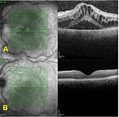
Fig. 2 shows an OCT scans of one the case.
Previous studies from other countries have similar findings regarding a significant reduction in CMT and BCVA improvement following the use of intravitreal bevacizumab injections for ME secondary to RVO [12-14]. Abegg et al. [12] reported that visual acuity was significantly better 4 to 6 weeks after bevacizumab treatment and the gain in visual acuity was accompanied by a significant decrease in retinal thickness from 454±117 µm to 305±129 µm. In another study, Gutiérrez et al. [13] reported that the mean BCVA improved from 1.32±0.24 logMAR at baseline to 0.8±0.15 logMAR (p=0.0003) at the 6-month follow-up and the CMT improved from 615±116 µm to 420±72 µm. Manayath et al. [14] present similar successful results in 2009.
Fig. 2. OCT scans. Before (A) and after the injections (B)
None of our patients had any adverse reactions. Some other studies reported adverse reactions such as raised intraocular pressure, intraocular inflammation and endophthalmitis [13,15]. This favourable adverse effect profile is also better than triamcinolone with one study reported that 26% of cases experienced an increase in intraocular pressure [16]. Hirashima et al. [17] compared bevacizumab alone or combined with macular laser photocoagulation for the treatment of macular edema secondary to BRVO. Niederhauser et al. [18] presented their results of bevacizumab and ranibizumab treatments of macular edema due to retinal vein occlusions. They reported that both treatments improved the visual acuity and the central foveal thickness at 3 months and a further improvement was not measured if the treatment was prolonged to 6 months [18]. They added that there were no significant differences measured between bevacizumab and ranibizumab [18].
The main limitation of the study is that there was no control group in order to make outcome comparisons. Also case series was small and follow-up time was only a year.
We presented our results and although the present study confirms the previous findings from other countries, it is important to demonstrate similar efficacy within the Turkish population. 0
100 200 300 400 500 600 700 800
4. CONCLUSION
Intravitreal Bevacizumab is an effective and safe for the treatment of macular edema secondary to retinal vein occlusion. Central macular thickness and visual acuity improves in first year of the treatment. CONSENT Not applicable. ETHICAL APPROVAL Not applicable. COMPETING INTERESTS
Authors have declared that no competing interests exist.
REFERENCES
1. Thapa R, Poudyal G. Short term results of intra-vitreal bevacizumab for the treatment of macular edema secondary to retinal vein occlusion. Nepal J Ophthalmol. 2013;5(9):63-8.
2. Gerding H, Monés J, Tadayoni R, Boscia F, Pearce I, Priglinger S. Ranibizumab in retinal vein occlusion: treatment recommendations by an expert panel. Br J Ophthalmol; 2014. PII: bjophthalmol-2014-305041. DOI: 10.1136/bjophthalmol-2014-305041.
3. Ford JA, Shyangdan D, Uthman OA, Lois N, Waugh N. Drug treatment of macular oedema secondary to central retinal vein occlusion: A network meta-analysis. BMJ Open. 2014;4(7):e005292. DOI: 10.1136/bmjopen-2014-005292.
4. Karia N. Retinal vein occlusion: pathophysiology and treatment options. Clin Ophthalmol. 2010;4:809–16.
5. Noma H, Funatsu H, Yamasaki M, et al. Pathogenesis of macular edema with branch retinal vein occlusion and intraocular levels of vascular endothelial growth factor and interleukin-6. Am J Ophthalmol. 2005;140:256–61.
6. Noma H, Minamoto A, Funatsu H, et al. Intravitreal levels of vascular endothelial growth factor and interleukin-6 are correlated with macular edema in branch retinal vein occlusion. Graefes Arch Clin Exp Ophthalmol. 2006;244:309–15.
7. Pielen A, Feltgen N, Isserstedt C, Callizo J, Junker B, Schmucker C. Efficacy and safety of intravitreal therapy in macular edema due to branch and central retinal vein occlusion: a systematic review. PLoS One. 2013;8(10):e78538. DOI: 10.1371/journal.pone.0078538.
8. Singer MA, Cohen SR, Groth SL, Porbandarwalla S. Comparing bevacizumab and ranibizumab for initial reduction of central macular thickness in patients with retinal vein occlusions. Clin Ophthalmol. 2013;7:1377-83.
9. Azad R, Vivek K, Sharma Y, Chandra P, Sain S, Venkataraman A. Ranibizumab as an adjunct to laser for macular edema secondary to branch retinal vein occlusion. Indian J Ophthalmol. 2012;60(4):263-6. 10. Thapa R, Paudyal G, Bernstein PS.
Demographic characteristics, patterns, and risk factors for retinal vein occlusion in Nepal: A hospitalbased case-control study. Clinical and Experimental Ophthalmology. 2010;38:583-90.
11. Iturralde D, Spaide RF, Meyerle CB, Klancnik JM, Yannuzzi LA, Fisher YL, Sorenson J, Slakter JS, Freund KB, Cooney M, Fine HF. Intravitreal bevacizumab (Avastin) treatment of macular edema in central retinal vein occlusion: A short-term study. Retina. 2006;26:279-84.
12. Abegg M, Tappeiner C, Schnurrbusch UW, Barthelmes D, Wolf S, Fleischhauer J. Treatment of Branch Retinal Vein Occlusion induced Macular Edema with Bevacizumab. BMC Ophthalmol. 2008;8:18.
13. Gutièrrez JC, Barquet LA, Caminal JM, Mitiana,Almolda SP, Domènech NP, Goita OP, Caso MR, Ginebreda JA. Intra-vitreal bevacizumab (Avastin) in the treatment of macular edema secondary to retinal vein occlusion. Clin Ophthalmol. 2008;2(4):787-91.
14. Manayath GJ, Narendran V, AI-Kharousi N, Wali UK. Bevacizumab therapy for macular edema in central retinal vein occlusion: Long-term results. Oman J Ophthalmol. 2009;2(2):73-8.
15. Badala’ F. The treatment of branch retinal vein occlusion with bevacizumab. Curr Opin Ophthalmol. 2008;19:234-38.
possible risk factors. Clin Ophthalmol. 2008;2(2):269-74.
17. Hirashima T, Chihara T, Bun T, Utsumi T, Hirose M, Oh H. Intravitreal Bevacizumab Alone or Combined with Macular Laser Photocoagulation for Recurrent or Persistent Macular Edema Secondary to Branch Retinal Vein Occlusion. J
Ophthalmol. 2014;2014:173084. Epub 2014 Jul 7.
18. Niederhauser N, Valmaggia C. Bevacizumab and Ranibizumab for macular edema due to retinal vein occlusions. Klin Monbl Augenheilkd. 2013;230(4):405-8.
© 2015 Yilmaz et al.; This is an Open Access article distributed under the terms of the Creative Commons Attribution License (http://creativecommons.org/licenses/by/4.0), which permits unrestricted use, distribution, and reproduction in any medium, provided the original work is properly cited.
Peer-review history: