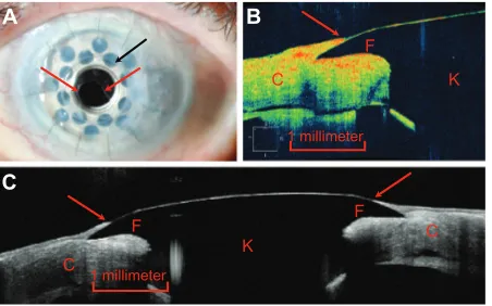
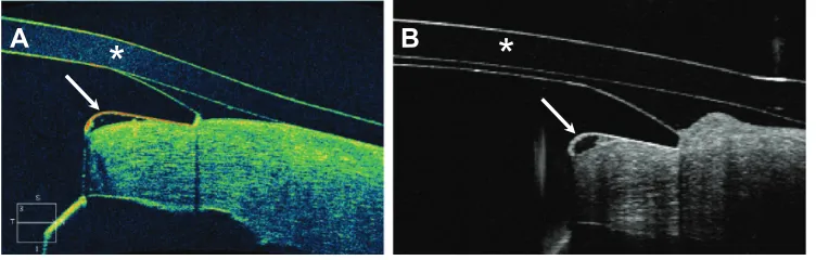
Boston type I keratoprosthesis-donor cornea interface evaluated by high-definition spectral-domain anterior segment optical coherence tomography
Full text
Figure
Related documents
Using feature extraction method 1 and ANN Backpropagation algorithm as a classifier of arm movements, the prosthetic hand movement was further examined for its
American Museum of Natural History , one of the largest and most celebrated museums in the world and a New York City icon (Exit Central Park at 81st Street/Central
It is also worth recalling Velho’s text in the introduction to Antropologia urbana: Cultura e sociedade no Brasil e em Portugal, where he restates his concern to avoid “reifying
Tool presentation: We present work in progress on a stand-alone implementation of Lagrangian reachability, a recently introduced over-approximation technique for nonlinear
We consider the use case of a vehicular environment, where drivers of vehicles are notified when entering a danger zone (DZ). In particular, a ME service running within a ME
In this study, we presented a unique case of severe HPE diagnosed via routine ultrasound in a neonate born in Zabol, Iran.. Based on the severity,
In a series of human fear conditioning studies in our lab, we convincingly demonstrated that administration of the β-adrenergic receptor antagonist pro- pranolol HCl before or
