_____________________________________________________________________________________________________
*Corresponding author: E-mail: [email protected];
www.sciencedomain.org
Squamous Cell CarcInoma of Gallbladder: A Case
Report
Hüseyin Pülat
1, Huseyin Eken
2*, Oktay Karaköse
1,
İ
smail Zihni
1,
Kazım Ça
ğ
lar Özçelik
1, Metin Çiri
ş
3and Hasan Erol Ero
ğ
lu
11
Department of Surgical Oncology, Suleyman Demirel University Medical Faculty, Isparta, Turkey.
2
Department of General Surgery, Erzincan University Medical Faculty, Erzincan, Turkey.
3
Department of Pathology, Suleyman Demirel University Medical Faculty, Isparta, Turkey.
Authors’ contributions
This work was carried out in collaboration between all authors. Author HP designed the study, wrote the protocol and wrote the first draft of the manuscript. Author HE managed the literature searches, analyses of the study performed the spectroscopy analysis. All authors read and approved the final manuscript.
Article Information
DOI: 10.9734/BJMMR/2016/23849 Editor(s): (1)Franciszek Burdan, Experimental Teratology Unit, Human Anatomy Department, Medical University of Lublin, Poland and Radiology Department, St. John’s Cancer Center, Poland.
Reviewers: (1) Salah Berkane, University of Bejaia, Algeria. (2)Ihsan Yıldız, Suleyman Demirel University, Turkey. (3) Anonymous, The Chinese University of Hong Kong, China. Complete Peer review History:http://sciencedomain.org/review-history/13696
Received 25th December 2015 Accepted 28th January 2016 Published 14th March 2016
ABSTRACT
Although gallbladder cancers are rarely seen, the mortality rates are high. Pure squamous cell cancer, which is one of the most rarely seen cancer types in the gallbladder, originates from the squamous metaplasia which forms following chronic irritation in the mucosa. The majority of cases are generally at the inoperable stage when diagnosed as they have been followed up for long-term cholelithiasis.
The case is here presented of a 59-year old female who underwent surgical and medical treatment for a bladder tumour 10 years previously and although gallbladder stones were determined at that time, as she was asymptomatic, surgery was not recommended, then with the progression of time, gallbladder squamous cell carcinoma developed.
Keywords: Gallbladder; squamous cell carcinoma; bladder tumor.
1. INTRODUCTION
Gallbladder squamous carcinoma comprise less than 2% of all gallbladder cancers. Metastasis has been reported at relatively low rates compared to adenocarcinoma [1]. The stage of the disease is the most important criteria in the prognosis and the only form of treatment presenting the possibility of a cure is surgery. This is possible in early stage tumours but the majority of cases are generally diagnosed when they are at the inoperable stage. However, in advanced and metastatic tumours, palliative procedures can be applied for complications [2]. An effective chemotherapy (CT) or radiotherapy (RT) protocol has been developed as yet [3,4].
In this paper, we present the case of a 59-year old female who was diagnosed with cholelithiasis 10 years previously but did not undergo surgery as she was asymptomatic, then over time gallbladder squamous cell carcinoma developed on the basis of chronic inflammation due to long-term cholelithiasis.
2. CASE REPORT
The 59-year old female had a 42-year history of smoking 2 packets of cigarettes per day. Transurethral bladder tumour resection (TUR-BT) and superficial bladder chemotherapy had
been applied 10 years previously for a bladder changing epithelial cell carcinoma (Mostofi, Grade 2) (Fig. 1) and on the abdominal computed tomography (CT) taken at that time, an appearance was observed consistent with gallbladder stones. However, no treatment was applied at that time as the patient was asymptomatic. It was then learned that the patient had undergone surgery 1 month previously at an external clinic for a diagnosis of cholelithiasis on the basis of the complaints of blunt abdominal pain, swelling and indigestion which had been ongoing for 6 months and in the last month, nausea and weight loss.
Fig. 1. Non-invasive low-grade, epithelial changing tumour of the bladder. (x100, HE)
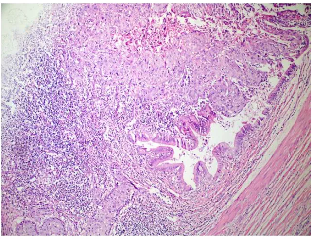
The operation, which was started laparoscopically went beyond the serosa in the gallbladder and was changed to open surgery on the visualisation of a mass showing adhesion to the common bile duct and a cholecystectomy was applied including part of the surrounding fatty tissue. In the pathological examination, diagnosis was made of squamous cell carcinoma of the gallbladder from a 4 x 4 x 3 cm diameter mass partially localised in the gallbladder neck, showing infiltration to perimuscular connective tissue in several areas, and perineural invasion in the tumoural tissue with clean surgical borders and which had infiltrated the serosa showing moderate level differentiation (Fig. 2). Serosa, vascular and lymphatic invasion of tumor was seen at histopathological examination.
On presentation at our clinic, the laboratory test results for the patient were determined as AST) 115 (0-31) U/L, alanine aminotransferase (ALT) 213 (0-34) U/L, lactate dehydrogenase (LDH) 321 (0-247) U/L, alkaline phosphatase (ALP) 189 (30-120) U/L, gamma glutamil transferase (GGT) 94 (0-38) U/L. Tumour markers such as AFP, CEA, CA and 19-9 were within normal limits. On the abdominal CT, 3 lesions smaller than 1cm consistent with metastasis were observed in segment 4 of the liver, and a mass with irregular borders and a necrotic centre 5 x 4 x 5 cm was observed in the liver hilus, adhering to the stomach antrum, the duodenum and the common bile duct (Fig. 3). The patient was admitted for planned radical surgery but as more widespread tumoral involvement was seen in the liver and adjacent organs in the exploration compared to on the CT image, it was accepted as unresectable. The postoperative period passed without problems so with no indications, neither CT nor RT were administered.
Fig. 3. Invasion of the liver and neighbouring organs
3. DISCUSSION
Although gallbladder cancers are seen rarely, the mortality rates are high. The biological behaviour of the tumour, the emergence of disease symptoms in the late stage and similarity to benign gallbladder diseases play a role in late diagnosis [5,6]. Incidence is seen to increase after the age of 50 years [2] and is seen in females 2-3 times more than in males [1]. This rate is in parallel with the incidence of gallbladder stones. The case presented here was female and 59 years old.
Gallbladder cancers associated with gallbladder stones (squamous cell, adeno-squamous cell and some adeno-carcinoma) are related to very long-term contact with the size of the stones [7]. Although 75-90% of cases have gallbladder stones, only 1% of caes with gallbladder stones are at risk of developing carcinoma [8]. In an autopsy study, 1%-2% of cholelithiasis cases were reported to have gallbladder cancer [9]. Chronic mechanic injury of the mucosa is the main triggering factor of this neoplastic transformation [10]. Anomalies in the main bile duct, porcelain gallbladder, adenomatous polyps and inflammatory intestinal diseases are other etiological factors [1].
A diagnosis of primary gallbladder cancer can only be made preoperatively in a third of cases [11]. Although tumour symptoms in gallbladder cancers may emerge such as severe pain spreading to surrounding tissue, jaundice, loss of appetitie and loss of weight, generally the symptoms are imperceptible and appear similar to benign diseases [1,12-14]. In the current case, after a 10-year asymptomatic period, there had been blunt abdominal pain, swelling and indigestion for the previous 6 months and complaints of nausea and weight loss in the previous month. On the determination of gallbladder stones on the abdominal USG, the patient was admitted for surgery and diagnosis was made intraoperatively.
duodenum, jejunum, colon, pancreas and abdominal wall [6]. In the current case there was long-term cholelithiasis and in the operation there was seen to be tumoural spread (grade 4B) in the liver and neighbouring organs.
In unresectable cases, it is attempted to eradicate jaundice with surgery, endoscopic or radiological drainage methods [8]. As an effecttive CT protocol has still not been developed, adjuvant CT is not generally recommended [3]. RT has been reported to be useful where the tumour has been resected but there are microscopic residual remains [4].
On 2015, palliative systemic chemotherapy must be indicated for patients. For example Gemciatbine alone or Gemcitabine with Cisplatine a good protocol. It was demonstrated since 15years ago. For example the tumour could be resectable after 3 or 4 cycles of this protocol. In differential diagnosis, there are pure chronic cholecytitis, gall bladder polyps and choledocholitiasis.
4. CONCLUSION
As gallbladder cancers may develop over time on the basis of cholecystitis with chronic stones, cholecystectomy should be recommended for patients with cholelithiasis when there is no concomitant disease such as cardiac or respiratory problems, even if they are asymptomatic. When a surgeon has gallbladder
cancer discovered during an operation, he must do the radical resection or send the patient to a specialized center, without doing a cholecystectomy which will worsen the prognosis of this disease. The message in this figure is to operate in the radical option at the first time.
CONSENT
All authors declare that ‘written informed consent was obtained from the patient (or other approved parties) for publication of this case report and accompanying images.
ETHICAL APPROVAL
All authors hereby declare that all experiments have been examined and approved by the appropriate ethics committee and have therefore been performed in accordance with the ethical standards laid down in the 1964 Declaration of Helsinki.
COMPETING INTERESTS
Authors have declared that no competing interests exist.
REFERENCES
1. Ünlü Y, Alimoğlu O, Uğurluoğlu C, Behzatoğlu K, Demiroğlu Z. Squamous cell carcinoma of gallbladder. Genel Tıp Derg. 2003;13:127-129.
2. Gençosmanoğlu R, Tahan V, Yapıcıer Ö. Gallbladder carcinoma: Up-to-date overview for etyopatogenezis, diagnostic methods, staging, therapeutic modalities and prognosis. Güncel Gastroenteroloji. 2003;7:157-169.
3. Donohue JH. Present status of the diagnosis and treatment of gallbladder carcinoma. J Hepatobiliary Pancreat Surg. 2001;8:530-534.
4. Houry S, Barrier A, Huguier M. Irradiation therapy for gallbladder carcinoma: Recent advances. J Hepatobiliary Pancreat Surg. 2001;8:518-524.
5. Küçük C, Erdoğan G, Güler İ, Avşaroğulları L, İkizceli İ. Gallbladder carcinomas. Çağdaş Cerrahi Dergisi. 2002;16:78–81.
6. Kaushik SP. Current perspectives in gallbladder carcinoma. J Gastroenterol Hepatol. 2001;16:848-854.
7. Eken H, Balci MG, Buyukakincak S, Isik A, Firat D, Cimen O. Rare tumors of the gallbladder: Clear cell carcinoma. Int J Surg Case Rep. 2015;9:65-8.
8. Orth K, Beger HG. Gallbladder carcinoma and surgical treatment. Langenbeck’s Arch Surg. 2000;385:501-508.
9. Kimura W, Shimada H, Kuroda A, Morioka Y. Carcinoma of the gallbladder and extrahepatic bile duct in autopsy cases of the aged with special reference to its relationship to gallstones. Am J Gastroenterol. 1989;84:386-390.
10. Moser AJ, Roslyn JJ. Gallbladder and biliary tree. In: Corson JD, Williamson RCN, editors. Surgery. London: Mosby. 2001;1-26.
11. Gürkan A, Kaçar S, Karaca C, Özdemir B, Varılsüha C, Zalluhoğlu N. Surgical approach to gallbladder cancer. Ulusal Cer Derg. 2006;22:49-53.
13. Isik A, Firat D, Peker K, Sayar I, Idiz O, Soytürk M. A case report of esophageal perforation: Complication of nasogastric tube placement. Am J Case Rep. 2014;15:168-71.
14. Isik A, Eryılmaz R, Okan I, Dasiran F, Firat D, Idiz O, Sahin M. The use of fibrin glue without surgery in the treatment of pilonidal sinus disease. Int J Clin Exp Med. 2014;7:1047-51.
_________________________________________________________________________________
© 2016 Pülat et al.; This is an Open Access article distributed under the terms of the Creative Commons Attribution License (http://creativecommons.org/licenses/by/4.0), which permits unrestricted use, distribution, and reproduction in any medium, provided the original work is properly cited.
Peer-review history: