_____________________________________________________________________________________________________
*Corresponding author: Email: [email protected];
www.sciencedomain.org
Mild Hyperhomocysteinemia, Decreased Vitamins
B
6
, B
12
and Folic Acid in Children with Sickle Cell
Disease
M. O. Ebesunun
1*, I. O. Nnaji
1and K. J. Adetunji
11Department of Chemical Pathology and Immunology, Faculty of Basic Medical Sciences,
Obafemi Awolowo College of Health Sciences, Olabisi Onabanjo University, Ago-Iwoye, Nigeria.
Authors’ contributions
This work was carried out in collaboration between all authors. Author MOE designed the study, supervised wrote the protocol, and wrote the final draft of the manuscript. Author ION collected, process the samples and managed the literature searches, author KJA wrote the first draft of the manuscript and managed the literature. All authors read and approved the final manuscript.
Article Information
DOI: 10.9734/BJMMR/2015/19623 Editor(s): (1) Chan-Min Liu, School of Life Science, Xuzhou Normal University, Xuzhou City, China. (2)Anonymous. Reviewers: (1) Vlachaki Efthymia, Aristotle University, Greece. (2)Hassan Ali Abdelwahid, Suez Canal University, Egypt. (3)Syed Ali Raza Kazmi, Institute of Biomedical and Genetic Engineering, Islamabad,
Pakistan. Complete Peer review History:http://sciencedomain.org/review-history/10599
Received 20th June 2015 Accepted 2nd August 2015 Published 19th August 2015
ABSTRACT
Background: Hyperhomocysteinemia has been identified as a risk factor for stroke and other vascular diseases in the general population, its role in sickle cell disease (SCD) has not been investigated in children with SCD in Nigeria.
Aim: This study was designed to evaluate plasma homocysteine, B-vitamins, folate and lipid profile in sickle cell disease (SCD) HbSS children in Nigeria.
Methods: Fifty (50) SCD children (12.04±4.17 years) consisting of 30 females and 20 males were selected from Sickle club center Abeokuta. Fifty non SCD (HbAA) children (12.62±4.28 years) consisting of 25 males and 25 females were included as controls. Anthropometric indices and plasma homocysteine, B12, B6, folic acid, lipids and lipoproteins were determined using standard procedures.
Results: The results showed significant decreases in body weight (29.84±10.68 kg) and height
(1.37±0.16 m), (p<0.045) in all SCD patients. Plasma total homocysteine (tHcy) (6.40±3.37 µmol/L) was significantly increased (p< 0.05), whereas plasma vitamins B6 (28.81±12.44 nmol/), B12 (184.16±35.13 pmol/L), folic acid (46.73±9.93 µmol/L), total cholesterol (102.42±28.62 mg/dl), low density lipoprotein cholesterol (24.45±5.25 mg/dl) (p< 0.01) and triglyceride (71.98±22.61 mg/dl) (p<0.04) were markedly decreased compared with the control values. Plasma high density lipoprotein cholesterol was however not significantly different from the control value. Plasma tHcy) did not correlate with any of the measured parameters.
Conclusion: Increased plasma total homocysteine level and reduced B vitamins as well as lipids profile obtained in this study are prominent features of sickle cell disease in this environment.
Keywords: Homocysteine; vitamins B12; B6;folic acid; sickle cell diseases.
1. INTRODUCTION
Available evidence has shown that increase in plasma homocysteine is a risk factor for venous thrombosis and arteriosclerosis [1,2]. The possibility that homocysteine, an important vascular risk factor, may contribute to the ischemic phenomena of sickle cell disease (SCD) has attracted some interest in plasma total homocysteine (tHcy) levels in patients with SCD.
Hyperhomocysteinemia has many causes [3]. Much attention has focused on folate, vitamin B12 also known as cobalamin and vitamins B6
deficiencies, whose treatment reverses
hyperhomocysteinemia.
Studies of paediatric patients supplemented with folic acid elsewhere [4] showed higher plasma tHcy level than in control subjects and suggesting that folate supplementation had a major influence on the plasma tHcy levels. Nutritional deficiencies of vitamin B12 and folic acid have been linked to an elevated plasma homocysteine concentration [5,6]. It thus suggests that SCD patients could have elevated plasma homocysteine concentration in vitamins B deficiencies state. Available, reports [6,7] have shown that SCD patients have the propensity to increased plasma tHcy and therefore ischemic heart disease.
Vitamins B12, B6 and folic acid are necessary for the remethylation and transsulfration in homocysteine metabolism pathway. Deficiencies of these vitamins are possible contributory risk factors to elevated plasma tHcy.
Also the variation in the concentrations of plasma lipids in SCD children are recognized as risk factors for cardiovascular disease (CVD) in some studies [4,7].
Studies on plasma homocysteine and B vitamins are scarce in Nigeria where 150,000 children
with sickle cell disease are born annually (Sickle Cell Foundation Nigeria) [8]. Sickle cell disease patients are very often susceptible to B vitamins deficiencies and in particular folic acid. The deficiencies of these vitamins could lead to raised plasma homocysteine in SCD patients.
This study was therefore designed to evaluate plasma tHcy, vitamins B6, B12 and folic acid as well as lipids and lipoproteins in SCD (HbSS) patients and non SCD (HbAA) controls.
2. MATERIALS AND METHODS
The study was designed to evaluate plasma tHcy, B vitamins as well as lipids and lipoproteins in sickle cell disease. Subjects were on their normal diet during the period. The duration of study was one year, field work was studied in February 2014 and ended in November same year. The study population was sickle cell disease patients attending Sickle Cell Club Abeokuta. A total of one hundred (100) children (4-18 years) comprising of fifty (50) SCD (HbSS) patients with mean age (12.04±4.17 years) diagnosed as having sickle cell disease based on haemoglobin electrophoresis and clinical findings were selected from Aglow Sickle Cell Club, Abeokuta Nigeria. Fifty (50) aged-matched non SCD (HbAA) with mean age (12.62±4.28 years) were included as controls. The controls
were selected based on haemoglobin
electrophoresis and clinical examinations. It was a cross sectional study.
2.1 Inclusion Criteria
All SCD patients attending this Sickle Cell Club who had not taken vitamin supplements for at least month and without renal disease and /or cardiovascular disease were included.
2.2 Exclusion Criteria
renal disease and/or cardiovascular disease were excluded.
Ethical approval was obtained from the Ethical Review Committee of State Hospital/sickle cell Centre, Abeokuta in accordance with Helsinki ethics. Written /oral informed Consent was obtained from each participant.
2.3 Sample Size
Fifty SCD patients were randomly selected out of seventy five Aglow Sickle Cell Club members. The patients were matched in age with fifty HbAA controls. Some participants who were enrolled in the study failed to turn up. Equal number of patients and controls were recruited to minimize confounding variables.
2.4 Anthropometric Measurement
The weight in kilograms and height in centimeters were measured. The body mass index (BMI) in kg/m2 of all subjects was calculated. Height was taken with the subjects in standing position without footwear and this was directly read on the calibrated meter rule. The weight was read on the scale (Salter weighing
scale by Golden Cross Tech). Waist
circumference was taken at the minimum circumference between the costal margins and the iliac crest, measured in the horizontal plane with the subject standing using non stretchable tape rule. Hip circumference was measured at the maximum circumference in the horizontal plane over the buttocks with non stretchable tape rule [9].
2.5 Blood Sample Collection
Breakfasts were provided for all subjects on the day of blood collection; 5mls of overnight fasting (10-12 hours) blood samples were collected by standard venipuncture without stasis and dispensed into Dipotassium Ethylene Diamine Tetra acetic Acid (K2EDTA) bottles and were placed immediately in ice pack in a dark container. These were processed within short time of collection. The blood samples were spun at 3500 rpm for 10 minutes using MSE centrifuge and the plasma was aliquot into clean plain bottles and stored at -20°C until analyzed.
2.6 Biochemical Analysis
Total cholesterol was determined using the method of Alain et al. [10] while the triglyceride was estimated using the enzymatic method of
David and Buccolo [11]. HDLC was estimated after LDLC and VLDLC were precipitated out by the addition of phosphotungstic acid in the
presence of magnesium ions. After
centrifugation, the HDLC cholesterol
concentration in the supernatant was determined with the method of Alain et al. [10].
The LDLC was calculated using the formula of Friedwald et al. [12]. LDLC=TC-(TG/5+HDLC).
2.7 Measurement of Plasma Total
Homocysteine
The method was based on quantitative
determination of serum total homocysteine by Axis® Homocysteine enzyme immunoassay (EIA) using ELISA.
2.7.1 Assay principle
Protein bound homocysteine (Hcy) is reduced to free Hcy and enzymatically converted to S-adenosyl-L-homocysteine (SAH) in a separate procedure prior to the immunoassay [13]. The homocysteine adenosyl deaminase is specific for the L-form of homocysteine which is the only form present in the blood.
2.8 Determination of B Vitamins
Analyses of folic acid, vitamins B12 and B6 were carried out using high performance liquid chromatography (HPLC) (International Institute of Tropical) - waters 616/626.
2.8.1 Principle
Separation by Liquid Chromatography was based on the distribution of solutes between a liquid mobile phase and a stationary phase. When particles of small diameter are used as stationary phase support, the technique is high performance liquid chromatography (HPLC). In HPLC, the stationary phase is composed of uniform, ultra-fine particles, which greatly increase its adsorptive area. The stationary phase is packed firmly into column. The resistance of flow in this column is high; therefore large pressures (500-5000 pounds per square inch) are required to deliver constant flow rates. Elute from the column was monitored by a variety of detectors such as ultra-violet or redox-potential electrode detectors.
2.9 Statistical Analysis
All results were subjected to statistical analysis using the SPSS software version16 and results were expressed as mean plus standard deviation. Student t-test was used to assess statistical differences between means and the differences were regarded as significant at p< 0.05. Pearson’s correlation coefficient was also used to assess the relationship between
biochemical parameters and biophysical
characteristics.
3. RESULTS
Table 1 shows the baseline characteristics of the study groups. The body weight (p<0.05 and height (p<0.035) were significantly reduced in
SCD patients when compared with the
corresponding control values. No significant changes were obtained in the other parameters.
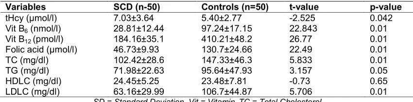
Table 2 shows the biochemical parameters of sickle cell disease patients and control group.
Plasma homocysteine was significantly
increased (p<0.042) in the sickle cell disease patients when compared with the corresponding control. On the other hand, significant decreases were obtained in plasma vitamins B6, B12, folic
acid, total cholesterol, LDLC (p<0.01) and triglyceride (p<0.032) respectively when compared with the control values. No significant change was obtained in plasma HDLC. When subjected to Post Hoc analysis, it revealed no significant changes. Data were normally distributed.
Table 3 shows the biochemical parameters of SCD males and control males. There were significant decreases in plasma vitamin B6, B12 folic acid, total cholesterol, LDLC (p<0.01) and triglyceride (p<0.05) when compared with the corresponding control values. Although the mean plasma tHcy was higher in the males SCD, the increase was however not significant. No significant changes were obtained in the other parameters.
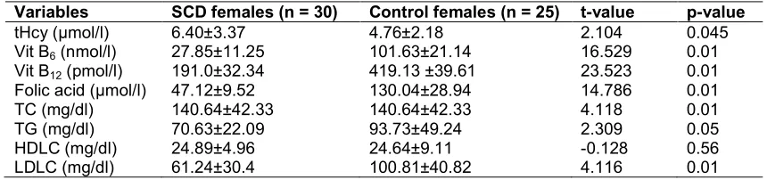
Table 4 shows the biochemical parameters of SCD females and control females. There were significant decreases in vitamin B6, B12, folic acid, total cholesterol, LDLC (p<.0.01) and triglyceride (p<0.05) when compared with the corresponding control values. A significant increase was obtained in total homocysteine (p<0.05) when compared with the female control value. HDLC was not significantly different compared with the corresponding control value.
Table 1. Baseline characteristics of the study groups (Mean±SD)
Variables SCD Controls t-value p-value
0.21
Weight (kg) 29.84±10.68 35.80±8.65 2.7796 0.045
Height (m) 1.37±0.16 1.49±0.20 3.222 0.05
WC (m) 0.25±0.03 0.25±0.04 0.284 0.21
HC (m) 0.27±0.038 0.28±0.041 0.911 0.11
WHR 0.93±0.05 0.91±0.06 -1.637 0.09
BMI (kg/m2) 15.36±2.39 15.68±1.31 0.836 0.610
SD = Standard Deviation, WC = Waist Circumference, HC = Hip Circumference, WHR = Waist Hip Ratio BMI = Body Mass Index, ns = Not Significant
Table 2. Biochemical parameters in the sickle cell disease and controls (Mean±SD)
Variables SCD (n-50) Controls (n=50) t-value p-value
tHcy (µmol/l) 7.03±3.64 5.40±2.77 -2.525 0.042
Vit B6 (nmol/l) 28.81±12.44 97.24±17.15 22.843 0.01
Vit B12 (pmol/l) 184.16±35.1 410.21±48.2 26.77 0.01
Folic acid (µmol/l) 46.73±9.93 130.7±24.66 22.49 0.01
TC (mg/dl) 102.42±28.6 147.33±46.3 5.833 0.01
TG (mg/dl) 71.98±22.63 95.64±47.93 3.157 0.05
HDLC (mg/dl) 24.45±5.25 23.48±7.81 -0.73 0.65
LDLC (mg/dl) 63.16±29.99 106.7±44.87 5.706 0.01
SD = Standard Deviation, Vit = Vitamin,TC = Total Cholesterol
Table 3. Biochemical parameters in sickle cell disease males and control males (Mean±SD)
Variables SCD males (30) Control males (25) t-value p-value
tHcy (µmol/l) 7.98±3.92 6.04±3.18 1.83 .0.23
Vit B6 (nmol/l) 30.24±14.22 92.85±10.64 16.9 0.01
Vit B12 (pmol/l) 173.88±37.4 401.3±54.98 16.9 0.01
Folic acid (µmol/l) 44.65±10.60 131.36±20.1 17.43 0.01
TC (mg/dl) 74.02±23.86 154.02±49.9 3.909 0.01
TG (mg/dl) 74.02±23.86 97.56±47.51 2.019 0.035
HDLC (mg/dl) 23.79±5.73 22.31±6.22 -0.82 0.63
LDLC (mg/dl) 66.05±29.90 112.6±48.71 3.744 0.01
SD = Standard Deviation, tHcy= Total Homocysteine, Vit = Vitamin, TC = Total Cholesterol TG = Triglyceride, HDLC = High Density Lipoprotein Cholesterol
LDLC = Low Density Lipoprotein Cholesterol, NS = Not Significant
Table 4. Biochemical parameters in sickle cell disease females and control females (Mean±SD)
Variables SCD females (n = 30) Control females (n = 25) t-value p-value
tHcy (µmol/l) 6.40±3.37 4.76±2.18 2.104 0.045
Vit B6 (nmol/l) 27.85±11.25 101.63±21.14 16.529 0.01
Vit B12 (pmol/l) 191.0±32.34 419.13 ±39.61 23.523 0.01
Folic acid (µmol/l) 47.12±9.52 130.04±28.94 14.786 0.01
TC (mg/dl) 140.64±42.33 140.64±42.33 4.118 0.01
TG (mg/dl) 70.63±22.09 93.73±49.24 2.309 0.05
HDLC (mg/dl) 24.89±4.96 24.64±9.11 -0.128 0.56
LDLC (mg/dl) 61.24±30.4 100.81±40.82 4.116 0.01
SD = Standard Deviation, tHcy= total homocysteine, Vit = Vitamin, TC = Total cholesterol, TG = Triglyceride;
HDLC = High density lipoprotein cholesterol; LDLC = Low density lipoprotein cholesterol; NS = Not significant
Pearson correlation analysis was conducted on the outcome variables of the present study. There were significant correlations between BMI, WC (r= 0.603, p<0.01) and HC (r= 100.608, p<0.01). Hip circumference was significantly correlated with waist circumference (r= 175 0.933, p<0.01) and inversely correlated with WHR (r= -0.581, p<0.01). Vitamin B6 was significantly correlated with vitamin B12 (r= 0.477, p<0.05) and inversely correlated with WHR (r= -0.329, p<0.01). TC was significantly correlated with LDLC (r= 0.950,178 p<0.01). TG was inversely correlated with WC (r= -0.292, p<0.05), hip circumference (r= -179 0.352, p<0.05) as well as vitamin B6 (r= -0.405, p<0.01). (Data are not illustrated in table).
4. DISCUSSION
Hyperhomocysteinemia is believed to damage endothelial cells and accelerate arterial vascular disease through several mechanisms [14]. The sickle cell disease children in this study were age matched with the controls. Patients were less bodily built, a very characteristic feature of the disease. This change in body weight could be as a result of the disease process. The results obtained showed significant increased plasma tHcy and decreased vitamins B6, B12 and folic
acid in all SCD patients. Earlier studies [15,16] showed that elevated plasma homocysteine level is a risk factor for early development of cardiovascular disease in adults. Some studies suggest that direct injury to vascular endothelial cells could contribute to vaso-occlusion, pain crisis and organ damage in sickle cell disease [16,17] in part. Evidence from other studies [18,19] indicated that plasma total homocysteine varies with age, sex and geographic region. The result of the present study showed higher mean tHcy in SCD males than female children, this was however, not statistically significant. Reports from various studies [15,16] have indicated that male gender exhibited a higher plasma tHcy value than their female counterparts. Age variation on plasma total homocysteine could not be determined in this study because the patients were age-matched with controls.
Remarkable significant decreased levels of vitamin B12, vitamin B6 and folic acid were obtained in sickle cell disease patients; reduced levels of these vitamins could be responsible for the increased plasma total homocysteine in part, since these vitamins are essential for the
remethylation and transulfuration of
Interestingly however, no association was obtained between homocysteine and these vitamins in the present study, an indication that increased plasma total homocysteine could be an independent risk factor for cardiovascular disease as reported in earlier studies [16,18 and 20].
Plasma total cholesterol, low density lipoprotein cholesterol and triglycerides were markedly reduced in sickle cell disease patients. Decreased plasma total cholesterol and low density lipoprotein cholesterol have been documented in virtually every study that examined lipids in sickle cell disease in adults [21,22] with slightly more variable results in sickle cell disease children. It could be suggested that hypercholesterolemia may result from increased cholesterol utilization during accelerated erythropoiesis in sickle cell anaemia. Cholesterol is largely conserved through the enterohepatic circulation, at least, in healthy individuals and biogenesis of new red blood cell membranes would probably use recycled cholesterol from haemolysed red blood cells [23]. On the other hand the mean plasma HDLC was not significantly different from the control value.
There was decreased plasma triglycerides in sickle cell patients compared with the control subjects, this is in contrast with previous studies where increased plasma triglycerides levels were reported [24,25]. Triglycerides have not been widely studied as cholesterol in sickle cell disease and earlier studies (24,26) have given inconsistent results. Reasons for inconsistencies between studies may include differences in diet, body weight, gender, sample size and probably severity of the disease [26,27].
5. CONCLUSION
Sickle cell disease children in this study have significant increased plasma homocysteine with corresponding decreases in vitamins B6, B12, folic acid.
Further studies to ascertain the role of vitamins B6, B12 and folic acid deficiencies as well as concurrent increase in tHcy in SCD patients are warranted in Nigerian African SCD children.
6. LIMITATIONS
Parents of the patients unwillingness to let their children participate in the study and the cost of reagents were limiting factors.
ACKNOWLEDGEMENTS
We wish to thank the entire Aglow Sickle Cell Club members for participating in this study. We also expressed our gratitude to Margaret Solomon for helping to the purchase the homocysteine reagents.
COMPETING INTERESTS
Authors have declared that no competing interests exist.
REFERENCES
1. Clark R, Daly L, Rodinson K, Nanghten E, Cahalene S, Fowler B, Grahem L. Hyperhomocysteinaemia: An independent risk factor for vascular disease. N Eng J Med. 1991;324:1149-1155.
2. Den Heijer M, Koster T, Blom HJ, Bos GM.
J, M Briet E, Reitman PH.
Hyperhomocysteinaemia as a risk factor in deep vein thrombosis. N Eng J Med. 1996;334:759-762.
3. Rodriguez-cortes HM, Greener JC, Hyland
K, et al. Homocysteine status
concentrations and folate status in children with sickle cell anaemia. J Paeiatr Haematol Oncol. 1999;21:219-223. 4. Segal JB, Miller ER, Brereton NH, et al.
Concentrations of B vitamins and
homocysteine in children with sickle cell anaemia. Southern Medical Journal. 2004; 97(2):149-155.
5. Stuart MJ, Negal RL. Sickle cell disease. Lancet. 2004;364:1343-1360.
6. World Health Organization (WHO) Sickle cell anaemia: Prevalence of sickle cell anaemia. 59th World Health Assembly. EB 117/2006/REC/2.
7. Hankey GH, Eikelboom JW. Homocysteine
and vascular disease. Lancet. 1999;354: 407- 413.
8. Madu AJ, Madu AK, Umar GK, Ibekwe K,
Duru A, Ugwu AO. Avascular necrosis in sickle cell (homozygous S) patients: Predictive clinical and laboratory indices. Nig. J. Clin. Practice. 2014;17(1):84-89. 9. Adrian J Cameron, Dianna J Magliano,
Jonathan E Shaw, Paul Z Zimmet, Bendix Carstensen, K George MM Alberti, Jaakko Tuomilehto, Elizabeth L M Barr, Vassen K Pauvaday, Sudhirsen Kowlessur, Stefan
Söderberg. The influence of hip
abdominal obesity and mortality. Int. J. Epidemiol. 2012;41(2):484-494.
10. Allain CC, Poon LS, Chan CS, Richmond W, Fu PC. Enzymatic determination of Total Plasma Cholesterol, Clinical Chemistry. 1974;20:470-475.
11. Buccolo G, David H. Quantitative
determination of serum triglycerides by the use of enzymes. Clin. Chem. 1973;19: 476-82.
12. Friedwald WT, Levy IR, Fredrickson DS. A method for estimating LDLC without centrifugation. Clin. Chem. 1972;18:499– 502.
13. Frantzer F, Faaren M, Atherm J, Nirdhil AK. Enzyme conversion immune assay for determination of total homocysteine in plasma or serum. Chin Chann. 1998;44:311–316.
14. Rees MM, Rodger GM.
Homocysteinaemia: Association of a metabolic disorder with vascular disease and thrombosis. Throm Res. 1993;71:337-359.
15. Ebesunun MO, Agbedana EO, Taylor GO, Oladapo OO. Plasma lipoprotein (a), Homocysteine, and other cardiovascular disease risk factors in Nigerians with CVD. Appl Physiol Nutri Metab. 2008;3(2):282-289.
16. Ebesunun MO, Obajobi EO. Elevated plasma homocysteine in type 2 diabetes mellitus: A risk factor for cardiovascular diseases. PAMJ. 2012);12:48.
17. Hebbel RB, Boogarets MAB, Eaton JW, et al. Erythrocyte adherence to endothelium in sickle cell anaemia. A possible determinant of disease severity. N Eng J Med. 1980;302:992-995.
18. Francis RB. Platelets, coagulation and fibrinolysis in sickle cell disease, their possible role in vascular occlusion. Blood Coag Fibrinolysis. 1991; 2:341-354. 19. Resfum H, Ueland PM, Nygard O, Vollset
SE. Homocysteine and cardiovascular disease. Annu Rev Med. 1999;49:31-62.
20. Bates CJ, Mansor MA, Gregory J.
Correlates of plasma homocysteine,
cysteine and cysteinyl-glycine in
respondent in the British national diet and nutrition survey of young people aged 4-18 years and a comparison with the survey of people aged 65 years and over. Br J Nutr. 2002;87:71-79.
21. Marzouki ZM, Khoja SM. Plasma and red cells membrane lipid concentration of sickle cell disease patients. Saudi Medical Jour. 2003;24:376-379.
22. Shores J, Peterson J, Vanderjagt D, Glew RH. Reduced cholesterol levels in African-American adult with sickle cell disease.
Journal of the National Medical
Association. 2003;95:813-817.
23. Zora S, Freeman L, Hildesheim M, Allen D, Remaley AT, Taylor JG, 6th, Kato GJ. Lipid levels in sickle-cell disease associated with haemolytic severity, vascular dysfunction and pulmonary hypertension. Br. J. Haematol. 2010; 149:436–445. 24 Morris, C. R. K., Poljakoric.
24. Wang M, Black V, Welder WC, et al. Dysregulated arginine metabolism
haemolysis-associated pulmonary
hypertension and mortality i sickle cell disease. Journal of the American Medical Association. 2005;294:81-90.
25. Buchowski MS, Swft LL, Akohue SA, Shankar SM, Flakoll PJ, Abunrad N.
Defects in post-absorptive plasma
homeostasis of fatty acids in sickle cell disease Journal of Parenteral and Eteral Nutrition. 2007;31:263-268.
26. Gotto AM, Pownall HJ. Manual of lipid disorders: reading the risk for coronary heart disease. Lippincott, Williams and Wikins, Philadelphia; 2003.
27. El-Hamzi MA, Jabbar FA, Warsy AS. Cholesterol and triglyceride levels in Patients with sickle cell anaemia. Scandinavian J Clin and Lab Invest. 1987; 47:351-354.
© 2015 Ebesunun et al.; This is an Open Access article distributed under the terms of the Creative Commons Attribution License (http://creativecommons.org/licenses/by/4.0), which permits unrestricted use, distribution, and reproduction in any medium, provided the original work is properly cited.
Peer-review history: