C A S E R E P O R T
A Rare Case of Moderately Differentiated
Adenocarcinoma With PD-L1 Overexpression
and a Heterogeneous LELC Component in the
Ascending Colon
This article was published in the following Dove Press journal:
OncoTargets and Therapy
Hideki Nagano1
Takeshi Watanabe1
Tamotsu Togawa 1
Kenji Ohnishi1
Toshihisa Kimura1
Atsushi Iida 1
Yasunori Sato2
Takanori Goi3
1Department of Surgery, National Hospital Organization Tsuruga Medical Center, Fukui 914-0195, Japan; 2Department of Human Pathology, Kanazawa University, Graduate School of Medicine, Kanazawa 920-8640, Japan; 3First Department of Surgery, Faculty of Medicine, University of Fukui, Fukui 910-1193, Japan
Background: Lymphoepithelioma-like carcinomas (LELCs), especially colorectal cancers (CRCs), are uncommon pathological phenotypes generally associated with poor aggressiveness and a preferable prognosis. However, PD-L1 overexpression in CRCs is associated with poor outcomes. We report a case of moderately differentiated adenocarcinoma with PD-L1
overexpres-sion, an LELC component, and Crohn’s-like lymphoid reaction (CLR) presenting with extreme
locoregional aggression and complete remission with chemotherapy after noncurative excision.
Case presentation:A 69-year-old man was referred to our hospital for abdominal fullness and pain. Computed tomography (CT) showed a circumferential tumor in the ascending colon, accompanied by bulky swollen lymph nodes. Under the preoperative diagnosis of T4N2M1 (lymph nodes) ascending colon cancer, we performed a right hemicolectomy;
however, paracaval and parailiac vein lymph nodes were abandoned. Pathologicalfindings
showed moderate to poorly differentiated adenocarcinoma invading the subserosa
accompa-nied by an LELC component invading the superficial muscularis propria. Lymph node
metastases were found in a neighboring tumor and in retroperitoneal lymph nodes with glandular differentiation. No relation to microsatellite instability (MSI) or Epstein-Barr virus (EBV) was observed. In the component with glandular differentiation, PD-L1 overexpression
was revealed. CLR findings were also observed. The tumor was diagnosed as T3N2M1
cancer that was moderately to poorly differentiated and had an LELC component. The patient was treated with chemotherapy, and the metastasized lymph nodes ultimately dis-appeared. He was alive without tumor recurrence 5-years post-operation.
Conclusion:This is a very rare case of moderately differentiated adenocarcinoma with PD-L1 overexpression and a heterogeneous LELC component that developed in the ascending colon. Unlike the previously reported weak aggressive properties of LELCs, the present case
showed an extremely aggressive locoregional extent, but complete remission was finally
achieved with chemotherapy. This type of LELC with CLR could be associated with a good response to chemotherapy and a good prognosis in CRC patients.
Keywords:lymphoepithelioma-like carcinoma, lymphoid stroma, adenocarcinoma, PD-L1,
Crohn’s-like lymphoid reaction, colon
Background
Lymphoepithelioma-like carcinoma (LELC) is defined as a tumor with histological
similarity to that of nasopharyngeal carcinoma, characterized histologically by
a syncytial growth pattern of undifferentiated malignant epithelial cells with ill-defined
Correspondence: Hideki Nagano Tel +81-776-25-1660
Fax +81-770-25-7409
Email naganohideki@nhofukui.org
OncoTargets and Therapy
Dove
press
open access to scientific and medical research
Open Access Full Text Article
OncoTargets and Therapy downloaded from https://www.dovepress.com/ by 118.70.13.36 on 25-Aug-2020
borders, prominent nucleoli, numerous mitoses, and prominent
stromal and intratumoral lymphoid infiltrates.1LELCs have
been described in several organs, including the thymus, sali-vary glands, lungs, vagina, tonsils, oral cavity, larynx, skin,
cervix, stomach, biliary tract, and urinary bladder.1However,
reports of LELCs derived from the colorectum are extremely rare, and we found only eight reports to date, all of which were
consistent with LELCfindings for the whole lesion.1–8LELCs
are also known to be relatively weakly aggressive in the clinic and have a high survival rate despite their poorly differentiated features,9,10and similar characteristics have also been reported in previous colorectal cancers (CRCs) with LELC. The inter-action between programmed death 1 (PD-1) and programmed death ligand-1 (PD-L1) is one of the most important costimu-latory signals; its mediated immunosuppression is an important part of the dynamic immune system balance because it limits
the immune response and prevents autoimmune disease.11
A recent meta-analysis revealed that PD-L1 overexpression in CRC was related to short overall survival (OS) and short
recurrence-free survival (RFS)/disease-free survival (DFS).11
Crohn’s-like lymphoid reaction (CLR), characterized by
peri-tumoral lymphoid aggregates and discrete lymphoid aggre-gates, is associated with a low incidence of locoregional recurrence, few distant metastases and prolonged cancer-specific survival and OS.12–15
Herein, we report a case of ascending colon cancer con-sisting of heterogeneous components of a moderately differ-entiated adenocarcinoma with PD-L1 overexpression and
LELC, and thefinding of CLR suggested extremely aggressive
locoregional spreading compared with that of ordinary CRC. After noncurative excision, complete remission of residual node metastases with chemotherapy was demonstrated.
This study was approved by the Ethics Committee of the National Hospital Organization Tsuruga Medical Center (registration number: 31-15), and written informed consent was obtained according to the Declaration of Helsinki.
Case Report
A 69-year-old Japanese man experienced a sensation of abdominal fullness and whole abdominal pain for 2 weeks. His body weight decreased by 3 kg over 3 months. He visited his family physician in September 2014 and was referred to our hospital the same day. He had received a colonoscopy and polypectomy 3 years prior. His past medical and family his-tories were noncontributory. A physical examination revealed
a palpablefist-sized, right-lower-quadrant mass accompanied
by tenderness. Laboratory tests, including a blood cell count (white blood cells (WBCs): 6300/µL; neutrophils: 70.6%;
lymphocytes: 20.6%), electrolytes, liver and renal function and nutritional values, showed no abnormalities. The serum levels of epithelial tumor markers, such as carcinoembryonic antigen (CEA; 1.7 ng/mL) and carbohydrate antigen 19-9
(CA19-9; 0.1 U/mL), and the lymphoma-specific marker
solu-ble interleukin-2 receptor (sIL-2R; 435 U/mL) were within normal limits.
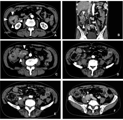
The patient underwent a computed tomography (CT) examination with contrast medium injection, and an extreme circumferential wall thickness in the ascending colon with a contrast effect, measuring 50 × 50 mm in size, was detected
in the right abdomen [Figure 1A]. According to the frontal
plane of the reconstructed view, the large mass was measured
to be 68 × 46 mm [Figure 1B]. Bulky swollen lymph nodes
involving ileocolic vessels were detected [Figure 1C].
Swollen lymph nodes were also observed in the
retroperito-neal space on the right side of the psoas muscle [Figure 1D],
attached to the right side of the inferior vena cava [Figure 1E], and on the right-to-posterior side of the right iliac vein,
com-pressing it [Figure 1F]. Distant metastases (ie, to the liver,
lungs or peritoneum) were not found. Colonoscopy did not reveal anything beyond a constriction of the tumor at the
ascending colon [Figure 2B]. The observable tumor border
was covered by a normal mucous membrane [Figure 2A], and
the diagnosis of carcinoma was not made by the biopsy
speci-mens. We conductedfluoroscopy using gastrografin
simulta-neously, revealing that the stenosis was irregular, measuring
62 mm in length and accompanied by a niche reflecting an
ulceration, which strongly suggested that the tumor consisted
of an epithelial neoplasm [Figure 3]. Considering these
ima-gingfindings, we made a preoperative diagnosis of stage IV
T4N2M1 (lymph nodes) ascending colon cancer.
During the surgery, which was performed in October 2014, liver or peritoneal metastasis was not revealed. The tumor of
the ascending colon was fixed to the neighboring swollen
lymph nodes. A right hemicolectomy accompanied by lymph node dissection along the superior mesenteric vein was con-ducted. After excision of the ascending colon tumor, the swol-len lymph nodes located at the neighboring psoas muscle were resected; however, the paracaval and parailiac lymph nodes were abandoned.
Pathological Findings
Gross Pathology
The circumferential ascending colon tumor was cut and
opened [Figure 4]. The resected specimen consisted of
a polypoid tumor accompanied by ulcer formation, measuring
OncoTargets and Therapy downloaded from https://www.dovepress.com/ by 118.70.13.36 on 25-Aug-2020
60 × 40 mm, located at the circumferential wall of the ascend-ing colon.
Microscopy
The tumor was comprised of a moderately to poorly (solid
type) differentiated adenocarcinoma component [Figure 5A,
×200]. In a portion of the tumor, a poorly differentiated carci-noma component accompanied by remarkable lymphocyte
permeation was found [Figure 5B, ×200]. In the component,
glandular formation was not observed, and the component was
arranged in cords or clusters. The individual tumor cells were large and pleomorphic with abundant eosinophilic cytoplasms, large vesicular nuclei, irregular nuclear profiles and prominent
nucleoli; however, thefindings of a syncytial growth pattern of
undifferentiated malignant epithelial cells and ill-defined
bor-ders were obscure [Figure 5C, ×400]. The lymphocytes
appeared small, round and mature. There were more infi
ltrat-ing lymphocytes than tumor cells. The glandular component
was accompanied by afibrous stroma; on the other hand, the
stroma was modest in the abundant lymphocyte permeation
A
B
C
E
F
D
*
*
Figure 1CTfindings with contrast medium injection. (A) The circumferential wall thickness in the ascending colon (arrowheads) and swollen lymph nodes is shown. (B) Frontal view of the multiplanar reconstruction shows a large mass measuring 68 × 46 mm (arrowheads). (C) Bulky lymph nodes involving ileocolic vessels were detected (arrowheads). (D–F) Swollen lymph nodes were detected on the right side of the psoas muscle (arrowhead) (D), attached to the right side of the inferior vena cava (arrowhead) (E), and on the right-to-posterior side of the compressed right iliac vein (arrowheads, *Right iliac vein) (F).
OncoTargets and Therapy downloaded from https://www.dovepress.com/ by 118.70.13.36 on 25-Aug-2020
component. Lymph node metastases were noted in the nodes neighboring the tumor and retroperitoneal lymph nodes; how-ever, metastasis was not observed in the nodes located in the root of the ileocolic artery or along the superior mesenteric vein. Intermediate degrees of lymphatic, venous, and
peri-neural infiltration were also noted. In the metastasized lymph
nodes, tumor cells exhibited both poorly and moderately dif-ferentiated histologies with gland formation, suggesting that the metastasis originated from the nonlymphocyte-rich
com-ponent [Figure 5D, ×400].
Microsatellite Instability (MSI) Assay
We performed a MSI assay (BML, Inc., Japan) after
obtain-ing the patient’s consent, and the result was negative.
Immunohistochemical staining (details of the antibodies
are shown inTable 1): Immunohistochemical staining using
an antibody for leukocyte common antigen (LCA; CD45, catalog number: M 0701; Dako, Japan) clearly showed a lymphocyte permeation department with an apparent demar-cation line [Figure 6A, loupe]. Additional staining with anα
-smooth muscle actin (αSMA, catalog number: M 851, Dako,
Japan) antibody clearly showed the muscularis propria. Interestingly, while the glandular component invaded the sub-serosa, the lymphocyte permeation-rich component invaded
A
B
Figure 2(A) Colonoscopy revealed the circumferential ascending colon tumor, and the observable tumor border was covered by a normal mucous membrane. (B) Colonoscopy did not reveal anything beyond a constriction. The diagnosis of carcinoma could not be made by biopsy.
Figure 3Secondary to colonoscopy, we conductedfluoroscopy using gastrografin, and irregular constriction was revealed. The niche was also observed (arrowhead), and we strongly doubted that the tumor consisted of an epithelial neoplasm.
OncoTargets and Therapy downloaded from https://www.dovepress.com/ by 118.70.13.36 on 25-Aug-2020
the superficial part of the muscularis propria [Figure 6B,
loupe]. The tumor-infiltrating lymphocytes were revealed to
have CD3-positive T lymphocyte predominance (CD3,
cata-log number: 413241, Nichirei, Japan) [Figure 6C, ×400]. In
addition, mild CLR consisting of CD20-positive lymphocytes was observed in the slightly remote colon wall from a tumor accompanied by lymphoid follicles (CD20, catalog number:
NCL-L-CD20-L26, LicaBiosystems, UK) [Figure 6D, ×40
and E, ×100]. The Ki67 labeling index showed a higher
value in the nonlymphocyte-rich component (42%) [Figure
6F, ×400] than in the lymphocyte-rich component (22%)
[Figure 6G, ×400]. We conducted immunohistochemical staining for PD-L1 (PD-L1, catalog number: ab205921, Abcam Japan), and overexpression was observed in the tumor cells of the nonlymphocyte-rich component, whereas very weak expression was observed in the lymphocyte-rich
component [Figure 6H, ×400 and I, ×400; the lower left of
Figure 6Hshows the positive control using tonsil tissue
sam-ples according to the manufacturer’s instructions, ×400]. We
also assayed single-stranded DNA (ssDNA, catalog number: 18731, IBL, Japan) to determine the levels of apoptosis, and overexpression was observed in the lymphocyte-rich compo-nent [Figure 6J, ×400; the arrows indicate the apoptotic cells;
the lower left ofFigure 6Jis the positive control using tissue
from the germinal center of the lymph follicle according to the
Figure 4Gross pathologicalfindings. Right hemicolectomy was performed. The resected specimen consisted of a polypoid tumor accompanied by an ulcer measur-ing 60 × 40 mm located at the circumferential wall of the ascendmeasur-ing colon.
A
B
C
D
Figure 5Microscopicfindings with hematoxylin-eosin staining. (A) The tumor was comprised of moderately to poorly differentiated adenocarcinoma (×200). (B) In a portion of the tumor, a poorly differentiated carcinoma component accompanied by remarkable lymphocyte permeation was found (×200). (C) In the lymphocyte-rich component, glandular formation was not observed, and the component was arranged in cords or clusters (×400). The individual tumor cells were large and pleomorphic with abundant eosinophilic cytoplasms, large vesicular nuclei, irregular nuclear profiles and prominent nucleoli; however, thefindings of a syncytial growth pattern and ill-defined borders were obscure. (D) In the metastasized lymph nodes, tumor cells presented gland formation, suggesting that the metastasis originated from the nonlymphocyte-rich component (×400).
OncoTargets and Therapy downloaded from https://www.dovepress.com/ by 118.70.13.36 on 25-Aug-2020
manufacturer’s instructions, ×400], whereas very weak expression was observed in the nonlymphocyte-rich
compo-nent [Figure 6K, ×400]. The opposite immunohistochemical
results were observed for Ki67.
In situ Hybridization
Epstein-Barr virus (EBV)-encoded small RNAs (EBERs) were negative in the tumor cells and positive in a small portion of interstitial lymphocytes (EBER RNA PNA Probe/Fluorescein, catalog number: Y5200, Dako, Japan) [Figure 6L, ×400].
Considering these results, the tumor was diagnosed as T3N2M1 cancer, not medullary carcinoma, with moderately to poorly differentiated carcinoma, PD-L1 overexpression and an LELC histology component inside the tumor.
The patient showed an uneventful recovery, and chemotherapy consisting of mFOLFOX6 was initiated 3 weeks after the operation. Bevacizumab was added to the mFOLFOX6 regimen beginning at the fourth course. After six courses of chemotherapy (three courses with the addition of bevacizumab), the patient underwent
fol-low-up CT, which revealed significantly reduced sizes of
the residual lymph nodes. Chemotherapy was continued, and after the patient had an allergic reaction to oxaliplatin in May 2015 (after 14 courses of mFOLFOX6), oxalipla-tin was removed from the regimen. In April 2016, after 31 courses of chemotherapy, chemotherapy was discon-tinued due to prolonged thrombopenia. During this per-iod, increased sizes of the residual lymph nodes were not observed with CT, and the residual lymph nodes
ulti-mately completely disappeared [Figure 7A and B]. The
patient was alive without tumor recurrence 5 years after the operation. The changes in the clinical parameters are
shown in Figure 8.
Discussion
Most carcinomas of the digestive tract are adenocarcinomas, while a small subset shows a solid growth pattern of malig-nant cells with the appearance of sheets or nests. Such tumors
are characterized by abundant tumor-infiltrating lymphocytes
and are divided into medullary carcinomas and LELCs. Medullary carcinomas are arranged in syncytial sheets and
have well-defined peripheral margins; the preponderance of
inflammation is peritumoral and associated with MSI.
LELCs do not tend to have continuous sheets of tumors that are broken up by large numbers of intratumoral
lympho-cytes and are associated with EBV.16
LELC was independently described in 1921 by Regaud
and Reverchon17 and Schminke18 as an undifferentiated
nasopharyngeal carcinoma (UNPC) characterized by large, polygonal tumor cells with vesicular nuclei and prominent nucleoli. The cytoplasmic borders are often indistinct and result in a syncytial pattern of growth. LELCs in the gastro-intestinal tract are mainly seen in the stomach, and gastric carcinomas with a lymphoid stroma constitute approximately
4% of all gastric carcinomas.7 Invariably, there are more
infiltrating lymphocytes than tumor cells.19However, to our
knowledge, regarding CRC, only eight cases of LELC have
been sporadically reported in the English literature.1–8
Colorectal LELCs as well as LELCs of other organs have been reported to be clinically less aggressive. To date, all colorectal LELCs have been reported to have homogenous histological features. However, it is histologically common that a mixture of different patterns composes a tumor. In gastric cancer cases, the histology has some overlap with gastric carcinoma with lymphoid stroma, and the absence of
massive lymphoid infiltrates is sometimes observed either in
whole or in part.20The present case showed a poorly
differ-entiated carcinoma component with remarkable lymphocyte permeation in a portion of the tumor, and the moderately to poorly differentiated adenocarcinoma component was seen on the background of glandular differentiation and sharp demarcation between these components. The lymphocytic component was mainly intratumoral, with a small peritu-moral presence. Although the ascending colon cancer was circumferentially large, regarding the lymphocytic
compo-nent, the tumor invasion remained in the superficial layer of
the muscularis propria, while the component with glandular differentiation without lymphocyte permeation reached the subserosal layer. The lymph node metastases exhibited glandular differentiation, suggesting that the metastasis ori-ginated from the nonlymphocytic component. Regarding the
Table 1Details of Used Antibodies
Antibody Clonotype Catalog
Number
Company Country
LCA (CD45)
Monoclonal M 0701 Dako Japan
αSMA Monoclonal M 851 Dako Japan
CD3 Monoclonal 413241 Nichirei Japan
CD20 Monoclonal
NCL-L-CD20-L26
LicaBiosystems UK
Ki67 Monoclonal 418071 Nichirei Japan
PD-L1 Monoclonal Ab205921 Abcam Japan
ssDNA Polyclonal 18731 IBL Japan
EBER Monoclonal Y5200 Dako Japan
OncoTargets and Therapy downloaded from https://www.dovepress.com/ by 118.70.13.36 on 25-Aug-2020
components with a lymphoid stroma, relatively shallow tumor permeation was maintained and showed relatively weakly aggressive features, as reported for colorectal LELCs. However, lymphocytic permeation was restricted to the poorly differentiated component demarcated from the
glandular component. The infiltrated abundant lymphocytes
did not show an inhibitory influence on the portion of the
cancer without lymphocyte infiltration.
PD-L1 overexpression was observed in the nonlymphocyte permeate component, whereas PD-L1 expression was very weak in the lymphocyte-rich component. A recent systemic
review and meta-analysis of colorectal cancer11revealed that
A
B
C
D
E
F
I
H
G
J
K
L
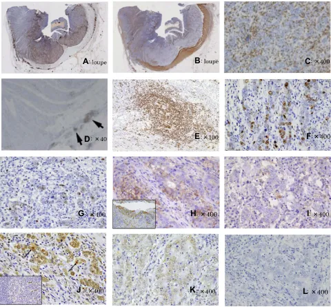
Figure 6Findings of immunohistochemical staining and in situ hybridization. (A) Immunohistochemical staining using an antibody for leukocyte common antigen (LCA; CD45) clearly showed a lymphocyte permeation department with an apparent demarcation line (loupe). (B) Additional staining with theα-smooth muscle actin (αSMA) antibody clearly showed the muscularis propria (loupe). While the glandular component invaded the subserosa, the lymphocyte permeation-rich component remained in the superficial part of the muscularis propria. (C) The tumor-infiltrating lymphocytes had CD3-positive T lymphocyte predominance (×400). (D, E) Mild CLR consisting of CD20-positive lymphocytes was observed in the slightly remote colon wall from a tumor ((D) arrows, ×40) accompanied by lymphoid follicles ((E), ×100). (F, G) The Ki67 labeling index value was higher in the nonlymphocyte-rich component (42%) ((F), ×400) than in the lymphocyte-rich component (22%) ((G), ×400). (H, I) PD-L1 overexpression was observed in the tumor cells in the nonlymphocyte-rich component (H, ×400), whereas very weak expression was observed in the lymphocyte-rich component (I, ×400). The lower left of (H) shows the positive control using tonsil tissue according to the manufacturer’s recommendation (×100). (J, K) Single-stranded DNA (ssDNA) overexpression was observed in the lymphocyte-rich component (J, ×400), suggesting apoptotic cells (arrows). The lower left of (J) shows the positive control using the tissue of the germinal center of the lymph follicle (×100). Weak ssDNA expression was observed in the nonlymphocyte-rich component (K, ×400). These results are opposite those of the Ki67 immunohistochemicalfindings. (L) In situ hybridization. EBV-encoded small RNAs (EBERs) were negative (×400).
OncoTargets and Therapy downloaded from https://www.dovepress.com/ by 118.70.13.36 on 25-Aug-2020
PD-L1 overexpression was related to short OS, short RFS/ DFS, a poor tumor stage and the absence of vascular invasion. PD-L1 overexpression in the nonlymphocyte component might play an important role in aggressive locoregional spread-ing without hematogenous metastasis by avoidspread-ing tumor immunity. We also conducted immunohistochemical staining
for ssDNA21in addition to Ki67 to clarify whether PD-L1 was
involved in apoptosis or cell proliferation during tumor growth. PD-L1 overexpression in the nonlymphocyte-rich component showed more Ki67 labeling and very weak ssDNA expression. These results suggest that PD-L1 overexpression is more extensively involved in cell proliferation than apoptosis.
Immunohistochemistry revealed that infiltrative lymphocytes
had a CD3-positive T lymphocyte predominance. The
lymphoid reaction in gastric LELCs is reported to be com-prised predominantly of T-cells, including numerous T-cell intracytoplasmic antigen (TIA-1)-positive activated cytotoxic lymphocytes and/or natural killer cells in close contact with
tumor cells.22 Of nine cases of reported colorectal LELCs,
including ours, a lymphocyte surface antigen assay was carried
out infive cases; three cases showed T lymphocyte
predomi-nance, one case showed the equivalent expression of B and
T lymphocytes; and the other case showed B-cell
predominance.
The individual tumor cells of the poorly differentiated carcinoma with abundant lymphocyte permeation were large and pleomorphic with abundant eosinophilic cytoplasms,
large vesicular nuclei, irregular nuclear profiles and
A
B
Figure 7CTfindings after chemotherapy. The lymph node attached to the right side of the inferior vena cava disappeared (A), the node at the right-to-posterior side of the right iliac vein disappeared, and the right iliac vein showed a normal thickness (B).
Figure 8Changes in various clinical parameters, including the timing of the operation, the therapeutic chemotherapy agent doses, the lymph node sizes and the platelet count values.
OncoTargets and Therapy downloaded from https://www.dovepress.com/ by 118.70.13.36 on 25-Aug-2020
prominent nucleoli; however, the findings of a syncytial growth pattern of undifferentiated malignant epithelial cells
and ill-defined borders were obscure and suggested that the
tumor was strictly but slightly different from typical UNPC
findings. However, some cases of LELC being reported for
CRC have shownfindings similar to those observed in our
case,2,8and diagnostic criteria regarding CRC may broaden.
Chetty16 reported that gastric medullary carcinoma
demonstrates a syncytial growth pattern, bears robust
peri-tumoral inflammation, and has a well-defined border,
while lymphoepithelioma-like carcinoma is formed by
small clusters of cells that “do not correspond to the
syncytial growth patterns seen in medullary carcinoma”
and has more prominent intratumoral inflammation and an
infiltrative border. Although we diagnosed the LELC of
CRC based on pathological findings similar to those of
nasopharyngeal LELCs, the diagnostic criteria for LELCs of CRC are not yet clear, and the disease entity has not been established.
In the present case, mild CLR was also observed. The
term CLR was coined by Graham and Appelman in 199023
and described as discrete lymphoid aggregates, some with germinal centers, mostly located in the muscularis propria and pericolonic adipose tissue beyond the advancing tumor
edge.24Several cancers of Lynch syndrome were noted to be
associated with dense Crohn’s-like lymphoid aggregates;
however, our case did not show MSI. In the present case, CLR consisted of CD20-positive B lymphocytes. In the early stages of CLR development, CD4+ T-cells cluster predomi-nantly with mature antigen-presenting dendritic cells. As CLR matures, increasing numbers of B-cells, as well as follicular dendritic cells, are recruited to create lymphoid
follicles.24Our CLR consisted of B-cells and had a mature
lymphoid structure with lymphoid follicle development. CLR has been associated with a low incidence of locor-egional recurrence, few distant metastases and prolonged
cancer-specific survival and OS.12–15Although locoregional
lymph node metastases were aggressive in our case, no hema-togenous metastases or lymph node metastases developed along the root of the main trunk of the feeding vessels, con-sistent with the reported CLR cases. Regarding immunologi-cal features, CLR has been consistently associated with
increased tumor intraepithelial and stromal lymphocytic infi
l-trates, which appears to be independent of the MSI status.14,24 CLR density is also correlated with peripheral immune mar-kers, such as the absolute peripheral lymphocyte count and the lymphocyte to neutrophil ratio, which also carry prognostic significance.15,24,25In addition, CLR-promoting chemokines
are inversely associated with the intratumoral expression of immune checkpoint molecules, including PD-1, PD-L1 and
cytotoxic T-lymphocyte-associated protein 4 (CTLA-4).24,26
CLR is reported to be predictive of the chemotherapy response in the context of metastatic CRC, suggesting a potential syner-gistic effect of cytotoxic chemotherapy with the adaptive
immune response to metastatic disease.24,27,28 Prominent
CLR can be found in over 30% of CRC cases.16,29Although
our case presented a relatively mild appearance of CLR, it
might have influenced complete remission with chemotherapy
after noncurative excision. It did not show an elevation in the absolute peripheral lymphocyte count (1300/µL) or the lym-phocyte to neutrophil ratio (29.2%); however, these immuno-logic indexes are thought to be independent of CLR. CRC with a lymphoid stroma can be divided into cases associated with EBV, MSI, or neither, as well as gastric cancer with a lymphoid stroma. Among the eight reported cases of
color-ectal LELC, the MSI status was investigated in only one case,7
and the result was high MSI. Our case presented no associa-tion with EBV infecassocia-tion or MSI. Generally, poorly differen-tiated carcinomas that are not related to EBV or MSI have a worse prognosis than related cases. This case interestingly presented both the advantages of the LELC phenotype and CLR and the disadvantages of PD-L1 overexpression and no relation between LELC and EBV or MSI for prognosis. It remains unclear what decisively contributed to the preferable outcome. It seems that these characteristics
contri-bute to a conflicting aggressive locoregional extent and
a complicated complete response to chemotherapy. Even if the LELC of CRC is not related to EBVor MSI, it is thought to be capable of a good response to chemotherapy and to have a good prognosis in cases of CLR. The accumulation and analyses of further cases are necessary.
Conclusion
We reported a very rare and interesting case of
moder-ately differentiated, PD-L1-overexpressing ascending
colon cancer with an LELC component accompanied by CLR presenting with extremely aggressive locoregional spreading and complete remission with chemotherapy after noncurative excision. Even if the LELC of CRC is not related to EBV or MSI and PD-L1 overexpression is found, a good response to chemotherapy and a good prognosis is achievable in cases of CLR. Further accu-mulation and analyses of cases of colorectal LELCs are necessary.
OncoTargets and Therapy downloaded from https://www.dovepress.com/ by 118.70.13.36 on 25-Aug-2020
Abbreviations
PD-L1, programmed death ligand-1; LELC, lymphoepithe-lioma-like carcinoma; CRC, colorectal cancer; PD-1, pro-grammed death 1; OS, overall survival; RFS, recurrence-free
survival; DFS, disease-free survival; CLR, Crohn’s-like
lym-phoid reaction; WBC, white blood cell; CEA, carcinoembryo-nic antigen; CA19-9, carbohydrate antigen 19-9; sIL-2R, soluble interleukin-2 receptor; CT, computed tomography; LCA, leukocyte common antigen; CD45, cluster of
differen-tiation 45; αSMA, α-smooth muscle actin; CD3, cluster of
differentiation 3; CD20, cluster of differentiation 20; ssDNA, single-stranded deoxyribonucleic acid; EBV, Epstein-Barr virus; EBER, EBV-encoded small RNAs; MSI, microsatellite instability; UNPC, undifferentiated nasopharyngeal carci-noma; TIA-1, T-cell intracytoplasmic antigen; CTLA-4, cyto-toxic T-lymphocyte-associated protein 4.
Ethics Approval and Consent to
Participate
All procedures used in this research were approved by the Ethical Committee of the National Hospital Organization Tsuruga Medical Center.
Consent for Publication
Written informed consent was obtained from the patient for the publication of this case report and any accompany-ing images. A copy of the written consent is available for review by the Editor-in-Chief of this journal.
Data Sharing Statement
All data generated or analyzed during this study are included in this published article.
Acknowledgment
The authors would like to sincerely thank Sarah Conte, Thomas Barbour and Kimberly Yasutis from American Journal Experts (AJE) for their English-language proof-reading of the manuscript.
Author Contributions
All authors made substantial contributions to the concep-tion and design of the study; the acquisiconcep-tion, analysis or interpretation of the data; and drafting or critical revision of the article for important intellectual content. All authors
approved of thefinal version to be published and agreed to
be accountable for all aspects of the work.
Disclosure
The authors have no competing interests to declare for this work.
References
1. Giovanni DP, Robert L, Daniel MQ, Paul RF, Janet UB, Kojo EJ. Lymphoepithelioma-like carcinoma of the colon in a patient with hereditary nonpolyposis colorectal cancer. Arch Pathol Lab Med. 1999;123:720–724.
2. Vilor M, Tsutsumi Y. Localization of Epstein-Barr virus genome in lym-phoid cells in poorly differentiated adenocarcinoma with lymlym-phoid stroma.
Pathol Int.1995;45:695–697. doi:10.1111/j.1440-1827.1995.tb03524.x 3. Palazzo JP, Mittal KR. Lymphoepithelioma-like carcinoma of the rectum
in a patient with ulcerative colitis.Am J Gastroenterol.1996;91:398–399. 4. Samaha S, Tawfik O, Horvat R, Bhatia P. Lymphoepithelioma-like carcinoma of the colon: report of a case with histologic, immunohis-tochemical, and molecular studies for Epstein-Barr virus.Dis Colon Rectum.1998;41:925–928. doi:10.1007/BF02235379
5. Kon S, Kasai K, Tsuzuki N, et al. Lymphoepithelioma-like carcinoma
of rectum: possible relation with EBV. Pathol Res Pract.
2001;197:577–582. doi:10.1078/0344-0338-00130
6. Kojima Y, Mogaki M, Takagawa R, et al. A case of
lymphoepithelioma-like carcinoma of the colon with ulcerative colitis.
J Gastroenterol.2007;42:181–185. doi:10.1007/s00535-006-1981-0 7. Delaney D, Chetty R. Lymphoepithelioma-like carcinoma of the
colon.Int J Clin Exp Pathol.2012;5:105–109.
8. Mori Y, Akagi K, Yano M, et al. Lymphoepithelioma-like carcinoma of the colon.Case Rep Gastroenterol.2013;7:127–133. doi:10.1159/000348765 9. Watanabe H, Enjoji M, Imai T. Gastric carcinoma with lymphoid
stroma. Its morphologic characteristics and prognostic correlations.
Cancer.1976;38:232–243. doi:10.1002/(ISSN)1097-0142
10. Nacopoulou L, Aaris P, Papacharalampous N, et al. Prognostic sig-nificance of histologic host response in cancer of the large intestine.
Cancer. 1981;47:930–936. doi:10.1002/1097-0142(19810301) 47:5<930::AID-CNCR2820470519>3.0.CO;2-1
11. Yang L, Xue R, Pan C. Prognostic and clinicopathological value of PD-L1 in colorectal cancer: a systematic review and meta-analysis.
Onco Targets Ther.2019;12:3671–3682. doi:10.2147/OTT.S190168 12. Buckowitz A, Knaebel HP, Benner A, et al. Microsatellite instability in
colorectal cancer is associated with local lymphocyte infiltration and low frequency of distant metastases.Br J Cancer.2005;92:1746–1753. doi:10.1038/sj.bjc.6602534
13. Ueno H, Hashiguchi Y, Shimazaki H, et al. Objective criteria for Crohn-like lymphoid reaction in colorectal cancer.Am J Clin Pathol. 2013;139:434–441. doi:10.1309/AJCPWHUEFTGBWKE4 14. Rozek LS, Schmit SL, Greenson JK, et al. Tumor-infiltrating
lym-phocytes, Crohn’s-like lymphoid reaction, and survival from color-ectal cancer.J Natl Cancer Inst.2016;12:108.
15. Posch F, Silina K, Leibl S, et al. Maturation of tertiary lymphoid structures and recurrence of stage II and III colorectal cancer.Oncoimmunology. 2018;7:e1378844. doi:10.1080/2162402X.2017.1378844
16. Chetty R. Gastrointestinal cancers accompanied by a dense lymphoid component: an overview with special reference to gastric and colonic medullary and lymphoepithelioma-like carcinomas. J Clin Pathol. 2012;65:1062–1065. doi:10.1136/jclinpath-2012-201067
17. Regaud C, Reverchon L. Sur un cas d’epitheliome epidermoide
developpe dams les massif maxillaire superieur.Rev Laryngol Otol Rhinol.1921;42:369–378.
18. Schminke A. Uber lympho-epitheliale Geschwulste. Beitr Pathol
Anat Allg Pathol.1921;68:161–170.
19. Cho HJ, Kim JY, Yoo J, Lee SS. Gastric carcinoma with lymphoid stroma: incidence of EBV and Helicobacter pyloriinfection.Appl Immunohistochem Mol Morphol. 2003;11:149–152. doi:10.1097/ 00129039-200306000-00010
OncoTargets and Therapy downloaded from https://www.dovepress.com/ by 118.70.13.36 on 25-Aug-2020
20. Matsuda I, Kan K, Doi S, Motoki Y, Onodera M, Hirota S. A case of gastric cancer with heterogenous components of EB virus (+)/TP53
(+) and EB virus (-)/TP53 (-). Int J Clin Exp Pathol.
2015;8:11766–11771.
21. Watanabe I, Toyoda M, Okuda J, et al. Detection of apoptotic cells in human colorectal cancer by two differentin situmethods: antibody
against single-stranded DNA and terminal deoxynucleotidyl
transferase-mediated dUTP-biotin nick end-labeling (TUNEL)
methods. Jpn J Cancer Res. 1999;90:188–193. doi:10.1111/cas. 1999.90.issue-2
22. Chapel F, Fabiani B, Davi F, et al. Epstein-Barr virus and gastric carcinoma in Western patients: comparison of pathological para-meters and p53 expression in EBV-positive and negative tumours.
Histopathology.2000;36:252–261. doi:10.1046/j.1365-2559.2000.00 843.x
23. Graham DM, Appelman HD. Crohn’s-like lymphoid reaction and
colorectal carcinoma: a potential histologic prognosticator. Mod Pathol.1990;3:332–335.
24. Maoz A, Dennis M, Greenson JK. The Crohn’s-like lymphoid reac-tion to colorectal cancer-tertiary lymphoid structures with immuno-logic and potentially therapeutic relevance in colorectal cancer.Front Immunol.2019;10:1884. doi:10.3389/fimmu.2019.01884
25. Sjoquist KM, Renfro LA, Simes RJ, et al. Personalizing survival predictions in advanced colorectal cancer: the ARCAD nomogram project. J Natl Cancer Inst. 2018;110:638–648. doi:10.1093/jnci/ djx253
26. Weinstein AM, Giraldo NA, Petitprez F, et al. Association of IL-36 gamma with tertiary lymphoid structures and inflammatory immune infiltrates in human colorectal cancer.Cancer Immunol Immunother. 2019;68:109–120. doi:10.1007/s00262-018-2259-0
27. Halama N, Michel S, Kloor M, et al. The localization and density of immune cells in primary tumors of human metastatic colorectal cancer shows an association with response to chemotherapy.Cancer Immun.2009;9:1.
28. Halama N, Michel S, Kloor M, et al. Localization and density of immune cells in the invasive margin of human colorectal cancer liver metastases are prognostic for response to chemotherapy.Cancer Res. 2011;71:5670–5677. doi:10.1158/0008-5472.CAN-11-0268 29. Alexander J, Watanabe T, Wu TT, Rashid A, Li S, Hamilton SR.
Histopathological identification of colon cancer with microsatellite instability. Am J Pathol. 2001;158:527–535. doi:10.1016/S0002-9440(10)63994-6
OncoTargets and Therapy
Dove
press
Publish your work in this journal
OncoTargets and Therapy is an international, peer-reviewed, open access journal focusing on the pathological basis of all cancers, potential targets for therapy and treatment protocols employed to improve the management of cancer patients. The journal also focuses on the impact of management programs and new therapeutic
agents and protocols on patient perspectives such as quality of life, adherence and satisfaction. The manuscript management system is completely online and includes a very quick and fair peer-review system, which is all easy to use. Visit http://www.dovepress.com/ testimonials.php to read real quotes from published authors.
Submit your manuscript here:https://www.dovepress.com/oncotargets-and-therapy-journal
OncoTargets and Therapy downloaded from https://www.dovepress.com/ by 118.70.13.36 on 25-Aug-2020