Original Research Article
Displaced intrauterine contraceptive device: a prospective study at
tertiary level hospital of Uttarakhand, India
Nidhi Kumari, Vineeta Gupta, Priyanka Chaudhari*, Shweta
Nimonkar, Archna Tandon
INTRODUCTION
Intrauterine contraceptive devices (IUCD) are safe effective reversible and reliable method of long term contraception. IUCD is the main stay of Contraception especially in developing countries, despite side effect and complication. Uterine perforation is rare but potentially serious complication with an incidence of less than one case per 1000 insertion. The large ongoing international prospective EURAS-IUD study (European Active Surveillance Study for Intrauterine Devices) revealed perforation rates of 0.68/1000 insertions for the LNG-IUS and 0.41/1000 insertions for Cu-IUCDs at 1 year of follow up (Heinmann 2015).1 The risk factor for uterine
perforation by IUCD is type of IUCD, position and size of uterus, congenital anomalies, infection, history of abortion and insertion in postpartum period.
A frequent clinical problem is loss of the filament at the external cervical os as the lost tail. This may be due to retracted or torn off tail, misplacement within the cavity, and growing uterus due to pregnancy causing retraction of threads, intramural penetration as extra uterine location. Proposed risk factors of uterine perforation include the immediate post-partum period and breast feeding, regardless of the timing of insertion. Both Andersson and van Haudenhoven have discussed the role of uterine involution and increased uterine contractility as potential contributing factors to IUCD perforation occurring in the postpartum period.2,3 Procedure for
retrieval of a misplaced device includes Cytobrush, IUCD hook artery forceps, and cylindrical brush. Once a patient comes with missing threads of IUCD, the device is located by X-ray or USG.
ABSTRACT
Background: The IUCD is a common method used for contraception. It is associated with complications like bleeding, perforation and migration to neighbouring organs as broad ligament, urinary bladder or omentum.
Methods: A prospective study was carried out at SGRRIM and HS, Dehradun over a period of two years between January 2014 to December 2015. A total of 38 patients with a diagnosis of displaced IUCD were included for their detailed demographic profile, presenting complaints, required diagnostic and therapeutic modalities.
Results: Ultrasound emerged as the preferred method to locate the displaced device. Majority of displaced IUCD were intrauterine (86.9%) and Hysteroscopy guided removal was the preferred method of removal regarding technique, safety, and cost and recovery time.
Conclusions: Responsibility of care provider does not end at insertion of IUCD. Follow up is equally important. Every case of missing IUCD must be investigated carefully to rule out the possibility of uterine perforation.
Keywords: Hysteroscopy, Intrauterine contraceptive device (IUCD), Perforation
Department of Obstetrics and Gynecology, SGRRIM and HS, Patel Nagar Dehradun, Uttarakhand, India
Received: 01 February 2018
Accepted: 07 March 2018
*Correspondence:
Dr.Priyanka Chaudhari,
E-mail: dishitapriya07@gmail.com
Copyright: © the author(s), publisher and licensee Medip Academy. This is an open-access article distributed under the terms of the Creative Commons Attribution Non-Commercial License, which permits unrestricted non-commercial use, distribution, and reproduction in any medium, provided the original work is properly cited.
Currently endoscopy has emerged as a preferred method for its removal. Use of hysteroscopy to remove a misplaced device helps in avoiding blind manipulation, which may cause haemorrhage, perforation and sometime visceral injury. On the other hand, complete removal is ensured in old fragmented and embedded devices without any associated complication.4
Extra uterine devices can be removed laparoscopically, in rare cases Laparotomy may be required. With this background the present study was conducted to raise awareness of the circumstances in which perforation of uterus by IUCD can occur, the consequences of such perforations and approaches to the management of these complications.
METHODS
The present prospective study was carried out in the department of Obstetrics and Gynecology at SGRRIM and HS, Dehradun after approval from research and ethical committee of the Institute.
This study was carried out to find out the incidence of displacement of IUCD in our region of Uttarakhand, it’s varied clinical presentations, investigations required to locate the IUCD and preferred method of intervention (D and C/ Hysteroscopy/ Laparoscopy/ Laparotomy) to remove it.
The study includes 38 patients, who reported to Obstetrics and Gynecology department with a diagnosis of misplaced IUCD over a period of two year from January 2014 to December 2015. All these patients had come as referred cases in which the routine procedure of IUCD retrieval had failed. Detailed demographic data, clinical presentation, physical examination, investigation findings and detailed procedure findings were noted. All women who did not have misplaced IUCD but voluntarily wanted IUCD removal or who had spontaneous expulsion of IUCD were excluded from the study. In this study we included only cases treated at a later date by surgical means. Thus, IUCD perforations occurring at the time of insertion and treated immediately by removal of device and asymptomatic perforations by IUCDs were not included in the present study.
In all the patients with displaced IUCD gynecological examination, USG evaluation and X-ray AP view (abdomen and pelvis) was done to locate the misplaced IUCD. In rare cases CT-scan and MRI imaging was required to find the device or locate the fistula tract made by displaced device.
If the device was found to be intrauterine, a 10mm operative hysteroscope with grasping forceps was used under anesthesia to locate and retrieve device. In those patients in whom the IUCD was confirmed to be extra uterine, diagnostic Laparoscopy was done to find and remove the IUCD. When Laparoscopic removal was not
possible or failed Laparotomy was done in the same sitting to remove the IUCD.
RESULTS
In India, where the population is rising very rapidly, family planning is the need of hour. It is therefore essential, that every effort should be made to bring down the failure and complication rates of the contraceptive measures, so that more couples can be drawn towards these services. Finally, as intrauterine contraception is one of the most widely used contraceptive method worldwide, a specific code for uterine perforation associated with an IUCD can be added in globally used diagnostic classifications in future.
[image:2.595.312.544.392.491.2]The present study was conducted on 38 patients who came with the diagnosis of misplaced IUCD. Total number of insertions during the period was not available because the patients came from different areas and the insertions were performed by trained doctors at private clinics as well as in a family planning centre. In our study group none of the patient had IUCD insertion by untrained or semiskilled personnel.
Table 1: Age and parity distribution with presenting complaints of patients.
Present study showed that the maximum number of patients (47.4%) were of 25-30 years of age group (Table 1). According to the time of insertion 81.6% patients had post placental IUCD (PPIUCD), 13.2% had interval IUCD insertion. Table 4 shows that 36.8% of cases had removal of IUCD between 6 to12 months of insertion.
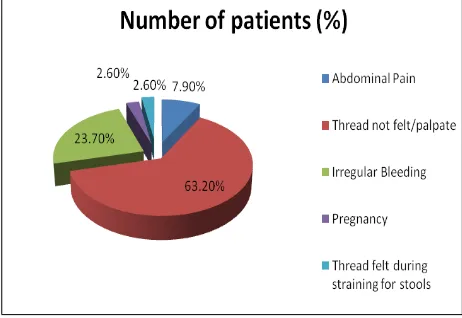
Figure 1: Presenting complaints.
Age(years)
Number of patients (%)
Parity
Number of patients (%)
20-25 8 (21.1) 1-2 10 (26.3)
25-30 18 (47.4) 3-4 22 (57.9)
30-35 7 (18.4)
>4 6 (15.8)
35-45 3 (7.9)
[image:2.595.312.543.585.744.2]Figure 1 show all the presenting complaints for which patient had come for consultation. Twenty-four (63.2%) of patient came with complaint of non-perception of threads during self-internal/vaginal examination. Nine (23.7%) of patient came with complaint of irregular bleeding per vagina, three (7.9%) patient present with pain in abdomen, one (2.6%) patient presented with complaint of perception of threads during straining for defecation. Among 38 patients, only One (2.6%) patient presented with advanced pregnancy of 6 month. After all required investigation diagnosis of misplaced IUCD and its location identified.
Table 2: Mode and time of insertion.
Time of insertion Number of patients (%)
After vaginal delivery 9 (23.7)
After 1st LSCS 15 (39.5)
After 2nd LSCS 7 (18.4)
Interval IUCD insertion 5 (13.2)
Post Abortion 2 (5.3)
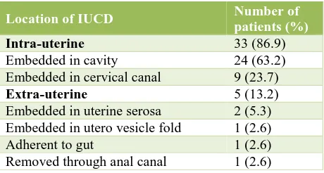
[image:3.595.53.285.243.318.2]The location of misplaced IUCD (Table 4). Thirty-three (86.9%) of patient had intrauterine and five (13.2%) of patient had extra uterine displacement of IUCD. Table- 5 showed the method used for removal of misplaced IUCD. Hysteroscopy was used to confirm the location and also for the removal in the same siting in twenty-four (63.2%) of patients.
Table 3: Time interval between time of insertion and removal of IUCD.
Time interval between insertion and removal
Number of patients (%)
<6 months 2 (5.3)
6-12 months 6 (15.8)
12-18 months 10 (26.3)
18-24 months 14 (36.8)
>24 months 6 (15.8)
Nine (23.7%) patients required D and C for intrauterine displaced IUCD. When hysteroscpy fails to identify IUCD in uterine cavity or USG and X-ray suggested that the misplaced IUCD is extra uterine then laproscopy is required. In current study five (13.2%) case had diagnosis of extra uterine displacement of IUCD.
In three (7.9%) patients with extrauterine IUCD, laparoscopic removal was done. In two (5.3%) cases, extra uterine IUCD was found to be embedded in the posterior wall of uterus at isthmus region which was removed by laparoscopy. One was embedded in sigmoid colon, adhesiolysis was done and IUCD was removed intact.
After that methylene blue dye test was done to check the integrity of sigmoid colon. In one case laparotomy was done because patient had history of previous three laparotomy operation. IUCD was embedded in the
uterovescical fold, abscess was drained and IUCD was removed. In the fourth case, we encountered a negative laparoscopy for misplaced IUCD.
Table 4: Location of IUCD.
Location of IUCD Number of
patients (%)
Intra-uterine 33 (86.9)
Embedded in cavity 24 (63.2)
Embedded in cervical canal 9 (23.7)
Extra-uterine 5 (13.2)
Embedded in uterine serosa 2 (5.3) Embedded in utero vesicle fold 1 (2.6)
Adherent to gut 1 (2.6)
Removed through anal canal 1 (2.6)
Only few flimsy adhesions were seen in pelvis and adenexa with gut but no IUCD seen in spite of its confirmed presence on X-ray and USG. Thereafter per rectal examination was done and very surprisingly IUCD was removed through anus without any sign of perforation of gastrointestinal tract, which was confirmed by laparoscopic examination.
DISCUSSION
IUCD for contraception were first introduced by Richter in 1909 and further developed by Grafenberg from 1929. For the last three decades, IUCD is the most accepted method of reversible contraception because of its high efficacy, low cost and low complication rates. The pelvic inflammatory disease, dysmenorrhea, irregular spotting, ectopic pregnancy, migration into bowel and adjacent organs, vesicouterine fistula and very rarely endometrial adenocarcinoma are the reported complication of IUCD.5
These complications are the common indications of IUCD removal.
Perforation of the uterus with IUCDs was first described in the 1930s.6,7 The mechanism of migration is not very
clear but thought to be the insertion procedure itself or the chronic inflammatory reaction with gradual erosion through the uterine wall. Any foreign body placed in the proximity of urinary bladder has the possibility to migrate into the bladder like any IUCD, vaginal diaphragm, surgical clips used in hernia repair, prosthetic slings and even bullet.8 IUCD may migrate to uterine serosa, urinary
bladder, gut, omentum and broad ligament.
The incidence ranges from 0.5-1/1000 insertions though exact incidence is not clear due to asymptomatic nature of these perforations.9 The incidence is influenced by
[image:3.595.51.284.452.539.2]may be due to distorted uterine cavity, adenomyosis, obesity or inexperienced clinician. Ultrasound guided insertions may reduce the incidence of malposition.
Ultrasound of lower abdomen and pelvis is the investigation of choice for displaced IUCD, but Partial perforation may not be detected by abdominal USG. Transvaginal-USG is recommended to know the extent of myometrial and bladder wall perforation. X-ray of abdomen erect view with or without uterine sound in uterine cavity may be required to locate the IUCD in the peritoneal cavity in relation to the uterus.
In our study twenty-four (63.2%) patients reported with complaint of non-palpable threads, while study done by N Elahi et al reported presentation with lost string in 32.14% of patients.10 Next frequent presenting complaint
was irregular bleeding in nine (23.7%) patients and above quoted study also reported 24.32% occurrence of menstrual irregularities in these patients. There was only one case (2.6%) of intrauterine pregnancy with IUCD in situ in our study as compared to 7.14% in the above study. Majority of patients i.e. Thirty-one (%) patients who had post placental IUCD insertion presented with misplaced IUCD and study done by Pandey S Jyoti et al also showed similar results (92%).11 Twenty-four
(63.2%) of patients presented with misplaced IUCD within 12 months to 24 months. Hysteroscopy has emerged out as preferred method of misplaced intrauterine IUCD removal. Twenty-four (63.2%) patients had hysteroscopic IUCD removal which is comparable to study done by Jyoti PS et al (60%).
According to the study done by Mishra S, removal rates are similar in clients having or not having complications (89.40% and 88.52% respectively) that emphasizes the importance of knowledge and motivation prior to insertion in continuing IUCD.12
Different reviews are present in literature regarding an attempt to remove or not any asymptomatic misplaced IUCD. Adoni and Benchetrit found no adhesions in 3 and 11 cases respectively.13
They suggested that surgery should be done in symptomatic cases only. According to majority of other authors, withdrawal of the migrated IUCD is advisable to reduce the chances of further complications as formation of adhesion and injury to bowel and urinary bladder or fistula formation.14
WHO also recommends removal of misplaced IUCD because of potential damage to adjacent organs and associated medico-legal problems.15 A regular follow up
of IUCD for visible thread would help in earlier detection of misplaced IUCD.
Limitation of this study should include large number of patients and controls for the study should be identified.
CONCLUSION
Responsibility of care provider does not end at insertion of IUCD. Follow up is equally important. Every case of missing IUCD must be investigated carefully to rule out the possibility of uterine perforation. Hysteroscopy is the preferred method in management of misplaced IUCD as it is performed under vision, causes minimum hospital stays, minimal invasive method and associated with early recovery.
Funding: No funding sources Conflict of interest: None declared
Ethical approval: The study was approved by the Institutional Ethics Committee
REFERENCES
1. Heinemann K, Reed S, Moehner S, Do Minh T. Risk of uterine perforation with levonorgestrel-releasing and copper intrauterine devices in the European active surveillance study on intrauterine devices. Contracept. 2015;91(4):274-9.
2. Andersson K, Ryde-Blomqvist E, Lindell K, Odlind V, Milsom I. Perforations with intrauterine devices: report from a Swedish survey. Contracept. 1998;57(4):251-5.
3. Houdenhoven VK, Kaam VKJ, Grootheest VAC, Salemans TH, Dunselman GA. Uterine perforation in women using a levonorgestrel-releasing intrauterine system. Contracept. 2006;73(3):257-60.
4. Mittal S, Kumar S, Roy KK. Role of endoscopy in retrieval of misplaced intrauterine device. Aus New Zealand J Obstet Gynaecol. 1996;36(1):49-51. 5. Sataa S, Sami BR, sabeur R, Karim C, Ali H.
Bladder Calculus resulting from the migration of an intrauterine contraceptive device: a report of ten cases. Nephro-Urology Monthly. 2011;3(1):54-61. 6. Murphy M. Migration of a Graefenberg ring. Lancet.
1933;222(5755):1369-70.
7. Andrews CJ. Migrating Gräfenberg contraception ring. JAMA. 1936;107:279.
8. Ratnam SS, Tow SH. Translocation of the loop. In: Zatuchni GI, editor. Post-partum Family Planning: A Report on the International Program. New York, NY: McGraw-Hill; 1970:371-384.
9. Alka K, Pradeep G, Sharma M, Agarwal N. Laproscopic removal of extrauterine IUCD using fluoroscopy guidance: a case report. J Gynaecol Surg. 2005;21(1):29-30.
10. Elahi N, Koukab H. Diagnosis and management of lost intrauterine contraceptive device. J Pak Med Assoc. 2002;52(1):18-20.
11. Jyoti PS, Geeta J, Vivek S. Misplaced IUCD: etiology and management: a retrospective study, Sch. J Med Sci. 2017;5(4F):1694-7.
(PPIUCD). J Obstet Gynecol India. 2014;64(5):337-43.
13. Adoni A, Chetrit AB. The management of
intrauterine devices following uterine perforation. Contracept. 1991;43(1):77-81.
14. Braaten KP, Goldberg AP. Malposition IUCD: when you should intervene (and when you should not). OBG Manag. 2012;24:38-46.
15. World Health Organization. Reproductive Health. Medical eligibility criteria for contraceptive use.
World Health Organization; 2010. Available at http://whqlibdoc.who.int/publications/2010/9789241 563888_eng.pdf.