Original Research Article
Prevalence of uterine lesions associated with leiomyomas and role of
endometrial biopsy in management
Minal J. Kumbhalwar*, Anuradha Konda
INTRODUCTION
Leiomyoma of uterus also called as fibromyoma or fibroid, fibromas, myofibromas, and myomas.
It is a well-circumscribed, benign tumour of uterus,
essentially composed of smooth muscle tissue
(myometrium) of the uterus and a variable amount of fibrous connective tissue i.e. extracellular matrix (collagen, proteoglycan, fibronectin).
It is the most common benign tumour of uterus, and is found in 20% of women in reproductive age group and occurrence tends to regress after menopause.1 It is more
common among older nulliparous and obese women, particularly the ones with family history of the disease. They are slow growing tumours and take 3-5 years to be clinically palpable, unlike ovarian tumours. The size of fibroid can vary from as small as a pea to as large as 5 to 6 inches.
They are classified by their location relative to the layers of the uterus (as subserous, intramural, or submucous) and can be single or multiple. Fibroids occur when smooth muscle and connective tissue cells from the uterine myometrium mutate and begin replicating abnormally until they form a mass separate from the normal myometrium.2 The most likely presentation of
ABSTRACT
Background: Present study highlights association between symptomatic patients of fibroids and its coexistence with pathologies like endometriosis, adenomyosis, polyp, endometrial hyperplasia and carcinoma. The aim was evaluating role of endometrial biopsy before surgery.
Methods: The study was observational cohort, conducted on women operated for fibroid or recently diagnosed with fibroid. 150 women were included. Histopathology reports of already operated were analysed for association between uterine pathology and fibroid. In prospective cases ultrasonography findings was noted and patients having leiomyoma underwent biopsy and reports studied for association. Chi-square test done to find association between qualitative variable and p value <0.05 considered significant.
Results: Out of 150, 24.6% had adenomyosis, 14% had endometrial hyperplasia in which 2% had atypia and 12% without atypia, 8.6% had cervical polyp, 5.33% had endometrial polyp, 4% had endometriosis while 42.7% had no association.
Conclusions: This study revealed increasing trend of coexistence of leiomyomas with uterine pathologies. Early identification of endometrial pathologies on clinical history and imaging helps in selection of high-risk patients who need biopsy to rule out malignancy thus avoiding routine D and C which is done for every case.
Keywords: Endometrial biopsy, Fibroid
DOI: http://dx.doi.org/10.18203/2320-1770.ijrcog20202717
Department of Obstetrics and Gynecology, Dr. Babasaheb Ambedkar Memorial Central Railway Hospital, Byculla East, Mumbai, Maharashtra, India
Received: 18 April 2020
Accepted: 29 May 2020
*Correspondence:
Dr.Minal J. Kumbhalwar,
E-mail: minal89igmc@gmail.com
fibroids is by their effect on the woman’s menstrual cycle or pelvic pressure symptoms.3
Incidence of fibroid in India
Nearly 20-30% women in reproductive age group have fibroid uterus. At any given time, nearly 15-25 million Indian women have uterine fibroids.1
Some estimates state that up to 30 to 77% of women will develop fibroids sometime during their childbearing years, although only about one-third of these fibroids are large enough to be detected during a physical examination. Uterine leiomyomas often are clinically occult, even when multiple. Their clinical detectability reflects their size and number, not just their presence.4
In more than 99% of fibroid cases, the tumours are benign (non-cancerous). These tumours are not associated with cancer and do not increase a woman's risk for uterine cancer.5
Epidemiology and etiology
Leiomyomas are the most common female reproductive tract tumours. They are probably of unicellular origin, and their growth rate is influenced by estrogen, growth hormone and progesterone. They are monoclonal tumours of the uterine smooth muscle cells and consist of large amounts of extracellular matrix that contain collagen, fibronectin, and proteoglycan.
The exact cause of uterine fibroids is not known, but it's thought that the ovarian hormones such as estrogen
(especially estradiol) and progesterone and the
individual's genes play a role in their development.2
Like other tumours of the female reproductive tract, fibroids are hormone-dependent and hormonally-responsive to the reproductive endocrine hormones of
estrogens (estradiol, estrone, and estriol), and
progesterone. In fact, fibroid tissue has more estrogen and progesterone receptors than normal uterine myometrium and therefore is more sensitive to alteration by these hormones throughout the various phases of the menstrual cycle, as well as during pregnancy, perimenopause, and menopause. Fibroids also contain as much as three times the levels of aromatase receptors and activity than normal
myometrium.6
Autocrine and paracrine hormones like prolactin, human placental lactogen, and various growth factors like transforming growth factor-β (TGF-β), basic fibroblast growth factor (bFGF), epidermal growth factor (EGF) also appear to play a role in the development of both fibroids and other abnormal growths like endometriosis.7
Fibroids are the commonest tumour found in the females. Many women undergo operative procedures like hysterectomy and myomectomy due to symptoms
occurring because of fibroid. Leiomyomas are an important health care concern because they are the most frequent indication for the performance of hysterectomy, accounting for nearly 240,000 such procedures in the United States.8
Pelvic pathologies commonly co-existent with fibroid uterus are endometrial hyperplasia, adenomyosis, endometriosis, endometrial polyp, cervical polyp and endometrial cancer pointing towards sharing of common etiological factor.1
Due to common etiological factor involved in other endometrial pathologies like endometrial hyperplasia, polyps, adenomyosis etc. all patients with fibroid should undergo prior investigation like ultrasonography and endometrial biopsy to rule out other lesions, so that both lesions are taken care of and are managed simultaneously in the same sitting. Due to the high coincidence of fibroid and endometrial hyperplasia, endometrial sampling should be performed in patients who are being considered for hysterectomy.
The present study was conducted to find out the association of uterine fibroid along with these coexistent pathologies and how doing hysteroscopic endometrial biopsy can alter and help in the management and prevent overtreatment and undertreatment of various pathologies concerned.
METHODS
This was an observational cohort study, designed to identify women with uterine fibroid and associated other uterine lesions. This study was conducted at Dr. Babasaheb Ambedkar Memorial Hospital, Central Railway, Byculla Mumbai, Maharashtra, India from June 2017 to April 2018.
Study was conducted on women operated earlier for fibroid and those who are recently diagnosed with fibroid at obstetrics and gynecology OPD in the study hospital.
A total 115 women with symptomatic fibroid uterus attending gynae OPD.
Justification of sample size
To calculate the sample size, following formula is used:
N = Z2 × P(1-P)/d2
Where;
Z=standard normal deviate=1.96 (at 95% confidence interval)
According to Khan AT et al study quoted uterine fibroids are the commonest benign uterine tumours, with an estimated incidence of 20%-40% in women during their reproductive years.3
Taking on average 30% prevalence in reproductive age group women, among these 25% of patients will be symptomatic so the p value comes out to be 8.
Applying above formula sample size = 113.04.
Inclusion criteria
• All reproductive age group women 25-55 years
attending gynae OPD presented as abnormal uterine bleeding and diagnosed as fibroid
• Women who underwent myomectomy and
hysterectomy for uterine fibroids.
Exclusion criteria
• Women with abnormal uterine bleeding not having
fibroid and other uterine pathologies as main causative factor
• Women who underwent hysterectomy for
gynecologic malignancy, ovarian tumours
• Women not willing to be a part of study
• Women with associated comorbidities like
dysmetabolic syndrome
• Women on anticancer drugs like tamoxifen
• Women with family history of HNPCC.
Methodology
As the study included both retrospective and prospective group two groups are formed. Those women who were
already being operated (hysterectomised or
myomectomised) their histopathology reports of
specimens were analysed for association between the uterine pathology and the fibroid.
While in case of prospective cases ultrasonography findings of symptomatic patients was checked and those
patients having uterine leiomyoma underwent
endometrial biopsy before getting operated and then the endometrial biopsy report was obtained.
Statistical analysis
Data was entered in Microsoft excel and was analysed using SPSS version 20.0. Qualitative data was analysed using frequency and %age and quantitative data was analysed using mean and standard deviation.
Chi-square test was done to find association between two qualitative variable and p value less than 0.05 was considered as significant.
Associations was documented on percentage basis as follows
• Prevalence of uterine fibroid with endometrial
hyperplasia and endometrial cancer
• Prevalence of uterine fibroid with endometrial polyp
• Prevalence of uterine fibroid with adenomyosis
• Prevalence of uterine fibroid with other uterine pathologies like cervical polyp and external endometriosis.
RESULTS
[image:3.595.315.544.375.489.2]Out of 150 study participants, 37 participants (24.6%) had adenomyosis, 21 participants (14%) had simple endometrial hyperplasia out of which 3 participants (2%) had endometrial hyperplasia with atypia and 18 participants (12%) had endometrial hyperplasia without atypia, 13 participants (8.6%) had cervical polyp,8 participants (5.33%) had endometrial polyp and 6 participants (4%) had external endometriosis while 64 participants (42.7%) had no association with fibroid uterus.
Table 1: Distribution according to diagnosis.
Associated pathologies Frequency Percentage
Adenomyosis 37 24.6%
External endometriosis 6 4.0%
Endometrial polyp 8 5.3%
Cervical polyp 13 8.6%
Endometrial hyperplasia 21 14.0%
Endometrial carcinoma 1 0.7%
No association 64 42.7%
Total 150 100.0%
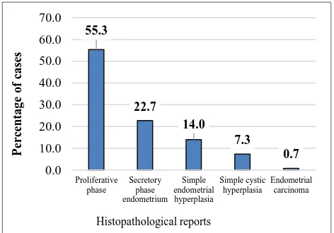
Out of 150 study participants, 21 participants (14%) had simple endometrial hyperplasia and only one participant (0.7%) had endometrial carcinoma on histopathological examination of endometrial biopsy.
Figure 1: Distribution according to histopathological reports.
55.3
22.7
14.0 7.3
0.7 0.0
10.0 20.0 30.0 40.0 50.0 60.0 70.0
Proliferative phase
Secretory phase endometrium
Simple endometrial hyperplasia
Simple cystic hyperplasia
Endometrial carcinoma
Per
ce
n
ta
g
e
o
f
ca
se
s
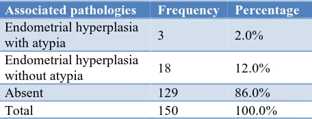
[image:3.595.310.546.556.721.2]Out of 150 study participants, 21 participants (14%) had simple endometrial hyperplasia out of which 3 participants (2%) had endometrial hyperplasia with atypia and 18 participants (12%) had endometrial hyperplasia without atypia on histopathological examination.
Table 2: Distribution of study participants according to endometrial hyperplasia with or without atypia.
Associated pathologies Frequency Percentage
Endometrial hyperplasia
with atypia 3 2.0%
Endometrial hyperplasia
without atypia 18 12.0%
Absent 129 86.0%
Total 150 100.0%
DISCUSSION
Fibroids are the commonest tumour found in the females. Many women undergo operative procedures like hysterectomy and myomectomy due to symptoms occurring because of the fibroid. Due to rising coexistence of endometrial pathologies like endometrial hyperplasia, polyps, adenomyosis etc., in patients with a fibroid, it is of paramount importance to take into account these lesions before starting treatment. The patients should undergo prior investigation like ultrasonography and endometrial biopsy to rule out other lesions so that both lesions are managed simultaneously. Due to the high coincidence of fibroid and endometrial hyperplasia, endometrial sampling should be performed in patients who are being considered for hysterectomy, especially in perimenopausal age group patients.
Various studies have been undertaken so far to determine
the coexistence specially regarding fibroid and
endometrial carcinoma and hyperplasia. This study aims to fill the lacunae in the literature and to add to authors existing knowledge about the association of various endometrial pathologies and significance of doing an endometrial biopsy in particular age group patient in the present-day scenario.
Authors did an observational cohort study (retrospective as well as prospective) on 150 women in the age group of 25-60 years from the railway population who came to OPD. A detailed history was taken and consent for the physical examination and for required investigations was taken. General examination with specific gynecological examination and ultrasonography was done and posted for diagnostic hysteroscopy and endometrial biopsy. Women who have already undergone surgical treatment were included in the study with complete data/discharge sheet. Histopathological reports were collected. Data were analysed using SPSS version 20.0.
In this study comprising of 150 study participants, the maximum number of the patients having Leiomyomas belong to perimenopausal age group 46-60 (50.7%)
followed by 31-45 age group (40%) thus confirming the higher incidence of leiomyomas in reproductive age group female. Studies by Cramer et al stated that 30 to 77% of women will develop fibroids sometime during their childbearing years.9
A total 82 participants in this study came with the complaint of heavy menstrual bleeding (54.7%) followed by 39 participants (26%) with postmenopausal bleeding and 27 participants (18%) with dysmenorrhea while only 2 came with a mass per vaginum. This suggests that majority of patients with fibroid comes with the complaints of heavy bleeding. Munusamy MM et al also in his study of 362 women with fibroids (66%) presented with severe anaemia due to menorrhagia followed by (33.4%) with palpable abdomen pelvic masses.10 This is
supported by other studies done by Dayal S et al, Muslina Akhter et al.11,12
Most of the participants in this study were multiparous (88.7%) and 8% were primiparous while only 3.3% of participants were nulligravida which is supported by a study done by Romanek K et al.13 but this finding is in
contrast with studies done by Sparic N et al, Wise LA et al, Okolo S et al.14-16
Several epidemiological studies found the increased risk of myomas in women with diabetes mellitus and hypertension. In this study 36.7% had hypertension while 18.7% had diabetes associated supported by studies done by Parker W et al, Lethaby A et al, He Y et al.17-19
Out of 150 study participants, 55 participants (36.6%)
underwent hysterectomy, 13 participants (8.7%)
underwent myomectomy indicating that all symptomatic fibroid patients do not require surgical management as primary treatment. Medical management is turning out to be bliss for the patients having contraindications for surgical treatment also supported by Wallach et al, Mas A et al, Moroni R et al, Gurusamy K et al.20-23
Authors found no relevance in any significant pap smear report in this study. All were negative for any cervical malignancy. Out of 150 study participants, majority 47.3% had the bulky uterus (6-8 weeks size) on PV examination, 36% had uterine size up to 12 weeks size and 7.3% had a uterine size in 13-16 weeks size. No significant correlation was found. No study is there in the literature comparing uterine size and coexistence of other pathologies.
Out of 150 study participants, 23 participants (15.3%) had fornceal fullness which can lead to bias in diagnosing uterine fibroid and ovarian or adnexal masses. However, the endometriotic cyst can be present in association with fibroid in various case of subfertility. Also, the patients belonging to this subgroup come with features of pelvic pain/dysmenorrhoea often. Uimari O et al, Nezhat C et al,
also supports the correlation between external
Out of 150 study participants, the majority of 92 participants (61.4%) had a bulky uterus on USG while 56
participants (37.3%) had normal uterine size.
Transvaginal ultrasonography is as efficient as magnetic resonance imaging in detecting myoma presence, but its capacity for exact myoma mapping falls short of that of magnetic resonance imaging, especially in large and multiple-myoma (>4) uteri as stated by Dueholm M.26
Out of 150 study participants, 52 participants (34.7%) had an endometrial thickness in range 7-10 mm, 20 participants (13.3%) had endometrial thickness >13 mm. this data could not be used for any analysis since many confounding factors were there as it was imaging modality and subjective variation was there. Also, endometrial thickness varies in the female as per the menstrual cycle changes. So, authors cannot use these data for any analysis.
Out of 150 participants in my study, 15 participants (10%) had an ovarian cyst. In 3-year, prospective study done by Gowri M et al on 259 hysterectomy patients’ leiomyomas, there were incidental concurrent lesions like granulosa cell tumour of ovary (0.4%), tubercular endometritis (0.4%), dermoid and chocolate cyst of ovary (0.8%), mucinous and serous cystadenoma of ovary (1.6%). More research is needed in this field.27
On diagnostic hysteroscopy of the study participants, authors found that 58 per cent had normal endometrium which included fluffy endometrium or tags. Other 20% had fibroid polyp, 16.7% had hyperplastic endometrium (thickened, undulating, edematous) while 5.3% had atrophic endometrium (the endometrium appeared flat, thin and fragile) (Figure 1). On further histopathological examination of an endometrial sample taken 14% had simple endometrial hyperplasia and only one participant (0.7%) had endometrial carcinoma. In this study participants, 2% had endometrial hyperplasia with atypia and 18 participants (12%) had endometrial hyperplasia without atypia on microscopy (Table 2). In this study out of 14% patients with endometrial hyperplasia associated comorbidities like hypertension and diabetes were present in 11 patients (52.38%) and 4 patients (19%) respectively.
Similar studies done by other authors support this study. The study done by Mangala et al, stated that microscopic examination of endometrium revealed 46.3% of the normal endometrial pattern while 22.8% of endometrial hyperplasia out of total 259 patients. This study is also supported by Sheetal et al.27,28
Out of 150 study participants, 37 participants (24.6%) had adenomyosis, 21 participants (14%) had simple endometrial hyperplasia out of which 3 participants (2%) had endometrial hyperplasia with atypia and 18 participants (12%) had endometrial hyperplasia without atypia, 13 participants (8.6%) had cervical polyp, 8 participants (5.33%) had endometrial polyp and 6
participants (4%) had external endometriosis while 64 participants (42.7%) had no association with fibroid uterus (Table 1).
In support of this study, various research articles are there in the literature. Priyadarshini P et al in their study of leiomyomas found that endometrial pattern commonly associated with fibroid were hyperplastic endometrium together accounting for 9% and associated adenomyosis (29%).29 Study similar to studies by Denligdish et al,
Rizvi et al, Nezhat C et al in his studies reported the close association between fibroids and endometriosis, about 90% of women who presented with fibroids turned out to have endometriosis.30-32
Camran et al also found out the association of adenomyosis with other uterine disorders in more than 80% of women 50% of patients have associated fibroids, 11% have endometriosis, and 7% have endometrial
polyps.33 Dual pathology of leiomyoma and adenomyosis
was noted in 29%.
Kanter, Klawans and Bauer reported hyperplasia of the endometrium in 53%, and adenomyosis in 52% of their fibromyoma cases.34
Kinay et al studied the prevalence of endometrial polyps coexisting with uterine fibroids and other uterine lesions.35 The prevalence of endometrial polyps in
women with uterine fibroid was 20.1%.
Table 3: Uterine lesions associated with fibromyoma.
Total cases of fibromyoma 100
Cases also showing hyperplasia of endometrium 17
Cases also showing benign endometrial polyp 16
Cases also showing adenomyosis of body of
[image:5.595.314.543.559.657.2]uterus 14
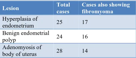
Table 4: Associated lesions observed during same period as 100 fibromyoma cases.
Lesion Total
cases
Cases also showing fibromyoma
Hyperplasia of
endometrium 25 17
Benign endometrial
polyp 24 16
Adenomyosis of
body of uterus 28 14
Light FW conducted a study on 100 fibroid cases, he found that 42 cases showed one or more lesions besides fibroid (Table 3 and 4).36
(34.57%). However, uterine myoma and endometrial polyp coexistence were less seen only in 2 cases (6.25%). Only in one case, the endometrial hyperplasia was seen together with uterine myoma.37 Shergill et al, involved
100 hysterectomy cases in their study and
histopathological examination revealed that 38 patients were diagnosed as leiomyoma with adenomyosis.38
Khoo CK et al in their study regarding an update on adenomyosis have mentioned the association of adenomyosis with other uterine pathologies. They found out that a large proportion of the women with adenomyosis have associated pathologies such as leiomyomas (80%), endometriosis (24%), endometrial polyps (14.7%), endometrial hyperplasia (13.6%), and
adenocarcinoma (5.3%).39
Participants with endometrial hyperplasia with/without atypia (n=19) were compared with the number of fibroids and whether multiple or single also size of the fibroid (cut off 6 cm) was compared, however, no significant association was made out. Also, authors did not find any statistically significant association between Participants having adenomyosis and external endometriosis (n=43) and the number of fibroids whether multiple or single or
size of fibroids. Participants with polyp
cervical/endometrial (n=19) were also compared for any association with the number of fibroids whether multiple or single and size of the fibroid, however, no statistically significant association was found.
In contrast to this study, Witherspoon opined that hyperestrinism first produces hyperplasia of the endometrium and later, if allowed to act over a period of time, causes the development of fibromyoma. He pointed out that additional lesions were twice as common in fibromyomas less than 5 cm in diameter (37 cases out of 62) as in association with more than 5 cm in diameter (11 out of 38). Witherspoon finds hyperplasia of the endometrium in practically all of his fibromyoma cases which is contradictory to this study.40
In the study done by Kinay et al endometrial polyps were more common in patients with ≥2 fibroids (p=0.023) and largest fibroid <8 cm (p=0.009) which contradicts this study.35
Thus, in the present study, authors found a significant association between coexistence of uterine fibroids and other uterine pathologies. Nearly 57.33% had one of the uterine pathologies associated with myomas. The strong association between the presence of adenomyosis 24.6% and hyperplasia 14% was obtained. Only a single case out of total 150 came to be positive for endometrial carcinoma that participant belongs to the postmenopausal group.
The study didn’t find any association of specific pathology when number and size of fibroids were taken into consideration which needs further research.
Limitations of the study was, in this study institute, only railway employees and their families were included so the result cannot be applied to the general population. The data collected from the majority of the retrospective patient which were included in the showed various lacunae due to inadequate availability of documents. Prospective patients who were included needs to be followed up on long-term aspects to observe for development of any uterine pathologies in future. Higher imaging modalities like MRI was not carried out due to limited resources.
CONCLUSION
With the increasing awareness in women regarding their health and menstrual bleeding pattern more and more cases has been reported to the hospital and with the advancement of imaging technologies accuracy of diagnosis of uterine pathologies have increased to a greater level.
This study revealed the increasing trend of coexistence of various uterine pathologies mainly adenomyosis and endometriosis with leiomyomas which parallels the trend seen in several studies. The present study, however, does not find any association depending upon the number and size of the fibroids which needs to be studied in future in a large population. However, authors found that endometrial hyperplasia with atypia and carcinoma is not so frequently found along with fibroid. So, every patient with leiomyoma need not undergo an operative procedure.
Majority of the women falling in adenomyotic and hyperplasia group belong to 46-60 age mainly perimenopausal and menopausal group. This points to the fact that prolonged exposure to estrogen is likely responsible for higher incidence of above two conditions alarming the need for streamlining the investigation and treatment modality on a priority basis. This will minimise study operative overload in government hospital. In Indian population where access to hospital facilities is difficult, selecting the patient who really needs to be evaluated for malignancy is extremely important.
To conclude, early identification of patient on basis of history and detailed clinical and imaging modality like USG will be helpful for selection of candidates who require diagnostic hysteroscopy and endometrial biopsy to rule out malignancy and other pathologies.
Recommendations
• Every case of fibroid uterus need not be subjected to
endometrial biopsy at the time of diagnosis.
• The incidence of endometrial hyperplasia with atypia
comorbidities like diabetes and hypertension and thickened endometrium by ultrasonography.
• Hence by judicially planning endometrial biopsy
only in few high-risk cases of fibroid in age group 46-60 years authors can avoid unnecessary operative interference and associated loss of man power valuable time of surgeons, discomfort to patients and financial implications.
Funding: No funding sources Conflict of interest: None declared Ethical approval: Not required
REFERENCES
1. Standard treatment guidelines obstetrics and
gynaecology Ministry of Health and Family Welfare;
1-102. Available at:
http://clinicalestablishments.gov.in/WriteReadData/4 571.pdf. Accessed on 20th March 2020.
2. Surgery MR. Specializing in multi-organ
reconstructive surgery uterine fibroid tumors - question about fibroids what are uterine fibroid tumors ? 2017;1-22.
3. Khan AT, Shehmar M, Gupta JK. Uterine fibroids: Current perspectives. Int J Womens Health. 2014;6(1):95-114.
4. Cramer SF, Patel A. The frequency of uterine
leiomyomas. Am J Clin Pathol. 1990;94(4):435-8.
5. Evans P, Brunsell S. Uterine fibroid tumors:
diagnosis and treatment. Am Family Phys.
2007;75(10):1503-8.
6. Cook JD, Walker CL. Treatment strategies for
uterine leiomyoma: the role of hormonal modulation. Semin Reprod Med. 2004;22(2):105-11.
7. Flake GP, Andersen J, Dixon D. Etiology and
pathogenesis of uterine leiomyomas: a review. Environ Health Perspect. 2003;111(8):1037-54.
8. Whiteman MK, Hillis SD, Jamieson DJ, Morrow B,
Podgornik MN, Brett KM, et al. Inpatient hysterectomy surveillance in the United States, 2000-2004. Am J Obstet Gynecol. 2008;198(1):34.e1-34.e7.
9. Cramer SF, Patel A. The frequency of uterine
leiomyomas. Am J Clin Pathol. 1990;94(4):435-8.
10. Munusamy MM, Sheelaa WG, Lakshmi VP. Clinical
presentation and prevalence of uterine fibroids: a 3-year study in 3-decade rural South Indian women. Int
J Reprod Contraception, Obstet Gynecol.
2017;6(12):5596.
11. Dayal S, Nagrath A. Clinicopathological correlation of endometrial, myometrial and ovarian pathologies with secondary changes in leiomyoma. J Pathol Nepal. 2016;6(11):937.
12. Akhter M, Salam A, Anwar K, Alam M, Shoma AK,
Salim M. Clinical Profile and Management Option of Fibroid Uterus Patient. Chattagram Maa-O-Shishu Hospital Med College J. 2015;14(2):48-51.
13. Romanek K, Bartuzi A. Risk factors for adenomyosis
in patients with symptomatic uterine leiomyomas. 2010;81(9):678-80.
14. Sparic R, Mirkovic L, Malvasi A, Tinelli A.
Epidemiology of uterine myomas: a review. Int J Fertil Steril. 2016;9(4):424.
15. Wise LA, Laughlin-Tommaso SK. Epidemiology of
uterine fibroids-from menarche to menopause. Clin Obstet Gynecol. 2016;59(1):2.
16. Okolo S. Incidence, aetiology and epidemiology of uterine fibroids. Best Pract Res Clin Obstet Gynaecol. 2008;22(4):571-88.
17. Parker WH. Etiology, symptomatology, and
diagnosis of uterine myomas. Fertil Steril.
2007;87(4):725-36.
18. Lethaby AE, Vollenhoven BJ. Fibroids (uterine
myomatosis, leiomyomas). BMJ Clin Evid.
2007;2007.
19. He Y, Zeng Q, Li X, Liu B, Wang P. The association
between subclinical atherosclerosis and uterine fibroids. Biondi-Zoccai G, editor. PLoS One. 2013;8(2):e57089.
20. Wallach EE, Vlahos NF. Uterine myomas: an
overview of development, clinical features, and management. Obstet Gynecol. 2004;104(2):393-406. 21. Mas A, Tarazona M, Dasí Carrasco J, Estaca G, Cristóbal I, Monleón J. Updated approaches for management of uterine fibroids. Int J Womens Health. 2017;9:607-17.
22. Moroni R, Vieira C, Ferriani R, Candido-Dos-Reis F,
Brito L. Pharmacological treatment of uterine fibroids. Ann Med Health Sci Res. 2014;4(Suppl 3):S185-92.
23. Gurusamy KS, Vaughan J, Fraser IS, Best LMJ,
Richards T. Medical therapies for uterine fibroids - a systematic review and network meta-analysis of
randomised controlled trials. PLoS One.
2016;11(2):e0149631.
24. Uimari O, Järvelä I, Ryynänen M. Do symptomatic endometriosis and uterine fibroids appear together? J Human Reprod Sci. 2011:4(1):34.
25. Nezhat C, Li A, Abed S, Balassiano E, Soliemannjad
R, Nezhat A, et al. Strong association between endometriosis and symptomatic leiomyomas. JSLS J Soc Laparoendosc Surg. 2016;20(3):e2016.00053.
26. Dueholm M, Lundorf E, Hansen ES, Ledertoug S,
Olesen F. Accuracy of magnetic resonance imaging and transvaginal ultrasonography in the diagnosis, mapping, and measurement of uterine myomas. Am J Obstet Gynecol. 2002;186(3):409-15.
27. Gowri M, Mala G, Murthy S, Nayak V.
Clinicopathological study of uterine leiomyomas in hysterectomy specimens. J Evol Med Dent Sci. 2013;2(46):9002-10.
28. Patil SG, Bhute SB, Inamdar SA, Acharya NS,
29. Priyadarshini P, Gomathy E. Clinicopathological study of uterine leiomyomas in hysterectomy specimens; a retrospective study. Int J Adv Res. 2018;6(62):2320-3407.
30. Deligdish L, Loewenthal M. Endometrial changes
associated with myomata of the uterus. J Clin Pathol. 1970;23(8):676-80.
31. Rizvi G, Pandey H, Pant H, Chufal SS, Pant P. Histopathological correlation of adenomyosis and leiomyoma in hysterectomy specimens as the cause of abnormal uterine bleeding in women in different age groups in the Kumaon region: a retro-prospective study. J Mid-life Health. 2013;4(1):27.
32. Nezhat C, Bhattacharya S, Kumari V, Arora M.
Endometriosis. Jaypee Brothers Medical Publishers; 2011:288.
33. Nezhat C, Balassiano E, Abed S, Nezhat CH,
Soliemannjad R, Nezhat FR. Coexistence of
endometriosis in women with symptomatic
leiomyomas-a reappraisal of the current literature. Obstet Gynecol. 2016;127:83S.
34. Kanter AE, Klawans AH, Bauer CP. A study of
fibromyomas of the uterus with respect to the
endometrium, myometrium, symptoms, and
associated pathology. Am J Obstet Gynecol. 1936;32(2):183-93.
35. Kınay T, Öztürk Başarır Z, Fırtına Tuncer S, Akpınar
F, Kayıkçıoğlu F, Koç S. Prevalence of endometrial
polyps coexisting with uterine fibroids and associated factors. Turkish J Obstet Gynecol. 2016;13(1):31-6.
36. Light FW. Uterine lesions associated with
fibromyoma. Am J Clin Pathol. 1939;9(4):483-90.
37. Turgut O, Yanık S, Ay AA, Gencay I, Nergiz S, Ay
A, Aydin O. The evaluation of preoperative and
histopathologic diagnosis in specimens of
hysterectomy performed for benign conditions. J Surg. 2014;2(1):11-3.
38. Shergill SK, Shergill HK, Gupta M, Kaur S.
Clinicopathological study of hysterectomies. J Indian Med Assoc. 2002;100(4):238-9.
39. Ching Hong S, Kiat Khoo C. An update on
adenomyosis uteri. Gynecol Minim Invasive Ther. 2016;5:106-8.
40. Witherspoon JT. The hormonal origin of uterine
fibroids: an hypothesis. Am J Cancer.
1935;24(2):402-6.