Formation of a functional morphogen gradient by a passive
process in tissue from the early Xenopus embryo
NATASHA MCDOWELL
1, JOHN B. GURDON*
,1and DAVID J. GRAINGER
21Wellcome/CRC Institute of Cancer and Developmental Biology, Cambridge and Department of Zoology, University of Cambridge, England and 2Department of Medicine, Addenbrooke’s Hospital, Cambridge, England
ABSTRACT In early development much of the cellular diversity and pattern formation of the embryo is believed to be set up by morphogens. However, for many morphogens, including members of the TGF-β superfamily, the mechanism(s) by which they reach distant cells is unknown. We have used immunofluorescence to detect, at single cell resolution, a morphogen gradient formed across vertebrate tissue. The TGF-β ligand is distributed in a gradient visible up to 7 cell diameters (about 150-200 µm) from its source, and is detectable only in the extracellular space. This morphogen gradient is functional, since we demonstrate activation of a high response gene (Xeomes) and a low-response gene (Xbra) at different distances from the TGF-β source. Expression of the high affinity type II TGF-β receptor is necessary for detection of the gradient, but the shape of the gradient formed only depends in part on the spatial variation in the amount of receptor. Finally, we demonstrate that the molecular processes that participate in forming this functional morphogen gradient are temperature independent, since the gradient forms to a similar extent whether the cells are maintained at 4°C or 23°C. In contrast, TGF-β1 internalisation by cells of the Xenopus embryo is a temperature-dependent process. Our results thus suggest that neither vesicular transcytosis nor other active processes contribute to a significant extent to the formation of the morphogen gradient we observe. We conclude that, in the model system used here, a functional morphogen gradient can be formed within a few hours by a mechanism of passive diffusion.
KEY WORDS:
Xenopus, morphogen, TGF-
β
, passive diffusion.
0214-6282/2001/$25.00
© UBC Press Printed in Spain
www.ijdb.ehu.es
*Address correspondence to: J.B. Gurdon. Wellcome/CRC Institute of Cancer and Developmental Biology, Tennis Court Road, Cambridge, CB2 1QR, England. FAX:- +1223-334-185. e-mail: [email protected]
Abbreviations used in this paper: TGF-β, Transforming Growth Factor-Beta.
Introduction
Across a range of species much of embryonic development appears to be dependent upon signals that act at a distance from their source. Many of these signals are thought to be morphogens, secreted molecules that form extracellular concentration gradients across tissue (for review see Neumann and Cohen, 1997). At any given position in the gradient, the cellular response is dependent upon the morphogen concentration at that position. A morphogen can thus induce a diversity of cell types in defined spatial relation-ships to one another and therefore contribute to the positional information required to pattern the embryo (Wolpert, 1969).
In vertebrates such as the frog, Xenopus laevis, much work on morphogens has focused on how members of the TGF-β superfamily, including activin, can act as morphogens in the induction of the mesoderm (for review see McDowell and Gurdon, 1999). This is the middle germ layer of the embryo and later gives rise to a range of tissue types including notochord, muscle and blood. In tissue of the
early embryo, such ligands have been shown to induce, at a distance of up to a few hundred µm, mesodermal response genes in a concentration-dependent manner (Gurdon et al., 1994). Further-more, use of dominant inhibitory receptors to eliminate signalling in response to TGF-β superfamily members has suggested that these molecules have an in vivo role in early development (Hemmati-Brivanlou et al., 1992; Dyson and Gurdon, 1997).
been identified as a mechanism of transcellular transport within the plane of the epithelium (as opposed to transport along the baso-apical axis) that is ATP-dependent and involves both endo- and exocytosis (Pfeiffer et al., 1999).
The first indication that Wg spreads from its source by vesicular transcytosis came from observations that Wg protein is localized to vesicular structures within the responding cells (van den Heuvel et al., 1989; Gonzalez et al., 1991). However, more recent observations that a mutation in shibire, the Drosophila homologue of the GTPase dynamin that is required for clathrin-coated vesicle formation, re-duces the range of Wg protein distribution provides direct evidence for a role of vesicular transcytosis in Wg movement (Bejsovec et al., 1995; Moline et al., 1999). Other genetic experiments have led to the proposal that a class of Wg receptor exists that is solely responsible
for transcytosing the Wg protein (Hays et al., 1997). Similar data has also been obtained for another morphogen Hedgehog (Hh). Immun-ofluorescence has located Hh in a punctate distribution in the wing disc (Tabata et al., 1994; Bellaiche et al., 1998) which is thought to reflect the localization of Patched, a Hh receptor and binding protein, to intracellular vesicles (Bellaiche et al., 1998).
In contrast, elucidation of the mechanism of passage of Dpp, a TGF-β superfamily member, has been hampered by the lack of an immunofluorescence detection method sufficiently sensitive to detect Dpp in the responding cells: an antibody against Dpp was able to detect the protein only in those cells in which it is synthe-sised (Lecuit and Cohen, 1998). Although the functional range of Dpp activity has recently been shown to be reduced in endocytosis-defective Drosophila wing-discs, the inability to detect the ligand
Fig. 1. Double immunofluorescence staining for TGF-β1 and TGF-βIIR. (A) A gradient of TGF-β1 protein, emanating from beads previously incu-bated in TGF-β1 protein, can be seen in animal caps previously injected with 4 ng of TGF-βIIR mRNA. Conjugates were fixed after 3 hours of culture [green arrowheads indicate TGF-β1 beads, or bead spaces (see text)]. The scale bar represents 50 µm. (B) The localization of TGF-βIIR protein in the section shown in (A). Injection of TGF-βIIR-mRNA results in highly mosaic expression of the protein. (C-E) Fluorescence due to ligand (FITC) and receptor (TRITC) co-localize. (C) Confocal image of the fluo-rescence due to TGF-β1 (FITC) in a pattern indica-tive of the cell surface. (D) TGF-βIIR (TRITC) stain-ing indicates the circumference of the cells in the section shown in (C). (E) When (C) and (D) are superimposed, a direct co-localization of fluores-cence due to ligand (C) and receptor (D) is observed (highlighted in yellow). (F-I) Tissue containing 4 ng TGF-βIIR mRNA, but BSA only beads (blue arrow-heads). (F) Confocal image of BSA bead section stained with anti-TGF-β1; the extracellular fluores-cence pattern seen in (C) is absent. (G) TGF-βIIR staining indicates the circumference of the cells in the section shown in (F). (H) In contrast to (E), the yellow extracellular co-localization is absent when (F) and (G) are superimposed. In all the confocal images (C-H) a background reticulate pattern can be observed. (I) No gradient of fluorescence is seen in TGF-βIIR tissue containing BSA only beads. (J) The background fluorescence observed in tis-sue containing both ligand and receptor, but lacking the anti-TGF-β1 primary antibody. (K) In wild-type tissue containing a TGF-β1 bead a gradient of fluorescence due to TGF-β1 is absent.
➤
➤
➤
➤
➤
➤
➤
➤
➤ ➤ ➤
➤
➤ ➤
➤
➤
A
B
C
D
E
F
G
H
makes it unclear whether endocytosis is required for Dpp signalling, transport or in receptor recycling (Gonzalez-Gaitan and Jackle, 1999).
In vertebrate embryos much less information is available on how molecules can reach distant cells. Probably the most data has been accumulated for molecules of the
TGF-β superfamily in the Xenopus embryo. For example, it has been shown that activin, as well as other TGF-β family members, can act directly on distant cells and that the release of other additional secreted signalling molecules is not required for their long-range signalling (McDowell et al., 1997). In 1996, Reilly and Melton proposed that movement of TGF-β family members through embryonic tissue may occur by a cell-to-cell relay of the same signal, where each cell along the gradient would induce production of TGF-β family members by its immediate neighbours. However, more recent data established that such a relay mechanism is unlikely to contribute to ligand spread across tissue (Jones et al., 1996; McDowell et al., 1997) since, for example, the long-range signal can pass through layers of cells unable to respond to the morphogen, and the discrep-ancies between the conclusions were accounted for (McDowell et al., 1997). In addition, the observation of 35
S-labelled activin molecules in cells distant from an implanted bead source also demonstrates that long-range signalling by activin does not necessitate a mechanism of relay
respect to either the direction or rate of signalling (McDowell et al., 1997). We have also investigated whether the molecular proc-esses that contribute to the formation of this functional morphogen gradient are temperature dependent, and therefore active proc-esses, as a first step to determining whether a gradient can be set up by passive diffusion or whether vesicular transcytosis is neces-sary for ligand movement across early embryonic tissue. In the absence of mutants in vesicular transport (such as the shibire mutant in Drosophila) and highly specific chemical inhibitors of endocytosis, the temperature dependence of morphogen gradient formation is likely to provide a useful tool to discriminate between the two models of ligand transport in the vertebrate embryo.
Results
A TGF-β ligand gradient
Animal cap cells from stage 8 Xenopus embryos do not contain the type II TGF-β receptor (TGF-βIIR) and cannot respond the TGF-β isoform. However, following injection of mRNA encoding TGF-βIIR, these ligands will induce mesodermal response genes in a manner analogous to endogenous mesoderm inducers (Bhushan et al., 1994; Reilly and Melton, 1996; McDowell et al., 1997). Conjugates were prepared as previously described (Gurdon et al., 1994; McDowell et al., 1997), in which TGF-β1 or TGF-β2 protein-loaded beads are sandwiched within either wild-type ani-mal cap tissue or within tissue that had been previously injected with TGF-βIIR mRNA. In order to optimise antigen preservation, such conjugates were shock frozen without fixation and cryostat sectioned. We then employed a highly sensitive immunofluores-cence procedure that has been optimized to quantitatively detect low concentrations of TGF-β (Grainger et al., 1995a; Mosedale et al., 1996). This protocol has been shown to increase specific staining up to fivefold over typical published protocols. A range of antibodies against TGF-β1 and TGF-β2 were tested; however, only (McDowell et al., 1997). However, although the 35S-labelled activin
formed a concentration gradient across the tissue, the path-length of the 35S nuclear decay made it impossible to determine whether the
ligand was intra- or extracellularly located. Hence, it is not known whether members of the TGF-β superfamily diffuse in the extracel-lular environment between cells or move through cells by vesicular transcytosis in the vertebrate embryo.
It has long been debated whether extracellular diffusion would be possible in the solid tissue of the Xenopus embryo. Kalt (1971) noted that the cells of the Xenopus embryo are tightly packed. Furthermore, one of the main arguments against diffusion is that there are too many molecules in the extracellular milieu with which a ligand might interact. For example, computer simulations have been used to propose that TGF-β diffusion would be dramatically hindered by the presence of its high affinity signalling receptors (Kersberg and Wolpert, 1998). In addition, the TGF-β family members bind proteoglycans, such as betaglycan (Lopez-Casillas et al., 1991): any interaction with these and other binding proteins (Yamaguchi et al., 1990) has also been proposed to limit the ability of TGF-β-related molecules to spread by passive diffusion in the extracellular environment (Reilly and Melton, 1996).
Technical difficulties currently preclude a definitive resolution of this debate. Localization of the morphogen, using antibodies, has so far failed to detect a functional morphogen gradient in vertebrate tissue. In addition, mutagenesis is not presently possible in Xeno-pus, preventing the use of the powerful experimental strategies that have begun to delineate the pathways involved in ligand distribution in Drosophila. In our study we have utilised a model system, involving the diffusion of TGF-β1 from a bead into animal cap tissue of the early Xenopus embryo, in order to exploit the exceptional sensitivity of an optimised immunofluorescence pro-cedure for the detection of TGF-β1 protein (Mosedale et al., 1996). In this way we have directly visualised a functional morphogen gradient of TGF-β1 in a solid tissue that appears unpolarized with
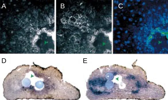
Fig. 2. The gradient of TGF-β1 is not an artefact resulting from mosaic receptor expression; green arrowheads indicate TGF-β1 bead space. When the fluores-cence due to (A) TGF-β1 is divided by (B) the fluorescence due to receptor,a gradient of fluorescence (C) can still be observed. The ratio of the FITC (TGF-β1) and TRITC
(TGF-βIIR) fluorescence signals is shown as a false colour image where red represents a high ratio and blue a low ratio. (D,E) The TGF-β1 gradient can elicit a multiple gene response in cells containing TGF-βIIR (green arrowheads indicate beads, or bead spaces). (D) Xeomes is activated by high concentrations of TGF-β1 adjacent to the bead. (E) Conversely, Xbra, a low response gene, is activated further away from the bead.
A
B
C
one, an antibody to TGF-β1 known to be of particularly high affinity (Grainger et al., 1995a) successfully visualized the ligand.
As shown in Fig. 1A, we visualised a gradient of TGF-β1 protein which has emanated from the beads (green arrowheads indicate the beads as well as the spaces from which the beads have been dislodged during sectioning) after only three hours of culture. The gradients (observed in approximately 20 conjugates) could often be seen to extend 150-200 µm across the tissue. Since the pattern of fluorescence we detect is consistent with the TGF-β1 being localised to the cell surface, we analysed some conjugates using double-label immunofluorescence with the antibody against
TGF-β1 detected with fluorescein, and an antibody directed against the extracellular portion of TGF-βIIR detected with rhodamine. We found that the FITC fluorescence due to TGF-β1 (Fig. 1A) and TRITC fluorescence due to TGF-βIIR (Fig. 1B) directly overlap. This co-localization was confirmed when the sections were viewed using confocal microscopy (TGF-β1 staining Fig. 1C; TGF-βIIR
staining Fig. 1D). When these channels are superimposed (Fig. 1E) yellow is seen indicating co-localization of green (TGF-β1) and red (TGF-βIIR).
When animal caps, injected with TGF-βIIR mRNA as above, are sandwiched around control beads loaded with BSA but no TGF-β1 (blue arrowheads), no gradient fluorescence signal following stain-ing for TGF-β1 was ever observed (Fig. 1 F,I; Fig. 1G shows the localization of TGF-βIIR in the section shown in 1F). Hence when the confocal images of TGF-β1 staining and TGF-βIIR staining (shown in Fig. 1 F,G) from a control conjugate are superimposed, no yellow colour (indicative of TGF-β1 co-localized with the
TGF-βIIR) is seen (Fig. 1H). A small amount of antibody-specific staining with the anti-TGF-β1 was detected within the nuclei (verified by Hoechst labelling, data not shown). This nuclear staining was present to the same extent (assessed by quantitative immunofluo-rescence analysis) in the BSA-treated control samples (Fig. 1 F,I) as in conjugates containing TGF-β1 beads (Fig. 1C). Staining of the nucleus with this antibody may be due either to non-specific binding of this primary antibody (but not any of the others used in this study) to the nuclei or, more likely, to specific binding of the antibody to a TGF-β1-like antigen that is constitutively present in the nuclei of animal cap cells of the early Xenopus embryo. The background fluorescence observed in tissue containing both lig-and lig-and receptor, but stained in the absence of the anti-TGF-β1 primary antibody (shown in Fig. 1J) was negligible. The back-ground fluorescence in an equivalent control for TGF-βIIR (i.e. lacking the anti-TGF-βIIR primary antibody) was also negligible (data not shown). Note that all of the antibodies used in this study bind non-specifically to the beads (Fig. 1 A,B,F,G,I,K); however, the fluorescence from the bead does not influence the fluores-cence detected from the nearby tissue (assessed by comparison of tissue fluorescence near beads which were either retained or lost from the section during sample preparation). Such bead fluorescence was excluded from all analysis.
When the analysis was repeated using wild-type animal caps (which were not injected with TGF-βIIR mRNA), no TGF-β1 gradi-ent was detected with either TGF-β1 loaded beads (Fig. 1k) or control beads. It has previously been shown that the related molecule TGFβ2 can pass across tissue lacking TGF-βIIR. We therefore conclude that the presence of the high affinity receptor sites is required to concentrate the ligand and thus allow visualisa-tion of the gradient by the method we are using, rather than being necessary to enable the ligand to diffuse.
Receptor distribution is not the sole determinant of gradient shape
In many of the sections analysed, the TGF-β1 gradient was found to be patchy across the tissue (as in Figs. 1A and 2A), an observation that was unexpected. However, double-labelling for TGF-βIIR indicates that the expression of TGF-βIIR is also highly mosaic across the tissue (Figs. 1B and 2B). It is known that injection of mRNA results in mosaic expression of the correspond-ing protein. Hence, if the presence of TGF-βIIR is necessary to enable detection of the ligand, then the patchy fluorescence due to TGF-β1 can be accounted for by the mosacism of receptor expression. We therefore investigated whether the shapes of the TGF-β1 gradients that we observe were due only to the mosai-cism of receptor distribution. Since the immunofluorescence protocol used here has previously been shown to yield a linear
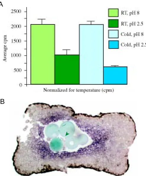
Fig. 3. (A) Ligand-receptor internalization is reduced when cells are cultured at 4°C. 125I-TGF-β1 was bound to cells at 4°C or at 23°C and the cells were
then washed to remove unbound ligand. After allowing 40 minutes for internalization to occur, they were washed twice more at either pH 2.5 or pH 8; all steps subsequent to binding were again performed at either 4°C or at 23°C. The radioactivity bound to the cells was then counted. Each sample was analysed in duplicate per experiment and the results combined from a total of three experiments. The histogram, normalized for temperature, indicates the 125I-TGF-β1 bound to the receptor-injected cells (in cpm)
following subtraction of the background binding of 125I-TGF-β1 to wild-type
cells. As can be seen, significantly more ligand (p=0.0092) is washed off with pH 2.5 at 4°C than with pH 2.5 at 23°C. (B) Xeomes expression is normal in tissue kept at 4°C. Conjugates were cultured for 3 h at 4°C, then cultured to stage 10.5 at 23°C (green arrowhead indicates TGF-β1 beads).
A
B
in a punctate pattern within the cells. This suggests that the TGF-β1 is not localized to intracellular vesicles, at least at high concentration. However, we cannot rule out that TGF-β1 is present in vesicles, but below our level of detection. We therefore adopted an alternative approach to distinguish extracellular diffusion from active transcytosis: diffusion is a passive process and hence relatively temperature independent (Burshtein, 1996), while transcytosis is an active, energy-requiring process which should be highly sensitive to varia-tions in temperature (Koenig and Edwardson, 1997).
It has been shown that low temperature (such as 4oC) is
sufficient to almost eliminate ligand-receptor internalization in mammalian cells (Koenig and Edwardson, 1997). If the same is true for Xenopus cells then a gradient established by transcytosis, which requires internalization, would not be formed across tissue kept at 4oC. Alternatively, if diffusion is responsible for the spread
of the ligand, then a gradient should still form at 4oC. Firstly, we
therefore needed to investigate whether the rate of TGF-β1 inter-nalisation by cells from the stage 8 Xenopus embryo is reduced at low temperature. Washing cells at a low pH can remove proteins bound to the cell surface whilst not releasing cytoplasmic proteins (Koenig and Edwardson, 1997; Dyson and Gurdon, 1998), thus allowing internalised ligand to be distinguished from ligand bound at the surface. For example, Massague and Kelly (1986) were able to remove approximately 90% of the 125I-TGF-β1 bound to the
surface of mammalian cells at 4°C by treatment with acidic me-dium, suggesting the majority of the ligand had not been internal-ised. In contrast, at 37°C the majority of the label could not be washed off with acidic medium suggesting internalisation had occurred. We therefore used this technique to examine TGF-β1 internalisation by amphibian cells. We bound 125I-TGF-β1 to cells
from stage 8 animal caps and then compared the amount of 125
I-TGF-β1 that could be removed by a low pH wash at 4°C to that which could be removed by low pH at 23°C (the binding, internali-zation and washing steps were all performed at either 4°C or 23°C; see Materials and Methods). If more 125I-TGF-β1 is removed by
washing with low pH at 4°C than at 23°C, this would indicate that the difference is due to ligand internalization occurring at 23°C. From Fig. 3A it can be seen that the acid-resistant (and therefore internalized) 125I-TGF-β1 associated with the cells is significantly
reduced (p=0.0092) when the cells are kept at 4°C as opposed to 23°C. These results are consistent with the report of Massague and Kelly (1986) indicating that TGF-β ligand internalisation is tempera-ture-sensitive. However, consistent with the observations of Dyson and Gurdon (1998), who found little internalisation of the related protein activin in amphibian cells, we found less internalisation of TGF-β1 by cells from the Xenopus embryo than by the mammalian cells lines examined previously.
As we do not know what happens to the ligand that is internal-ized, we cannot use the above data as support either for or against transcytosis. For example, the internalized TGF-β1 may be di-rected towards lysosomes for degradation. However, since we have shown that low temperature significantly decreases (by about 50%) the rate of TGF-β1 ligand internalization, we can ask whether the spread of the gradient is also decreased by a similar amount following a reduction in temperature. For example, if a reduction in gradient spread were to be seen at 4°C then it would suggest that the ligand that is internalized at 23°C is actually involved in ligand movement and hence we could conclude that the gradient is likely to be formed by a mechanism of transcytosis. relationship between antigen concentration and fluorescence, we
were able to divide the fluorescence due to the ligand (Fig. 2A) by the fluorescence due to the receptor (Fig. 2B), in order to obtain an estimate of the ligand concentration corrected for variations in the TGF-βIIR concentration (Fig. 2C). After correction for
TGF-βIIR levels, a gradient of TGF-β1 can still be observed (Fig. 2C). We conclude that, while the high affinity TGF-βIIR sites may be involved in the formation and/or detection of the gradient, the receptor distribution is not the sole determinant of the shape of the gradient.
The TGF-β1 gradient is functional as a morphogen gradient The above results indicate that TGF-β1 forms a concentration gradient as it moves away from a localized, exogenous source. However, since the experimental system used is a model system, it is important to show that under the conditions of our experiments this model gradient can act as a functional morphogen gradient. The concentration of TGF-βIIR mRNA we injected was the same as that previously documented to transduce the TGF-β signal and induce mesodermal gene response in Xenopus cells (Bhushan et al., 1994; McDowell et al., 1997), indicating that such a concentra-tion of TGF-βIIR mRNA can provide a physiologically normal response. To provide the ligand source, we have used beads incubated in 100 nM solution of TGF-β1. Although this is a non-physiological concentration, it is important to note that we do not know what fraction of TGF-β1 actually loads onto the beads. Furthermore, previous work has indicated that only 5% of the ligand that actually loads onto the beads is released from the bead into the tissue (Mc Dowell et al., Curr. Biol. 1997). However, in order to ascertain that the concentration range of TGF-β1 in the tissue is physiological, we analysed gene expression response in the vicinity of the gradient. In order to describe a molecule as a morphogen it must be shown that it can elicit at least two gene responses, in addition to the nil response, from the responding cells (Gurdon et al., 1998). We found that TGF-β1 can activate, in a manner similar to activin, the high response gene Xeomesodermin (Xeomes) (Ryan et al., 1996) in a ring close to the bead (Fig. 2D), while Xbrachury (Xbra) (Smith et al., 1991) a low response gene, is turned on further away from the bead (Fig 2E). As would be expected from the mosaic pattern of receptor expression (and ligand localization), the gene expression response is also some-what patchy. In addition, TGF-β1 seems to be a weaker inducer of mesoderm than activin, as both gene expression responses ap-pear closer to the ligand source than is the case for activin (Gurdon et al., 1994). Nonetheless, in cells containing TGF-βIIR it is clear that the TGF-β1 gradient we are observing is physiologically relevant and that TGF-β1 can act as a morphogen in a manner similar to activin under our experimental conditions.
Alternatively, if no reduction in the spread of the gradient were to be seen at 4°C, then it would suggest that the ligand internalized at 23°C does not significantly contribute to gradient formation and
instead suggest that the gradient is formed by ligand diffusion in the extracellular milieu.
Conjugates were therefore incubated at either 4°C or 23°C, then sectioned and double-labelled with
anti-TGF-β1 and anti-TGF-βIIR as before. To show that Xenopus tissue kept at 4°C is still viable, some conjugates were returned to 23°C after three hours at 4°C, cultured for a further two hours to allow gene expression to occur then fixed and analysed for Xeomes and Xbra expression at stage 10.5. The expression of Xeomes in such conjugates (Fig. 3B) is similar to that observed in conjugates cultured continuously at 23°C (compare Fig. 2D). Equivalent data (not shown) was obtained for Xbra expression. In addition, the embryos transiently incubated at 4°C underwent the elongation movements associated with gastrulation (data not shown). It would therefore appear that culture at 4°C does not adversely effect on the tissue.
In the conjugates cultured at 4°C and then analysed by immunofluorescence, the concentration of receptor present generally appears weaker than in conjugates cultured at 23°C, probably because cells kept at 4°C are limited in their ability to translate the injected TGF-βIIR mRNA. The cells are also slightly larger when conjugates are kept at 4°C, presumably because they have undergone less rounds of cell division at the lower temperature. Nevertheless, a gradient of TGF-β1 ligand can still be observed after 3 hours of culture at 4°C (Fig. 4A and at high magnification in E). Fig. 4 B,F shows the localization of TGF-βIIR in same section. As expected, in conjugates containing control beads without TGF-β1 and cultured at 4°C, no fluores-cence due to TGF-β1 is observed (Fig. 4G), although
TGF-βIIR is still detected (Fig. 4H).
Importantly, in the conjugates containing beads loaded with TGF-β1, the distance to which the ligand has spread across the tissue is similar in conjugates cultured at 4°C to those cultured at 23°C (compare Fig. 4A to Fig. 4C and 1A; Fig. 4D shows the localization of TGF-βIIR in the section analysed in 4C). A similar result was observed in 4 other samples. Hence, in contrast to ligand internalization, the extent of the gradient formed is not significantly reduced at 4°C. This suggests that whatever processes are involved in movement of the TGF-β1 molecules to sites distant from the exogenous source, they are not active, energy requir-ing processes such as vesicular transcytosis which re-quires ligand internalisation. The best candidate mecha-nism to establish such a concentration gradient in a temperature-independent manner is passive diffusion through the extracellular milieu.
Discussion
We have used a high sensitivity immunofluorescence technique to visualize a model gradient of TGF-β1 in the solid tissue of the early Xenopus embryo. To our knowl-edge, our detection of this gradient is the first visualization of a morphogen gradient across vertebrate tissue at a subcellular resolution. The gradients we observe can extend a substantial distance (150 µm-200 µm) from their source in only three hours of culture and are functional since mesodermal response genes are induced in their vicinity.
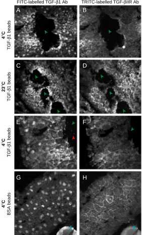
Fig. 4. In contrast to TGF-β1 internalization, the extent of the gradient is not significantly reduced at 4°C. Conjugates were prepared and cultured for 3 h at either 4°C or 23°C (green arrowheads indicates TGF-β1 bead spaces; blue arrowheads indicate BSA bead). (A) In tissue injected with 4 ng TGF-βIIR mRNA and cultured at 4°C, FITC fluorescence due to TGF-β1 can be observed at a distance from the beads. (B) The localization of TGF-βIIR protein in the section shown in (A). (C) The extent of the gradient when tissue containing TGF-βIIR and TGF-β1 is cultured at 23°C (D) Image showing the localization of TGF-βIIR protein in the section shown in (C). (E) High magnification image of the section shown in (A). A few random specks of fluorescence can be seen both on the tissue and in the bead space (indicated by the red arrowhead); as these also occur on the surrounding slide it is likely that they are artefacts. (F) High magnification image of the section shown in (B). (G) No fluorescence due to TGF-β1 is seen when tissue containing a BSA bead (blue arrowhead) and TGF-βIIR is cultured at 4°C. (H) The localization of TGF-βIIR in the section shown in (G).
FITC-labelled TGF-β1 Ab TRITC-labelled TGF-βIIR Ab
In order to visualise a morphogen gradient, however, it has been necessary to resort to a model system using a ligand/receptor complex that is not endogenous to the cells of the early Xenopus embryo. The use of a model system such as this has both advantages and disadvantages. On the positive side, we were able to utilise a well-validated, high sensitivity detection method that has been used extensively to look at TGF-β1 in mammalian systems (Grainger et al., 1995a; Grainger et al., 1995b; Mosedale et al., 1996; Lawn et al., 1996; Reckless et al., 1997; Grainger et al., 1998). To date, we have been unsuccessful in our attempts to detect activin (which has been extensively studied in Xenopus embryos) either as an endogenous gradient in the embryo, or in a similar model system using activin-loaded beads. Several factors may account for this failure: firstly, there are relatively few antibod-ies available for activin and none of these may be of sufficient affinity. Secondly, although present in wild-type tissue, the activin receptor cannot be over-expressed to the same levels as
TGF-βIIR, possibly because activin is a more powerful mesoderm inducer than the TGF-βs. Injection of greater than 2 ng ActRIIB mRNA is toxic to the embryo, while our experiments were per-formed with 4 ng of TGF-βIIR mRNA. Since we demonstrated that no TGF-β1 gradient was detectable in the absence of TGF-βIIR receptors, it seems likely that the lower levels of ActRIIB necessary to avoid toxicity limited our ability to detect activin gradients using our methods.
However, since our experiments were performed using TGF-β1, it is not possible to be certain that other potentially more physiologi-cal TGF-β superfamily members behave in the same fashion. Although unlikely, it is also possible that the mechanism of gradient formation in our model system is substantially different from that which occurs in the undisrupted embryo. For example, dissection of the animal caps into culture might induce alterations in the tissue structure, although we obtained no evidence for such a change. In particular, we see no histological evidence for any disruption of the cell:cell interactions in our conjugates compared with whole em-bryos. In addition, at present it is unclear whether mesoderm induction, in vivo, involves short-range induction rather than the release of range signals. However, during gastrulation, long-range inhibitors are thought to diffuse from a localized source, the Spemann-Mangold Organizer, to create an activity gradient of a locally produced agonist. Since this agonist is soluble and extracel-lular, and not a transmembrane receptor, then establishing the mechanism of passage of the antagonists may prove technically difficult. The use of bead sources to generate model morphogen gradients has already provided powerful clues as to the mecha-nisms of tissue patterning in the vertebrate embryo, and it is likely that the results presented here will be similarly useful. In particular, the work we describe, while for technical reasons necessitating a model system, establishes that passage through the solid tissue of the early Xenopus embryo is possible by a passive process, most likely by diffusion through the extracellular milieu.
It is interesting to consider why the passage of TGF-β across Xenopus tissue may occur differently to the passage of Wg, Dpp and possibly Hh in Drosophila. Unlike the solid tissue of the Xenopus embryo, both the Drosophila embryonic epidermis and wing epithelium consist of a sheet of polarised tissue only one cell thick. It is unlikely that in such single cell sheets an extracellular diffusion mechanism could function; for example, in the wing disc there would be too much risk of the ligand diffusing into the void between the disc epithelium and overlying peripodial cells. The
polarisation of these cell sheets could also provide directionality to a mechanism of transcytosis or to long-range signalling by means of cell projections termed cytonemes (Ramirez-Weber and Kornberg, 1999). Since the tissue of the Xenopus embryo is solid and apparently unpolarized with respect to either the direction or rate of signalling (McDowell et al., 1997), extracellular diffusion could provide an efficient means of transporting ligands. Further-more, the speed with which the gradient is formed suggests that the underlying mechanisms responsible for its formation may be different in the Xenopus tissue. We can detect TGF-β1 at a distance of 150-200 µm in only 3 hours. In contrast, the Wg and Dpp gradients form over a period of 2 days, leading to the proposal that unhindered diffusion is unlikely to be responsible for Wg and Dpp transport (Lecuit and Cohen 1998).
Our observations provide no indication as to the precise mecha-nisms by which the ligand is transported. We have shown that the dominant process(es) are temperature independent, and therefore passive, and that high concentrations of ligand are not detected within the cells. The simplest mechanism consistent with these observations would be free diffusion. Alternatively, diffusion of a ligand to distant cells may be facilitated by the diffusion in the plasma membrane of cell surface glycosaminoglycans (GAGs), subsequent to their being bound by the ligand in question. This mechanism has been proposed for Hh and Wg, as an alternative to transcytosis, due to their close association with the plasma membrane and from data generated following the mutation of a Drosophila gene, tout-velu, which is involved in GAG synthesis (Bradley and Brown, 1990; Reichsman et al., 1996; Porter et al., 1996; Bellaiche et al., 1998; The et al., 1999). However, in contrast to Wg and Hh, both TGF-β1 and activin show no such association with the cell surface and are readily secreted from both blastula cells and oocytes (McDowell et al., 1997; Reilly and Melton, 1996). Thus, although we cannot rule out that the TGF-β ligands undergo a mechanism of facilitated diffusion mediated by GAGs, in contrast to Wg and Hh free diffusion of these ligands in the extracellular milieu is entirely feasible.
Whatever the details of the transport processes that are occur-ring, we can conclude that, at least under the experimental condi-tions of our model system, neither vesicular transcytosis nor any other temperature sensitive process is playing a major role in establishing the functional morphogen gradient we have observed.
Materials and Methods
Embryo injections, manipulations and bead preparation
Embryos were injected at the 4-cell stage in 1 x MBS, 4% Ficoll with 4 ng TGF-βIIR. Synthetic capped mRNA was produced using Mega-Script in vitro transcription kits (Ambion) and cap analogue (New England Biolabs). Animal caps were dissected at stage 8 and conjugates made as described in the text. Beads (Affi-gel blue, 100 mesh, Bio-Rad) were washed 4 times in Ca2+ /Mg2+
free 1 x MBS. TGF-β1 (R and D Systems) was diluted to 100 nM concentra-tion in 1 x MBS containing 0.1% BSA. Beads were then added to 10% final volume (Gurdon et al., 1994). Control beads were prepared similarly by incubation in 1 x MBS + 0.1% BSA in the absence of TGF-β1.
Immunofluorescence analysis
antibod-ies. Primary antibodies were detected using FITC (Jackson Immunoresearch; 703-095-155) and TRITC (Jackson Immunoresearch; 711-025-152) labelled secondary antibodies. Some of the images (but not the confocal images) were processed using Adobe Photoshop in order to improve contrast for presentation, but all analysis was performed on the raw image data collected by the Quantitative Immunofluorescence software (Improvision).
In situ hybridisation
In situ hybridisations, using either Xbra or Eomes antisense probes, were performed on sections as previously described (Lemaire et al., 1995).
125I TGF-β1 binding to cells
The following procedure is based on that described by Dyson and Gurdon (1998). Animal caps from uninjected embryos or embryos previ-ously injected with TGF-βIIR mRNA at the 4 cell stage were dissected at stage 8. Cells were dissociated in Buffer A (Ca2+ /Mg2+ free 1 x MBS
containing 0.1% BSA and 0.5 mM EDTA), then placed in polyhema-treated eppendorfs. Buffer A was also used throughout the procedure for the incubation and washing steps. Each eppendorf contained either TGF-βIIR– injected or wild-type cells from approximately seven animal caps. The cells were microfuged and then resuspended in Buffer A containing 200 pM 125
I-TGF-β1 (Amersham Life Sciences) (at either 4°Cor23°C) for approxi-mately 20 minutes, with gentle dispersal of the cells every few minutes. For this binding step, and all subsequent steps, half the samples were treated at 4°C and half at 23°C. The cells were then washed once at the respective temperatures, by dispersal and centrifugation in 1 ml of solution, in order to remove unbound ligand. They were then left 40 minutes to allow any internalization to occur. This was followed by two subsequent washes at either pH 2.5 or pH 8, and the radioactivity associated with the cells counted. For all samples, the background binding of 125I-TGF-β1 to
wild-type cells was subtracted from the radioactivity bound to the receptor-injected cells incubated under the same conditions. Each condition was analysed in duplicate in each of three separate experiments, and statistical analysis was performed on the normalised data from all three experiments.
Acknowledgements
We thank R. Derynck for providing the construct encoding TGF-βIIR. We especially thank Daniel St. Johnston for helpful discussions, David Mosedale for technical advice, Julia Kaltschmidt for help with confocal microscopy and Ken Ryan, Srinath Sampath, Oliver Grimm and Kazuya Shimizu for comments on the manuscript. This work was supported by the Cancer Research Campaign and The Wellcome Trust. N.McD. is supported by a Magdalene College Manifold Trust Research Fellowship and D.J.G. by the Royal Society.
References
BEJSOVEC, A. and WIESCHAUS, E. (1995). Signalling activities of the Drosophila wingless gene are separably mutable and appear to be transduced at the cell surface. Genetics 139: 309-320.
BELLAICHE, Y., THE, I. and PERRIMON, N. (1998). Tout-velu is a Drosophila homologue of the putative tumour suppressor EXT-1 and is needed for Hh
diffusion. Nature 394: 85-88.
BHUSHAN, A., LIN, H.Y., LODISH, H.F. and KINTNER, C.R. (1994). The transforming
growth factor β type 2 receptor can replace the activin type 2 receptor in inducing
mesoderm. Mol. Cell. Biol. 14: 4280-4285.
BRADLEY, R.S. and BROWN, A.M. (1990). The proto-oncogene int-1 encodes a secreted protein associated with the extracellular matrix. EMBO. J. 9: 1569-1575.
BURSHTEIN, A.I. (1996). Introduction to thermodynamics and kinetics of matter. John Wiley and Sons, Inc.
CARDIGAN, K.M., FISH, M.P., RULIFSON, E.J. and NUSSE, R. (1998). Wingless repression of Drosophila frizzled 2 expression shapes the wingless morphogen gradient in the wing. Cell 93: 767-777.
DYSON, S. and GURDON, J.B. (1998). The interpretation of position in a morphogen gradient as revealed by occupancy of activin receptors. Cell 93: 557-568.
DYSON, S. and GURDON, J.B. (1997). Activin signalling has a necessary function in Xenopus early development. Curr. Biol. 7: 81-84.
GONZALEZ, F., SWALES, L., BEJSOVEC, A., SKAER, H. and MARTINEZ-ARIAS, A. (1991). Secretion and movement of the Wingless protein in the epidermis of the Drosophila embryo. Mech. Dev. 35: 43-54.
GONZALEZ-GAITAN, M. and JACKLE, H. (1999). The range of spalt-activating Dpp signalling is reduced in endocytosis-defective Drosophila wing discs. Mech. Dev. 87: 143-151.
GRAINGER, D.J., METCALFE, J.C., GRACE, A.A. and MOSEDALE, D.E. (1998). Transforming growth factor-beta dynamically regulates vascular smooth muscle differentiation in vivo. J. Cell. Sci. 111: 2977-88.
GRAINGER, D.J., MOSEDALE, D.E., METCALFE, J.C., WEISSBERG, P.L. and KEMP, P.R. (1995a). Active and acid-activable TGF-beta in human sera, platelets and plasma. Clin. Chim. Acta 235: 11-31.
GRAINGER, D.J., WITCHELL, C.M. and METCALFE, J.C. (1995b). Tamoxifen elevates transforming growth factor-beta and suppresses diet-induced formation of lipid lesions in mouse aorta. Nat. Med. 1: 1067-73.
GURDON, J.B., DYSON, S. and St JOHNSON, D. (1998). Cells’ perception of position in a concentration gradient. Cell 95: 159-162.
GURDON, J.B., HARGER, P., MITCHELL, A. and LEMAIRE, P. (1994). Activin signalling and response to a morphogen gradient. Nature 371: 487-492.
HAYS, R., GIBORI, G.B. and BEJSOVEC, A. (1997). Wingless signalling generates pattern through two distinct mechanisms. Development 124: 3727-3736.
HEMMATI-BRIVANLOU, A. and MELTON, D.A. (1992). A truncated activin receptor inhibits mesodermal induction and formation of axial structures in Xenopus embryos. Nature 359: 609-614.JONES, C.M., ARMES, N. and SMITH, J.C.
(1996). Signalling by TGF-β family members; short-range effects of Xnr-2 and
BMP-4 contrast with long-range effects of activin. Curr. Biol. 6: 1468-147.
KALT, M.R. (1971). The relationship between cleavage and blastocoel formation in Xenopus laevis: light microscope observations. J. Embryol. Exp. Morphol. 26: 37-49.
KERSBERG, M. and WOLPERT, L (1998). Mechanisms for positional signalling by morphogen transport: a theoretical study. J. Theor. Biol. 191: 103-114.
KOENIG, J.A. and EDWARDSON, J.M. (1997). Endocytosis and recycling of G-protein coupled receptors. Trends Pharmocol. Sci. 18: 276-287.
LAWN, R.M., PEARLE, A.D., KUNZ, L.L., RUBIN, E.M., RECKLESS, J., METCALFE, J.C. and GRAINGER, D.J. Feedback mechanism of focal vascular lesion forma-tion in transgenic apolipoprotein(a) mice. Biol. Chem. 271: 31367-71.
LECUIT, T. and COHEN, S.M. (1998). Dpp receptor levels contribute to shaping the Dpp morphogen gradient in the Drosophila wing imaginal disc. Development 125: 4901-4907.
LEMAIRE, P., GARRETT, N. and GURDON, J.B. (1995). Expression cloning of Siamois, a Xenopus homeobox gene expressed in dorsal-vegetal cells of blastu-lae and able to induce a complete secondary axis. Cell 81: 85-94.
LOPEZ-CASILLAS, F., CHEIFETZ, S., DOODY, J., ANDRES, J.L., LANE, W.S. and MASSAGUE, J. (1991). Structure and expression of the membrane proteoglycan betaglycan, a component of the TGF-beta receptor system. Cell 67: 785-95.
MASSAGUE, J. and KELLY, B. (1986). Internalization of transforming growth factor-beta and its receptor in BALB/c 3T3 fibroblasts. J. Cell Physiol. 128: 216-22
MASSAGUE, J., ATTISANO, L. and WRANA, J.L. (1994). The TGF-β family and its
composite receptors. Trends Cell Biol. 4: 172-178.
McDOWELL, N. and GURDON, J.B. (1999). Activin as a morphogen in Xenopus mesoderm induction. Seminars Cell Dev. Biol. 10: 311-317.
McDOWELL, N., ZORN, A.M., CREASE, D.J. and GURDON, J.B. (1997). Activin has direct long-range signalling activity and can form a concentration gradient by diffusion. Curr. Biol. 7: 671-681.
MOLINE, M.M., SOUTHERN, C. and BEJSOVEC, A. (1999). Directionality of Wing-less protein transport influences epidermal patterning in the Drosophila embryo. Development 126: 4375-4384.
MOSEDALE, D.E., METCALFE, J.C. and GRAINGER, D.J. (1996). Optimization of immunofluorescence methods by quantitative image analysis. J. Histochem. and Cytochem. 44: 1043-1050.
NEUMANN, C. and COHEN, S. (1997). Morphogens and pattern formation. BioEssays 19: 721-729.
Drosophila. Seminars Cell Dev. Biol. 10: 303-309.
PORTER, J.A., YOUNG, K.E., BEACHY, P.A. (1996). Cholesterol modification of hedgehog signalling proteins in animal development. Science 274: 255-259.
RAMIREZ-WEBER, F-A. and KORNBERG, T.B. (1999). Cytonemes: Cellular proc-esses that project to the principal signalling center in Drosophila imaginal discs. Cell 97: 599-607.
RECKLESS, J., METCALFE, J.C. and GRAINGER, D.J. (1997). Tamoxifen de-creases cholesterol sevenfold and abolishes lipid lesion development in apolipoprotein E knockout mice. Circulation 95: 1542-8.
REICHSMAN, F., SMITH, L. and CUMBERLEDGE, S. (1996). Glycosaminoglycans can modulate extracellular localization of the wingless protein and promote signal transduction. J. Cell. Biol. 135: 819-827.
REILLY, K.M. and MELTON, D.A. (1996). Short-range signalling by candidate
morphogens of the TGFβ family and evidence for a relay mechanism of induction.
Cell 86: 743-754.
RYAN, K., GARRETT, N., MITCHELL, A. and GURDON, J.B. (1996) Eomesodermin,
a key early gene in Xenopus mesoderm differentiation. Cell 87: 989-1000.
SMITH, J.C., PRICE, B.M., GREEN, J.B.A., WEIGAL, D. and HERRMANN, B.G. (1991). Expression of a Xenopus homolog of brachury (T) is an immediate early response to mesoderm induction. Cell 67: 79-87.
TABATA, T. and KORNBERG, T. (1994) Hedgehog is a signalling protein with a key role in patterning Drosophila imaginal discs. Cell 76: 89-102.
THE, I., BELLAICHE, Y. and PERRIMON, N. (1999) Hedgehog movement is regu-lated through tout velu-dependent synthesis of a heparan sulfate proteoglycan. Molecular Cell 4: 633-639.
VAN DEN HEUVEL, M., NUSSE, R., JOHNSTON, P. and LAWRENCE, P.A. (1989). Distribution of the wingless gene product in Drosophila embryos: a protein involved in cell-cell communication. Cell 59: 739-749.
WOLPERT, L. (1969). Positional information and the spatial pattern of cellular
differentiation. J. Theor. Biol. 25: 1-47.
![Fig. 1. Double immunofluorescence staining forTGF-previously injected with 4 ng of TGF-spaces (see text)]](https://thumb-us.123doks.com/thumbv2/123dok_us/1023231.1127097/2.609.42.393.83.551/fig-double-immunofluorescence-staining-fortgf-previously-injected-spaces.webp)