Differentiation of mouse primordial germ cells into female or
male germ cells
NORIO NAKATSUJI* and SHINICHIRO CHUMA
Department of Development and Differentiation, Institute for Frontier Medical Sciences, Kyoto University, Japan
ABSTRACT Mouse primordial germ cells (PGCs) migrate from the base of the allantois to the genital ridge. They proliferate both during migration and after their arrival, until initiation of the sex-differentiation of fetal gonads. Then, PGCs enter into the prophase of the first meiotic division in the ovary to become oocytes, while those in the testis become mitotically arrested to become prospermatogonia. Growth regulation of mouse PGCs has been studied by culturing them on feeder cells. They show a limited period of proliferation in vitro and go into growth arrest, which is in good correlation with their developmental changes in vivo. However, in the presence of multiple growth signals, PGCs can restart rapid proliferation and transform into pluripotent embryonic germ (EG) cells. Observation of ectopic germ cells and studies of reaggregate cultures suggested that both male and female PGCs show cell-autonomous entry into meiosis and differentiation into oocytes if they were set apart from the male gonadal environments. Recently, we developed a two-dimensional dispersed culture system in which we can examine transition from the mitotic PGCs into the leptotene stage of the first meiotic division. Such entry into meiosis seems to be programmed in PGCs before reaching the genital ridges and unless it is inhibited by putative signals from the testicular somatic cells.
KEY WORDS:
primordial germ cells, oocytes, meiosis, sex-differentiation, EG cells.
0214-6282/2001/$25.00
© UBC Press Printed in Spain www.ijdb.ehu.es
*Address correspondence to: Prof. Norio Nakatsuji. Department of Development and Differentiation, Institute for Frontier Medical Sciences, Kyoto University, 53 Kawaracho, Shogoin, Sakyo-ku, Kyoto 606, Japan. FAX: +81-75-751-3890. e-mail: nnakatsu@frontier.kyoto-u.ac.jp
Abbreviations used in this paper: dpc, days post coitum; EG, embryonic germ; LIF, leukemia inhibitory factor; PGC, primordial germ cells.
Introduction
Mouse primordial germ cells (PGCs) are first recognizable in the posterior region of the extraembryonic mesoderm by their surface alkaline phosphatase activity at around 7.25 days post coitum (dpc) (Chiquoine, 1954; Ginsburg et al., 1990). They are localized at the base of allantois and surrounded by the extraembryonic mesoderm cells and the basal surface of the visceral endoderm layer. Prior to the first identification of PGCs, it is known that their precursor cells originate in the proximal part of the epiblast (Tam and Zhou, 1996) near the extraembryonic tissues. They are supposed to involute through the primitive streak and migrate posteriorly toward the allantois. It is still under investigation how epiblast cells are determined to become PGCs. A recent study indicates that BMP-4 from the extraembryonic region is required for the generation of PGCs from proximal epiblast (Lawson et al., 1999). It may be possible that they are also influenced by a local signal from the surrounding extraembryonic mesoderm cells or visceral endoderm layer at the base of allan-tois.
From around 8.5 dpc, PGCs migrate into the embryonic meso-derm, through the hindgut endoderm and along the dorsal
mesen-tery, and finally into the genital ridge at 10.5 - 11.5 dpc. PGCs continue to proliferate mitotically during the migration and after arrival at the genital ridge until 12.5 - 13.5 dpc. At 12.5 dpc, developing gonads show the first signs of sexual dimorphism with testicular cord formation in the males. PGCs at this stage take different fates according to the sex of embryos. They enter into the prophase of the first meiotic division in the ovary to become oocytes, while those in the male gonads are mitotically arrested to become prospermatogonia (Hilscher et al., 1974). Previous stud-ies have indicated that the sex-differentiation of the germ cells is directed by the sex of the gonadal somatic cells and not by that of the germ cells themselves (reviewed in McLaren, 1994).
for the survival of PGCs in vitro as well as in vivo (Dolci et al., 1991; Godin et al., 1991). Leukemia inhibitory factor (LIF) (De Felici and Dolci, 1991; Matsui et al., 1991) and oncostatin M (OSM) (Hara et al., 1998), both of which belong to the interleukin-6 (IL-6) cytokine family, were shown to promote the growth and/or survival of PGCs in vitro. Signaling from their receptor subunit gp130 was essential for the derivation of pluripotential embryonic germ (EG) cells from PGCs in vitro (Koshimizu et al., 1996).
Observation of ectopic germ cells in the mesonephroi or adrenal cortex (Zamboni and Upadhyay, 1983; Francavilla and Zamboni, 1985) suggested that both male and female PGCs show cell-autonomous entry into meiosis if they were set apart from the male gonadal environments. Studies of reaggregated cultures of embryonic gonads (McLaren and Southee, 1997) also demonstrated that both female and male PGCs at 10.5-11.5 dpc, shortly after arriving at genital ridges, enter into meiosis when they were surrounded by lung somatic cells. Compared with studies of growth regulation, however, relatively little is known about the regulative mechanisms that are involved in the differen-tiation of PGCs into oocytes or prospermatogonia in fetal gonads. In this review, we describe development and differentiation of fetal germ cells by focusing on our studies related to the differen-tiation from PGCs into female or male germ cells (Fig. 1). Dr. Anne McLaren has been always the leading person in this field, and we would like to acknowledge that we have received numerous suggestions and inspiration from her.
Cell-autonomous growth regulation in PGCs
Our studies (Kawase et al., 1994; Ohkubo et al., 1996) and others (De Felici and McLaren, 1983; Donovan et al., 1986; Matsui et al., 1992) indicated that proliferation and growth arrest, as well as morphological changes of PGCs, are cell-autono-mously programmed, and they are well correlated with their differentiation in embryos at corresponding stages. PGCs ob-tained from embryos at 8.5 dpc increase in number for 3 days and then start to decrease on day 4, while 11.5 dpc PGCs stop proliferation immediately in vitro (Fig. 2). Thus, PGCs in vitro seem to mimic the pattern of proliferation and growth arrest in vivo.
To obtain more detailed information about the proliferation pattern of PGCs, single PGCs from 8.5 dpc embryos were cultured separately on feeder cells (Ohkubo et al., 1996). Counts of PGCs in each well showed weakly synchronized cell division for
4 days, followed by a decrease in number on day 5. It was noticed that PGCs in culture change their morphology according to their age and culture period. We divided the morphology of PGCs into three categories and counted each number separately in the clonal culture of 8.5 dpc PGCs (Fig. 3). The first type, «polarized» shows polarization and characteristics of motile cells. The second type, «spread» is well spread but shows no polarization. The last type, «round» is rounded up on the feeder cells. Ratios of the three types of PGCs started to change on day 3. The polarized type decreased, while the spread and round types increased. Such change simulates the developmental progress in vivo when PGCs arrive at the genital ridge and differentiate into nonmotile germ cells. It is worth to note that the three types of PGCs appeared in single PGC colonies very frequently on day 3 and 4. These results are consistent with the hypothesis that they are stochastically regulated events but not strictly determined by the age or number of cell division.
Growth factors for PGCs
Several studies have shown that the number of PGCs in-creases rapidly from 30-50 cells at 7.5 dpc toward around 3,000 Fig. 1. Differentiation of PGCs into
female or male germ cells. PGCs pro-liferate during migration into fetal go-nads. In the ovary, they enter into meio-sis and become oocytes, which are ar-rested in the prophase of the first mei-otic division. In the testis, PGCs are mitotically arrested in the G1/G0 phase and become prospermatogonia, which later restart mitosis as spermatogonia and produce meiotic spermatocytes postnatally.
cells at 11.5 dpc (Snow et al., 1981; Tam and Snow, 1981; Ginsburg et al., 1990). They imply that PGCs proliferate at a cell cycle rate of 16 hours. Investigation of growth factors during this period has shown that SLF and LIF are both effective to support survival and proliferation of PGCs in culture (De Felici and Dolci, 1991; Dolci et al., 1991; Godin et al., 1991; Matsui et al., 1991). Receptors for SLF, c-kit, are expressed on PGCs (Matsui et al., 1990; Manova and Bachvarova, 1991). Even after addition of these growth factors to culture media, however, it is still not possible to obtain PGC proliferation at a rapid rate similar to that in vivo.
In search for other growth factors, we showed that tumor necrosis factor (TNF)-α stimulates proliferation of PGCs in culture (Kawase et al., 1994). Such effect of TNF-α was evident for PGCs at earlier stages during migration (7.5-8.5 dpc), but it becomes ineffective at later stages (10.5-12.5 dpc). Then, we found that retinoic acid acts as a growth activator of mouse PGCs in vitro (Koshimizu et al., 1995). Retinoic acid is known to induce differen-tiation of embryonic stem (ES) cells and thus inhibit proliferation of stem cells. However, unexpectedly, it increases growth of PGCs in culture and promotes derivation of EG cells from PGCs (Koshimizu et al., 1996).
We also found that the conditioned medium made with Buffalo rat liver cells (BRL-CM) promotes proliferation of PGCs obtained from embryos at 7.5 to 11.5 dpc (Kawase et al.,1996).It is known
that BRL cells secrete many growth factors and cytokines includ-ing soluble SLF (Zsebo et al.1990)and LIF (Smith et al.1988), both of which stimulate proliferation/survival of mouse PGCs. How-ever, BRL-CM was much more effective than combination of soluble SLF and LIF to the culture medium.Moreover,a combina-tion of BRL-CM,forskolin, and Sl/Sl4-m220 feeder cells that express the membrane-bound form of SLF stimulated rapid proliferation of PGCs at a rate similar to that in vivo (Kawase et al., 1996).
Role of gp130 signaling in PGCs
Effects of LIF and LIF-related cytokines on PGC growth in vitro were examined in detail (Koshimizu et al., 1996). Addition of LIF or OSM in culture prolonged survival of 10.5 dpc PGCs and slowed down the disappearance of PGCs isolated from 11.5 dpc genital ridges. In addition, the IL-6/sIL-6R complex retarded the depletion of PGCs effectively, confirming that these ligands acted on PGCs through gp130. The IL-6/sIL-6R complex induces homodimerization of gp130 and directly activates its downstrean signaling (Taga et al., 1989; Yasukawa et al., 1990; Yoshida et al., 1994; Nichols et al., 1994).
The functional contribution of gp130 to PGCs was further examined by using the neutralizing antibody against gp130 (Fig. 4). Its addition drastically inhibited the survival of PGCs isolated from fetal gonads. Effects on PGCs before arrival at genital ridges were less apparent. Thus, the functional contribution of gp130-mediated signaling seems to be more significant in the gonadal PGCs than the PGCs during migration.
De-regulation of growth arrest in PGCs and derivation
of EG cells
EG cells were first reported to appear from the culture of PGCs in the presence of LIF and bFGF (Matsui et al., 1992; Resnick et al., 1992). We unexpectedly found that combination of LIF with forskolin or retinoic acid (RA) also causes the derivation of EG cells in the absence of bFGF (Koshimizu et al., 1996). They were morphologi-cally indistinguishable from EG cells produced by combination of LIF and bFGF.
The number of colonies in the presence of LIF plus forskolin or LIF plus RA was higher than that in LIF plus bFGF. In addition, a further increase of the colony number was observed in combination Fig. 3. Morphological change of PGCs in culture. Changing ratios of the
three types of PGCs in the clonal culture of PGCs obtained from 8.5 dpc embryos (Ohkubo et al., 1996).
Fig. 4. Effects of anti-c-KIT or anti-gp-130 neutral-izing antibodies on PGC growth in culture. PGCs obtained from 11.5 dpc (A) or 8.5 dpc (B) embryos were cultured on feeder cells for 2 days in the absence (-) or presence (+) of 103 U/ml LIF (Koshimizu
of three factors, namely, LIF, bFGF, and forskolin (or RA). These results indicated that bFGF and forskolin (or RA) activate inde-pendent signaling pathways and that they can work additively for the EG cell formation. However, ligands for gp130 was always indispensable for the appearance of EG cell colonies (Koshimizu et al., 1996). We established several EG cell lines under this culture condition (Fig. 5). They produced cystic embryoid bodies and also differentiated into teratoma by transplantation into mouse testes (Chuma and Nakatsuji, unpublished results).
Thus, a combination of multiple signals for cell growth causes a small population of PGCs to escape from the growth arrest and re-initiate rapid proliferation. However, such EG cells seem to have lost many characteristics of germ cells and de-differentiate into pluripotent stem cells akin to the ES cells. Derivation of such EG cells is possible from the migratory PGCs and also from gonadal PGCs up to 12.5 dpc. It is interesting that EG cells derived from the gonadal germ cells showed the erased status of the genomic imprinting (Labosky et al., 1994; Tada et al., 1998). Thus, such EG cell lines possess both the important characteris-tics of the fetal germ cells and also the pluripotency to produce chimeras.
Cell-autonomous programming for transition into meiosis
Observation of ectopic germ cells (Zamboni and Upadhyay, 1983; Francavilla and Zamboni, 1985) and studies of reaggregated cul-tures of embryonic gonads (McLaren and Southee, 1997) suggested that both male and female PGCs show cell-autonomous entry into
meiosis if they are set apart from the male gonadal environments. However, precise examination of the meiotic transition and identifi-cation of its regulative factors have been impeded by the lack of a dispersed culture system for fetal germ cells, in which the meiotic transition could be detected and analyzed in detail.
Female PGCs cease mitosis and enter into the prophase of the first meiotic division at around 13.5 dpc, progressing through the preleptotene, leptotene, zygotene, pachytene, diplotene stages and to diakinesis before arresting after birth (Borum, 1961; Speed, 1982). At the leptotene stage, each pair of sister chromatids forms a meiosis-specific longitudinal axial core to which the chromatin loops are attached. The Scp3 (Cor1/Sycp3) protein is one of the components of these axial cores (Heyting et al., 1988; Dobson et al., 1994; Klink et al., 1997). Scp3 is a good marker to detect the meiotic transition, since its expression is highly specific for meiosis and it functions from the initial step of the first meiotic division (Yuan et al., 2000). Another good marker for meiosis is Dmc1 that is a meiosis-specific mammalian RecA homologue (Yoshida et al., 1998). We tried to devise a culture system of PGCs, in which patterns of meiotic transition and its regulative factors could be examined precisely (Chuma and Nakatsuji, 2001).
In the female gonads, expression of both Scp3 and Dmc1 were detected by RT-PCR from 12.5 dpc. Unexpectedly, male gonads also showed the expression of both genes at the same stages at lower levels. Using a rabbit polyclonal antibody that we raised against a synthetic polypeptide of the mouse Scp3 protein (Klink et al., 1997), Scp3 was detected inside the nuclei of germ cells in the 12.5 dpc ovary. Then, formation of axial cores became visible as Fig. 5. EG cells derived from
PGC culture. (A) EG cell colo-nies obtained by culturing PGCs in the presence of LIF and forskolin or retinoic acid. (B) A histological section of the ter-atoma formed by transplanta-tion of EG cells into the adult testis of W mutant mice.
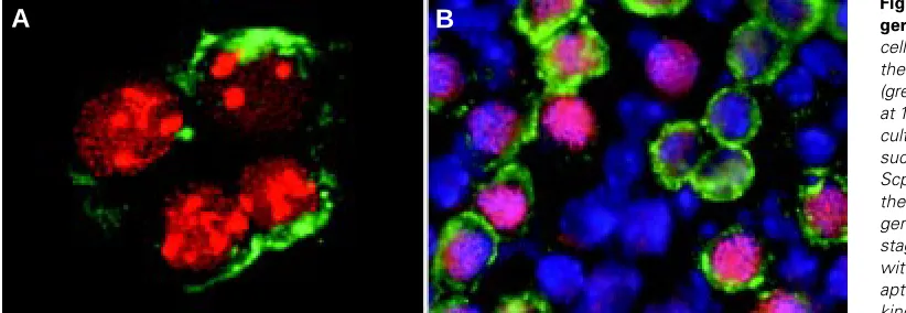
Fig. 6. Entry into meiosis by fetal germ cells in vitro. (A) Meiotic germ cells in dispersed culture, stained with the anti-Scp3 (red) and anti-SSEA-1 (green) antibody. Female genital ridges at 11.5 dpc had been dissociated and cultured for 3 days on feeder cells. In such culture, PGCs expressed the Scp3 protein and later began to form the axial core structures. (B) Meiotic germ cells at the zygotene-pachytene stage in the fragment culture, stained with the antibody raised against syn-aptonemal complex proteins (red, kindly provided by Dr. Moens), anti-SSEA-1 antibody (green) and with Hoechst 33258 dye (blue). Fragments of the 11.5 dpc female genital ridge had been cultured for 4 days.
B
A
fine fibrous structures inside nuclei at 13.5 dpc. In the male, no signal was observed at 12.5 dpc, but fetal testes at 13.5 - 15.5 dpc contained germ cells that showed weak Scp3 staining, as also reported by Di Carlo et al. (2000).
We next examined the expression of both genes by female PGCs in dispersed culture. Female urogenital ridges at 10.5 dpc were dissociated and cultured on Sl/Sl4 m220 feeder cells. RT-PCR products from both Scp3 and Dmc1 genes increased after 3 days of culture. Immunostaining confirmed that female PGCs obtained from urogenital ridges or genital ridges at 10.5-11.5 dpc started to express the Scp3 protein and later began to form axial cores (Fig. 6A), indicating that female PGCs entered into the leptotene stage of the first meiotic division after dissociation and cultivation on feeder cells. In addition, not only the PGCs isolated from the fetal gonad, migratory PGCs obtained from mesenteries at 10.5 dpc also expressed Scp3 and some of them formed axial cores after cultivation for several days. This observation supports the hypothesis that PGCs acquire meiotic competence before reaching the gonadal environment. (Chuma and Nakatsuji, 2001). We seldom observed progression of meiosis to the zygotene-pachytene stages in this dispersed culture condition. Since germ cells at these progressed stages were frequently observed in fragmented or reaggregated cultures of female genital ridges (Fig. 6B), further progression of meiosis probably require three-dimen-sional structures with surrounding somatic and supportive cells.
Previous studies have shown that male PGCs also enter into meiosis if they had been isolated at 10.5 or 11.5 dpc and cultured in reaggregates in the absence of male gonadal environments (McLaren and Southee, 1997). Consistent results were obtained that male germ cells at 10.5 or 11.5 dpc formed axial cores when dissociated and cultured on feeder cells for several days, but those
from the 12.5 or 13.5 dpc testes showed only weak expression of the Scp3 protein. Thus, an irreversible determination for the sex-differentiation of fetal germ cells seems to occur at around 12.5 dpc. The unexpected expression of these genes in male gonads in vivo also raises the possibility that male PGCs may start to prepare for meiosis until around 12.5 dpc but the male somatic cells produce intercellular signals such as putative meiosis-preventing substances (MPS) (Dolci and De Felici, 1990) to stop meiosis.
Signaling mechanisms involved in the sex
differentia-tion of fetal germ cells
At 12.5 dpc, genital ridges show the first signs of sexual dimorphism with testicular cord formation in the males. During this period, several genes, such as Sry, Sox9, Dax-1, WT1 and Ad4BP/ SF-1, were shown to play important roles in the differentiation of somatic cells (reviewed in Swain and Lovell-Badge, 1999). How-ever, there is little information about the signaling molecules involved in the somatic-germ cell interaction that directs the sex-differentiation of germ cells.
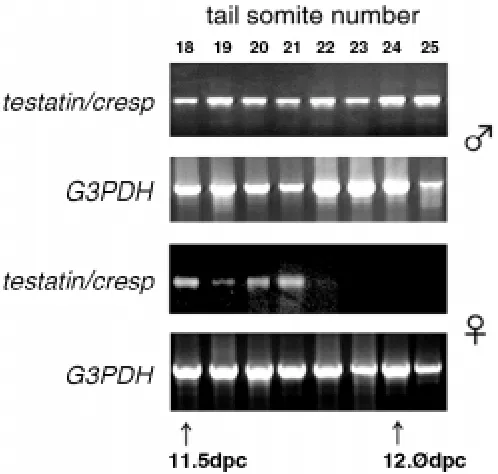
We attempted to isolate genes involved in the sex-differentiation of fetal germ cells by using subtraction and differential screening between male and female fetal gonads at 13.5 dpc (Tamura and Nakatsuji, unpublished results). One of novel genes found in the screening showed specific expression in the testis from the early stages of sex-differentiation (Kanno et al., 1999). It was related to type II cysteine proteinase inhibitors, the cystatin family, and it had the highest similarity with the CRES (cystatin-related epididymal specific) gene (Barrett et al., 1986; Cornwall et al., 1992). After we named it cresp, a paper reporting the same gene named testatin, was also published by another laboratory (Töhönen et al., 1998). The cystatins are present in most tissues and biological fluids (Abrahamson et al., 1986; Tavera et al., 1990). In the testis, the cysteine protease cathepsin L (Elicson-Lawrence et al., 1991) and its inhibitor cystatin C (Tavera et al., 1990) are secreted from Sertoli cells. Expression of the testatin/cresp mRNA was almost confined exclusively to the male gonad and the expression increased immediately after the initiation of testis differentiation at 11.5 - 12.5 dpc, while it decreased to an undetectable level in the female during the same period (Kanno et al., 1999) (Fig. 7). Töhönen et al. (1998) reported the expression of testatin/cresp in Sertoli cells and its precursors from 11.5 dpc, and they suggested that this gene is located downstream of Sry or Sox9 during early events of the sex-differentiation in testicular somatic cells. In contrast, our results of Fig. 7. RT-PCR analysis of testatin/cresp. Stages of the development of
embryos from 11.5 to 12.5 dpc were determined by counting the tail somites. Expression in the female gonad decreased to an undetectable level in the middle of these stages (Kanno et al., 1999). An embryo with 18 tail somites corresponds to the 11.5 dpc, and 24 somites to 12.0 dpc.
TABLE 1
CHANGES IN THE NUMBER OF PGCs AFTER TRANSFECTION OF
SV40LT, E1B 19K, BCL-2 OR BCL-XL IN CULTURE
(Watanabe et al., 1997)
Expression vector Relative changes in PGC number (%)#
pSV-LT 100.5 ± 7.6 (n=15) P=0.49
pEFBOS-E1B19k 184.9 ± 17.2 (n=18) P<0.001 pCAGGS-hbcl-2 202.3 ± 25.0 (n= 4) P<0.001 pCAGGS-c bcl-xL 195.1 ± 24.0 (n=11) P<0.001
#The number of PGCs in the pBSSK-transfected well was used as the control (100%).
in situ hybridization and immunohistochemical analyses demon-strated that the testatin/cresp mRNA was localized both in the germ and Sertoli cells in the fetal and adult testes (Fig. 8). (Kanno et al., 1999). Germ cells take different developmental pathways depend-ing on the sex of the gonadal somatic cells. The upregulation of testatin/cresp expression in the male gonad and its downregulation in the female takes place immediately before such sex-differentia-tion of the germ cells, thus suggesting its important funcsex-differentia-tion.
Regulation of apoptosis in fetal germ cells
Another aspect of germ cell development is regulation of apoptosis. It is well known that a large population of germ cells goes into apoptosis during development of female and male gonads, although its biological significance is unknown. PGCs obtained from the genital ridge undergo apoptosis in culture (Pesce et al., 1993) and those in the extragonadal sites and within developing gonads show the hallmarks of apoptosis (Coucouvanis et al., 1993).
Enforced expression of the adenovirus E1B 19K gene in cul-tured somatic cells can block apoptosis induced by various agents such as TNF-α and Fas antigen (Gooding et al., 1991; Hashimoto
et al., 1991; White et al., 1992). Also, the overexpression of bcl-2 or bcl-XL gene can prevent apoptosis caused by various stimuli (Vaux et al., 1988; Hockenbery et al., 1990; Nunez et al., 1990). Therefore, we tested if the forced expression of E1B 19K, bcl-2, or bcl-XL prevents the apoptosis of PGCs in culture (Watanabe et al., 1997). The transient expression of these genes in PGCs obtained Fig. 8. Expression of testatin/cresp in a 12.5 dpc testis. A section of a
male gonad at 12.5 dpc was double-stained with digoxigenin-labeled testatin/cresp cRNA and anti-SSEA-1 monoclonal antibody and FITC-labeled second antibody, counter stained with methylgreen (Kanno et al., 1999). Photographs of (A) bright-field microscopy, (C) fluorescence microscopy and (B) composite images, are shown. Arrows indicate germ cells labeled with the anti-SSEA-1 antibody but not with the hybridization signals. Black arrowheads indicate the double-labeled germ cells. In the insets showing magnified views, arrows, black arrowheads and white arrowheads indicate the germ cells labeled only with SSEA-1, the double-labeled germ cells, and somatic cells double-labeled only with the hybridization signals, respectively. A scale bar indicates 50 µm.
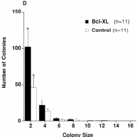
Fig. 9. Effects of the forced expression of bcl-xL in PGCs. Histogram showing the distribution of the colony size of PGCs transfected with pCAGGS-cbcl-xL or pBSSK as control (Watanabe et al., 1997). PGCs obtained from 11.5 dpc embryos were cultured for 1 day, transfected and cultured for an additional 2 days.
A
B
from the genital ridge at 11.5 dpc resulted in a remarkable increase of the surviving PGCs in culture (Table 1). Such effects of E1B 19K and bcl-xL were more pronounced in case of the round-shaped PGCs that were present as single or pairs of cells, than the polarized or spread type PGCs that made larger colonies (Fig. 9). Thus, the disappearance of the rounded PGCs that represent more advanced stages can be more effectively inhibited by the expres-sion of these apoptosis inhibiting genes.
In good accordance with these results, a recent study using mice with hypomorphic mutation (Rucker et al., 2000) of the bcl-x gene reported a large loss of germ cells in the fetal gonad and adult testis and ovary. Our study (Kasai et al., unpublished results) also indicates a decrease of spermatogenic cells in the testis when gene dosage was halved in the heterozygotes for the bcl-x null mutation (Motoyama et al., 1995).
As described in this review, many aspects of molecular mecha-nisms that regulate differentiation into female or male germ cells still remain unknown. Utilization of in vitro culture systems would facilitate such studies. Intracellular and intercellular signaling in the regulation of sex-differentiation of germ cells into oocytes or prospermatogonia remains to be one of interesting and unsolved problems of the mammalian germ-line development.
References
ABRAHAMSON, M., SALVESEN, G., BARRETT, A. J. and GRUBB, A. (1986). Isolation of six cysteine proteinase inhibitors from human urine. J. Biol. Chem. 261: 11282-11289.
BARRETT, A. J., FRITZ, H., GRUBB, A., ISEMURA, S., JARVINEN, M., KATUNUMA, N., MACHLEIDT, W., MULLER-ESTERI, W., SASKI, M. and TURK, V. (1986). Nomenclature and classification of the proteins homologous with the cysteine proteinase inhibitor cystatin. Biochem. J. 236: 312-317.
BORUM, K. (1961). Oogenesis in the mouse. A study of the meiotic prophase. Exp. Cell. Res. 24: 495-507.
CHIQUOINE, A. D. (1954). The identification, origin, and migration of the primordial germ cells of the mouse embryo. Anat. Rec. 118: 135-146.
CHUMA, S.and NAKATSUJI, N. (2001). Autonomous transition into meiosis of mouse fetal germ cells in vitro and its inhibition by gp130-mediated signaling. Dev. Biol. 229: 468-479.
CORNWALL, G. A., ORGEBIN-CRIST, M.-C. and HANN, S. R. (1992). The CRES gene: a unique testis-regulated gene related to cystatin family is highly restricted in its expression to the proximal region of the mouse epididymis. Mol. Endocrinol. 6: 1653-1664.
COUCOUVANIS, E. C., SHERWOOD, S. W., CARSWELL-CRUMPTON, C., SPACK, E. G. and JONES, P. P. (1993). Evidence that the mechanism of prenatal germ cell death in the mouse is apoptosis. Exp. Cell Res. 209: 238-247.
DE FELICI, M. and MCLAREN, A. (1983). In vitro culture of mouse primordial germ cells. Exp. Cell. Res. 144: 417-27.
DE FELICI, M. and DOLCI, S. (1991). Leukemia inhibitory factor sustains the survival of mouse primordial germ cells cultured on TM4 feeder layers. Dev. Biol. 147: 281-284.
DI CARLO, A.D., TRAVIA, G. and DE FELICI, M. (2000). The meiotic specific synaptonemal complex protein SCP3 is expressed by female and male primordial germ cells of the mouse embryo. Int. J. Dev. Biol. 44: 241-244.
DOBSON, M. J., PEARLMAN, R. E., KARAISKAKIS, A., SPYROPOULOS, B. and MOENS, P. B. (1994). Synaptonemal complex proteins: occurrence, epitope mapping and chromosome disjunction. J. Cell Sci 107: 2749-2760.
DOLCI, S., WILLIAMS, D. E., ERNST, M. K., RESNICK, J. L., BRANNAN, C. I., LOCK, L. F., LYMAN, S. D., BOSWELL, H. S. and DONOVAN, P. J. (1991). Requirement for mast cell growth factor for primordial germ cell survival in culture. Nature 352: 809-811.
DOLCI, S. and DE FELICI, M. (1990). A study of meiosis in chimeric mouse fetal gonads. Development 109: 37-40.
DONOVAN, P. J., STOTT, D., CAIRNS, L. A., HEASMAN, J. and WYLIE, C. C. (1986). Migratory and postmigratory mouse primordial germ cells behave differently in culture. Cell 44: 831-838.
ELICSON-LAWRENCE, M., ZABLUDOFF, S. and WRIGHT, W. W. (1991). Cyclic protein 2, a secretory product of rat Sertoli cells, is the proenzyme form of cathepsin L. Mol. Endocrinol. 5: 1789-1798.
FRANCAVILLA, S. and ZAMBONI, L. (1985). Differentiation of mouse ectopic germinal cells in intra- and perigonadal locations. J. Exp. Zool. 233: 101-109.
GINSBURG, M., SNOW, M. H. and MCLAREN, A. (1990). Primordial germ cells in the mouse embryo during gastrulation. Development 110: 521-528.
GODIN, I., DEED, R., COOKE, J., ZSEBO, K., DEXTER, M. and WYLIE, C. C. (1991). Effects of the steel gene product on mouse primordial germ cells in culture. Nature 352: 807-809.
GOODING, L. R., AQUINI, L., DUERLSEN-HUGHES, P. J., DAY, D., HORTON, T. M., YEI, S. and WOLD, W. S. M. (1991) The E1B 19,000-molecular-weight protein of group C adenoviruses prevents tumor necrosis factor cytolysis of human cells but not of mouse cells. J. Virol. 65: 3083-3094.
HARA, T., TAMURA, K., DE MIGUEL, M. P., MUKOUYAMA, Y., KIM, H. J., KOGO, H., DONOVAN, P. J. and MIYAJIMA, A. (1998). Distinct roles of oncostatin M and leukemia inhibitory factor in the development of primordial germ cells and Sertoli cells in mice. Dev. Biol. 201: 144-153.
HASHIMOTO, S., ISHII, A. and YONEHARA, S. (1991) The E1b oncogene of adenovirus confers cellular resistance to cytotoxicity of tumor necrosis factor and monoclonal anti-Fas antibody. Int. Immunol. 3: 343-351.
HEYTING, C., DETTMERS, R. J., DIETRICH, A. J., REDEKER, E. J. and VINK, A. C. (1988). Two major components of synaptonemal complexes are specific for meiotic prophase nuclei. Chromosoma 96: 325-332.
HILSCHER, B., HILSCHER, W., BULTHOFF-OHNOLZ, B., KRAMER, U., BIRKE, A., PELZER, H. and GAUSS, G. (1974). Kinetics of gametogenesis. I. Comparative histological and autoradiographic studies of oocytes and transitional prospermatogonia during oogenesis and prespermatogenesis. Cell Tissue Res. 154: 443-470.
HOCKENBERY, D., NUNEZ, G., MILLIMAN, C., SCHREIBER, R. D. and KORSMEYER, S. J. (1990) Bcl-2 is an inner mitochondrial membrane protein that blocks programmed cell death. Nature 348: 334-336.
KANNO, Y., TAMURA, M., CHUMA, S., SAKURAI, T., MACHIDA, T. and NAKATSUJI, N. (1999). A cystatin-related gene, testatin/cresp, shows male-specific expression in germ and somatic cells from the initial stage of murine gonadal sex-differentia-tion. Int. J. Dev. Biol. 43: 777-784.
KAWASE, E., SHIRAYOSHI, Y., HASHIMOTO, K. and NAKATSUJI, N. (1996). A combination of buffalo rat liver cell-conditioned medium, forskolin and membrane-bound stem cell factor stimulates rapid proliferation of mouse primordial germ cells in vitro similar to that in vivo. Devel. Growth Differ. 38: 315-322.
KAWASE, E., YAMAMOTO, H., HASHIMOTO, K. and NAKATSUJI, N. (1994). Tumor necrosis factor-β (TNF-β) stimulates proliferation of mouse primordial germ cells in culture. Dev Biol 161: 91-95.
KLINK, A., LEE, M. and COOKE, H. J. (1997). The mouse synaptonemal complex protein gene Sycp3 maps to band C of chromosome 10. Mamm. Genome 8: 376-377.
KOSHIMIZU, U., TAGA, T., WATANABE, M., SAITO, M., SHIRAYOSHI, Y., KISHIMOTO, T. and NAKATSUJI, N. (1996). Functional requirement of gp130-mediated signaling for growth and survival of mouse primordial germ cells in vitro and derivation of embryonic germ (EG) cells. Development 122: 1235-1242.
KOSHIMIZU, U., WATANABE, M. and NAKATSUJI, N. (1995). Retinoic acid is a potent growth activator of mouse primordial germ cells in vitro. Dev. Biol. 168: 683-685.
LABOSKY, P. A., BARLOW, D. P. and HOGAN, B. L. M. (1994). Mouse embryonic germ (EG) cell lines: transmission through the germline and differences in the methylation imprint of insulin-like growth factor 2 receptor (Igf2r) gene compared with embryonic stem (ES) cell lines. Development 120: 3197-3204.
LAWSON, K. A., DUNN, N. R., ROELEN, B. A., ZEINSTRA, L. M., DAVIS, A. M., WRIGHT, C. V., KORVING, J. P. and HOGAN, B. L. M. (1999). Bmp4 is required for the generation of primordial germ cells in the mouse embryo. Genes Dev 13: 424-436.
MATSUI, Y., ZSEBO, K. and HOGAN, B. L. M. (1992). Derivation of pluripotential embryonic stem cells from murine primordial germ cells in culture. Cell 70: 841-847.
MATSUI, Y., TOKSOZ, D., NISHIKAWA, S., NISHIKAWA, S., WILLIAMS, D., ZSEBO, K. and HOGAN, B. L. M. (1991). Effect of Steel factor and leukemia inhibitory factor on murine primordial germ cells in culture. Nature 353: 750-752.
MATSUI, Y., ZSEBO, K. M. and HOGAN, B. L. M. (1990). Embryonic expression of a haematopoietic growth factor encoded by the Sl locus and the ligand for c-kit. Nature 347: 667-669.
McLAREN, A. (1994). Germline and soma: interactions during early mouse develop-ment. Seminars in Dev. Biol. 5: 43-49.
McLAREN, A. and SOUTHEE, D. (1997). Entry of mouse embryonic germ cells into meiosis. Dev. Biol. 187: 107-113.
MOTOYAMA, N., WANG, F., ROTH, K. A., SAWA, H., NAKAYAMA, K., NAKAYAMA, K., NEGISHI, I., SENJU, S., ZHANG, Q., FUJII, S. and Loh, D. Y. (1995).Massive cell death of immature hematopoietic cells and neurons in Bcl-x-deficient mice. Science 267: 1506-1510.
NICHOLS, J., CHAMBERS, I. and SMITH, A. (1994). Derivation of germ line competent embryonic stem cells with a combination of interleukin-6 and soluble interleukin-6 receptor. Exp. Cell Res. 215: 237-239.
NUNEZ, G., LONDON, L., HOCKENBERY, D., ALEXANDER, M., MCKEARN, J. P. and KORSMEYER, S. J. (1990). Deregulated Bcl-2 gene expression selectively pro-longs survival of growth factor-deprived hemopoietic cell lines. J. Immunol. 144: 3602-3610.
OHKUBO, Y., SHIRAYOSHI, Y. and NAKATSUJI, N. (1996). Autonomous regulation of proliferation and growth arrest in mouse primordial germ cells studied by mixed and clonal cultures. Exp. Cell. Res. 222: 291-297.
PESCE, M., FARRACE, M. G., PIACENTINI, M., DOLCI, S., and DE FELICI, M. (1993). Stem cell factor and leukemia inhibitory factor promote primordial germ cell survival by suppressing programmed cell death (apoptosis).Development 118: 1089-1094.
RESNICK, J. L., BIXLER, L. S., CHENG, L. and DONOVAN, P. J. (1992). Long-term proliferation of mouse primordial germ cells in culture. Nature 359: 550-551.
RUCKER III, E. B., DIERISSEAU, P., WAGNER, K-U, GARRETT, L., WYNSHAW-BORIS, A., FLAWS, J. A. and HENNIGHAUSEN, L. (2000). Bcl-x and Bax regulate mouse primordial germ cell survival and apoptosis during embryogenesis. Molecu-lar Endocrinology 14: 1038-1052.
SMITH, A. G., HEATH, J. K., DONALDSON, D. D., WONG, G. G., MOREAU, J., STAHL, M. and ROGERS, D. (1988). Inhibition of pluripotential embryonic stem cell differentiation by purified polypeptides. Nature 336: 688-690.
SNOW, M. H.L.,TAM, P. P.L. and McLAREN,A.(1981). On the control and regulation of size and morphogenesis in mammalian embryos. In Levels of Genetic Control in Development (Eds. S. Subtleny and U. K. Abbott), pp. 201-217. Alan R. Liss, New York.
SPEED, R. M. (1982). Meiosis in the foetal mouse ovary. I. An analysis at the light microscopic level using surface-spreading. Chromosoma 85: 427-437.
SWAIN, A. and LOVELL-BADGE, R. (1999). Mammalian sex determination: a molecu-lar drama. Genes Dev. 13: 755-767.
TADA, T., TADA, M., HILTON, K., BARTON, S. C., SADO, T., TAKAGI, N. and SURANI, M. A. (1998). Epigenetic switching of imprintable loci in embryonic germ
cells. Dev. Genes Evol. 207: 551-561.
TAGA, T., HIBI, M., HIRATA, Y., YAMASAKI, K., YASUKAWA, K., MATSUDA, T., HIRANO, T. and KISHIMOTO, T. (1989). Interleukin-6 triggers the association of its receptor with a possible signal transducer, gp130. Cell 58: 573-581.
TAM,P. P. L. and SNOW, M. H. L. (1981). Proliferation and migration of primordial germ cells during compensatory growth inmouseembryos. J.Embryol. Exp. Morph. 64: 133-147.
TAM, P. P. L. and ZHOU, S. X. (1996). The allocation of epiblast cells to ectodermal and germ-line lineages is influenced by the position of the cells in the gastrulating mouse embryo. Dev. Biol. 178: 124-132.
TAVERA, C., PROVOT, D., GIROLAMI, J.P., LEUNG-TACK, J. and COLLE, A. (1990). Tissue and biological fluid distribution of cysteine proteinase inhibitor: rat cystatin C. Biol. Chem. Hoppe-Seyler. 371: 187-192.
TÖHÖNEN, V., _STERLUND, C. and NORDQVIST, K. (1998). Testatin: a cystatin-related gene expressed during early testis development. Proc. Natl. acad. Sci. USA. 95: 14208-14213.
VAUX, D. L., CORY, S. and ADAMS, J. M. (1988). Bcl-2 gene promotes haemopoietic cell survival and cooperates with c-myc to immortalize pre-B cells. Nature 335: 440-442.
WATANABE, M., SHIRAYOSHI, Y., KOSHIMIZU, U., HASHIMOTO, S., YONEHARA, S., EGUCHI, Y., TSUJIMOTO, Y. and NAKATSUJI, N. (1997). Gene transfection of mouse primordial germ cells in vitro and analysis of their survival and growth control. Exp. Cell Res. 230: 76-83.
WHITE. E., SABBATINI, P., DEBBAS, M., WOLD, W. S. M., KUSHER, D. I. and GOODING, L. (1992) The 19-kilodalton adenovirus E1B transforming protein inhibits programmed cell death and prevents cytolysis by tumor necrosis factor α. Mol. Cell Biol. 12: 2570-2580.
WYLIE, C. C. (1999). Germ cells. Cell 96: 165-174.
YASUKAWA, K., SAITO, T., FUKUNAGA, T., SEKIMORI, Y., KOISHIHARA, Y., FUKUI, H., OHSUGI, Y., MATSUDA, T., YAWATA, H., HIRANO, T., TAGA, T. and KISHIMOTO, T. (1990). Purification and characterization of soluble human IL-6 receptor expressed in CHO cells. J. Biochem. 108: 673-676.
YOSHIDA, K., CHAMBERS, I., NICHOLS, J., SMITH, A., SAITO, M., YASUKAWA, K., SNOYAB, M., TAGA, T. and KISHIMOTO, T. (1994). Maintenance of the pluripotential phenotype of embryonic stem cells through direct activation of gp130 signaling pathways. Mech. Dev. 45: 163-171.
YOSHIDA, K., KONDOH, G., MATSUDA, Y., HABU, T., NISHIMUNE, Y. and MORITA, T. (1998). The mouse RecA-like gene Dmc1 is required for homologous chromo-some synapsis during meiosis. Mol. Cell 1: 707-718.
YUAN, L., LIU, J. G., ZHAO, J., BRUNDELL, E., DANEHOLT, B. and HOOG, C. (2000). The murine SCP3 gene is required for synaptonemal complex assembly, chromo-some synapsis, and male fertility. Mol. Cell 5: 73-83.
ZAMBONI, L. and UPADHYAY, S. (1983). Germ cell differentiation in mouse adrenal glands. J. Exp. Zool. 228: 173-193.