Developmental gene network analysis
ROGER REVILLA-I-DOMINGO and ERIC H. DAVIDSON*
Division of Biology 156-29, California Institute of Technology, Pasadena, CA, USA
ABSTRACT The developmental process is controlled by the information processing functions executed by the cis-elements that regulate the expression of the participating genes. A model of the network of cis-regulatory interactions that underlies the specification of the endomesoderm of the sea urchin embryo is analyzed here. Although not all the relevant interactions have yet been uncovered, the model shows how the information processing functions executed by the cis-regulatory elements involved can control essential functions of the specification process, such as transforming the localization of maternal factors into a domain-specific program of gene expres-sion; refining the specification pattern; and stabilizing states of specification. The analysis suggests that the progressivity of the developmental process is also controlled by the cis-regulatory interactions unraveled by the network model. Given that evolution occurs by changing the program for development of the body plan, we illustrate the potential of developmental gene network analysis in understanding the process by which morphological features are maintained and diversify. Comparison of the network of cis-regulatory interactions with a portion of that underlying the specification of the endomesoderm of the starfish illustrates how the similarities and differ-ences provide insights into how the programs for development work and how they evolve.
KEY WORDS:
Gene network, genetic program, evolution and development, genomic regulatory system, sea urchin
0214-6282/2003/$25.00 © UBC Press
Printed in Spain www.ijdb.ehu.es
*Address correspondence to: Dr. Eric H. Davidson. Division of Biology 156-29, California Institute of Technology, Pasadena, CA 91125. Fax: +1-626-793-3047. e-mail: [email protected]
The genetic programs that control the processes by which the body plans of animals are built were invented, and shaped, by the evolutionary process. How these programs work, if nothing else, is a matter of great curiosity. Because gene networks constitute the control systems for development, analysis of such networks ex-plains both the process of development and the process by which development has evolved (Davidson, 2001).
Ultimately, development is the process by which the body plans of animals are laid down. Distinct cell types are produced in particular spatial domains, each with particular structural proper-ties given by the distinct programs of gene expression that the cells execute. Through the process of specification each domain in the embryo obtains its developmental identity. Once specified, each domain will run through a progression of states of regulatory gene expression, leading to the establishment and ultimately the stabi-lization of the terminal programs of gene expression that give each cell type its unique properties.
Spatial cues are always required in order to trigger specifica-tion in development. These spatial cues sometimes consist of localized maternal regulatory factors that are distributed to par-ticular cells with the egg cytoplasm, and are partitioned during cleavage. Alternatively they can also consist of signaling ligands produced by other cells, in consequence of their own prior state of specification. Ultimately, these spatial cues affect the course of
events in development by causing the activation (or repression), in a certain region of the embryo, of particular genes encoding transcription factors. Through this process, new, more refined, domains of specification are created, and the complexity of the embryo increases. But although it is the spatial cues that trigger the events of spatial specification, the locus of programmatic control for each developmental event is the sequence of the particular cis-regulatory elements that respond to the inputs presented (Davidson, 2001).
The experiments reviewed here represent a step towards the goal of determining the complete network of DNA-based inter-actions that underlie one particular major process of develop-ment, namely, the specification of the endomesoderm of the sea urchin embryo. Given that evolution occurs by changing the program for development of the body plan, we also illustrate briefly how developmental gene network analysis sheds light on the process by which morphological features are maintained and diversify.
Unraveling the gene regulatory network that underlies
the process of endomesoderm specification in the sea
urchin embryo
The armature of the network
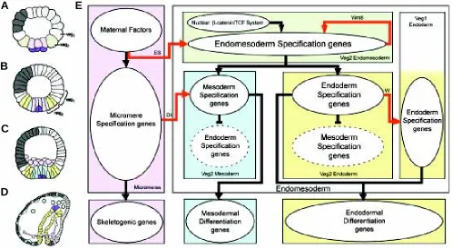
Figure 1 illustrates the process of endomesoderm specification in the sea urchin embryo (Fig. 1 A-D), and it shows a diagram (Fig. 1E) that describes the specification events and the genetic functions that underlie this process.
Ultimately, the endomesoderm consists of the endodermal gut, the skeletogenic mesenchyme and several other mesodermal cell types, including pigment cells (Fig. 1D).
By the seventh cleavage cycle (Fig. 1A), the cell lineages of typical sea urchin embryos have been segregated into a canonical set of territories, each of which is destined to give rise to certain distinct cell types (Hörstadius, 1939; Cameron et al., 1987, 1991), and in each of which a distinct set of genes is already running (reviewed by Davidson et al., 1998; Davidson, 2001). The upper or animal pole half of the embryo now consists of blastomeres that produce only the cell types ultimately found in the oral and aboral ectoderm. The lower half consists of the veg1 ring, their sister cells of the veg2 ring immediately below, and the large and small micromeres at the vegetal pole. In the undisturbed embryo, the large micromeres (the population of cells colored lavender in the diagram) will produce all the cells of the skeletogenic mesenchyme lineage, and the progeny of veg1 and veg2 will produce the rest of the endomesoderm. At the ciliated swimming-blastula stage (Fig. 1B), the veg2 lineage has been segregated into two distinct domains: the inner veg2 ring
Fig. 1.Endomesoderm specification in the sea urchin embryo.(A-D) Schematic diagrams of sea urchin embryos displaying specified domains, from Davidson et al. (2002b). The color coding shows the disposition of specified endomesoderm components: Lavender indicates skeletogenic lineage; dark purple indicates small micromere precursors of adult mesoderm; green indicates endomesoderm lineage that later gives rise to endoderm, yellow, and mesoderm, blue; light grey indicates oral ectoderm; dark grey indicates aboral ectoderm; white indicates regions yet to be specified at the stages shown. (A) 7th cleavage embryo (about 10 h after fertilization). (B) Blastula stage embryo at about 9th cleavage (about 15 h after fertilization). (C) Mesenchyme
blastula stage embryo (about 24 h after fertilization). (D) Late gastrula stage embryo (about 55 h after fertilization). The drawing shows the later disposition of all the endomesodermal cell types about midway through embryonic morphogenesis. (E) Process diagram describing endomesoderm specification events in the sea urchin embryo. Boxes represent domains of specification according to the color of their background. The color coding represents the same endomesoderm components as in the schematic diagrams A-D. Ovals in the boxes represent sets of genes that execute certain developmental functions. Arrows indicate that the set of genes in the oval where the arrow originates, triggers the developmental function executed by the genes in the oval where the arrow ends. In particular, red arrows represent signaling events. Barred lines indicate repression of the developmental function executed by the genes in the oval where the barred line ends. Developmental time in the process diagram runs from top to bottom in accordance with the stages represented by the schematic diagrams A-D. Abbreviations: ES, Early Signal; Dl, Delta; W, Wnt8. Evidence is reviewed in Davidson et al. (2002a), and from P. Oliveri, A. Ransick, D.R. McClay and E.H. Davidson, unpublished data.
A
B
C
D
consists of cells that will give rise to mesodermal cell types, including pigment cells; and the rest of the veg2 domain will give rise to endodermal cells (Ruffins and Ettensohn, 1993, 1996). At the mesenchyme blastula stage (Fig. 1C), the skeletogenic mesen-chyme cells have ingressed into the blastocoel, leaving behind a now fully specified central disc of prospective mesodermal cell types, and peripheral to them, the endodermal precursors (reviewed by Davidson
et al., 1998). After this, the adjacent veg1 progeny will become specified as endoderm as well (Logan and McClay, 1997), and gastrular invagination ensues.
The mechanisms that trigger each one of the specification events that are symbolized by the colors in Fig. 1 A-D are now reasonably well understood. The micromere lineage is autonomously specified as soon as these cells are formed at fourth cleavage (reviewed by Davidson et al., 1998). The spatial cues that trigger their specification are maternally localized. As soon as they are born, the micromeres emit a signal that, together with spatial cues that are autonomously localized, triggers the specification of the surrounding veg2 cells to endomesodermal fate (Ransick and Davidson, 1993, 1995). The segregation of veg2 between mesodermal and endodermal domains depends on a second signaling event from the micromeres that takes place at 7th-9th cleavage, and is executed by the ligand Delta (Sherwood and McClay, 1999; Sweet et al., 1999; McClay et al., 2000; Sweet et al., 2002). The cells in the inner veg2 ring, which are exposed to the Delta signal from the micromeres, are specified as mesoderm. The rest of the veg2 cells will acquire endodermal fate. The result is that the initial crude pattern of specification, which defines veg2 as endomesoderm, has now been refined into two distinct specification states. Finally, another signaling event from the veg2 endoderm triggers the specification of the surrounding veg1 also as endoderm (Logan and McClay, 1997; Ransick and Davidson, 1998).
The knowledge summarized in Fig. 1E provides us with the armature on which the network of gene interactions is subsequently built. It tells us what specification functions must be executed by the genes in each domain: for example we know that the genes in the lavender box (Fig. 1E) must be able to translate the maternally localized spatial cues into a skeletogenic program of differentiation, and they must also be able to cause expression of the ligand Delta; and that the genes in the blue box must be able to listen to the spatial information given by the Delta signal in order to create a state of specification on which the mesodermal differentiation program is then installed.
The process diagram of Fig. 1E also serves another purpose. It tells us how we can interfere specifically with a certain specification event or domain, which is an essential tool in the enterprise of building the regulatory network, as we see below.
Useful as the knowledge contained in Fig. 1E might be, it should be made clear that this knowledge by itself does not provide us with any real understanding of the developmental process. Figure 1E by itself fails to show us the explicit mechanisms of specification, the instructions followed by each cell on its way to becoming specified. These instructions are encoded in the genomic DNA. It is the goal of the following to unravel the network of DNA-based interactions from which the instructions for development can be read.
Building the network of cis-regulatory interactions
In order to clothe with real genes the armature of interactions indicated in Fig. 1E, a major gene discovery effort was undertaken,
by performing several differential macroarray screens (Rast et al., 2000). The goal of each of these screens was to isolate cDNA transcripts that are differentially expressed in a given domain of the endomesoderm. To this end, different specification events were interfered with so as to generate populations of RNA transcripts lacking given classes of endomesodermal sequence, and these populations were compared to normal embryo RNA or to RNA from embryos in which the RNA populations contained larger amount of endomesodermal sequences than normal. By using a very sensi-tive subtracsensi-tive hybridization technology on these populations of transcripts, probes were created in which sequences differentially expressed in the chosen endomesodermal domain were greatly enriched. These probes were then used to screen large-scale arrays of ~105 clone cDNA libraries (macroarrays) (Rast et al., 2000).
In order to determine the interactions among the different genes, a large-scale perturbation analysis was carried out, in which the expression of many genes was individually altered experimen-tally, and the effect on all other relevant genes in the network was then measured by quantitative polymerase chain reaction (QPCR) (Davidson et al., 2002a). Given the cis-regulatory interactions predicted by the QPCR experiments, direct cis-regulatory analysis is used to test the predicted network linkages, and in certain instances to unravel the key information processing functions executed by the relevant cis-regulatory elements.
The cis-regulatory network: the control system for the
specification process
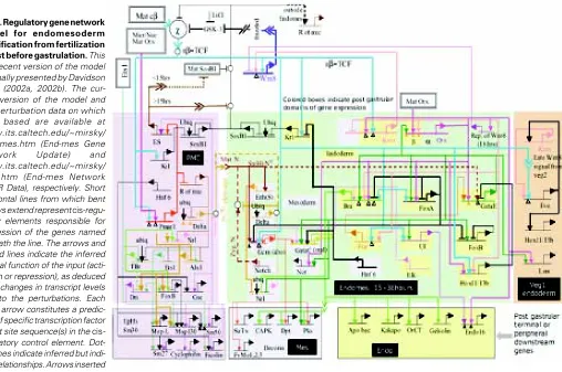
A model for the process of endomesoderm specification is shown in Fig. 2 in the form of a network diagram that combines all significant perturbation data; information on time and place of gene expression, as determined by whole mount in situ hybridization (WMISH) and QPCR measurements; cis-regulatory data where available; and all the underlying information of experimental em-bryology.
At each cis-regulatory element in the model predicted regulatory interactions with the products of other genes in the network are indicated. Therefore each one of these predicted interactions can be experimentally tested by determining the presence and function of the relevant binding sites in the relevant cis-regulatory elements. The importance of this point is worth emphasizing. It means that eventually the cis-regulatory network can be turned into a solid, experimentally confirmed structure.
Even though not all the cis-regulatory interactions that underlie the specification of the endomesoderm of the sea urchin embryo have yet been identified, and even though not all the identified interactions have yet been tested, the model of Fig. 2 allows us to see how the network of cis-regulatory interactions controls the specification process. The model shows how the cis-regulatory interactions control the specification functions that need to be executed for the different domains of the endomesoderm of the sea urchin to become what they become.
Interpreting the spatial cues: specification of the micromeres
The genes tbr, alx and ets, are all known to activate a number of genes that are responsible for the differentiation of the mi-cromere lineage into skeletogenic cells [Kurokawa et al., 1999; Fuchikami et al., 2002; Ettensohn et al., 2003 and www.its.caltech.edu/~mirsky/qpcr.htm (End-mes Network QPCR Data)]. Early in development, these three skeletogenic regulators are all kept silent everywhere in the embryo by a repressor gene (r of mic). At this time, delta, which is responsible for executing one of the micromespecific developmental functions, is also re-pressed everywhere in the embryo by the same repressor gene. Immediately after the micromeres are born at 4th cleavage, the
pmar1 gene is activated specifically in these cells. This gene has a repressor function that shuts down the expression of ‘r of mic’. Now, delta, and the skeletogenic regulators tbr, alx and ets are allowed to be expressed exclusively in the micromeres, and as a result the skeletogenic program is set in train (Oliveri et al., 2002). The mechanism just described ensures that once the pmar1 is activated, the micromere specification program will be installed without the need for any further spatial cues. If pmar1 is ectopically expressed everywhere in the embryo, the skeletogenic regulator
tbr, the signaling ligand Delta, and the skeletogenic differentiation gene sm50 are all also expressed everywhere, and the whole
embryo is now expressing the functions normally executed only by the cells of the micromere lineage (Oliveri et al., 2002, 2003). The fact that pmar1 is sufficient to establish the skeletogenic program, together with the fact that pmar1 is activated by factors that are all either maternally present or autonomously localized in the mi-cromere nuclei, tells us why the mimi-cromeres are autonomously specified. The most important general point is that the explanation of this embryological phenomenon is now provided in terms of the genomically encoded map of cis-regulatory interactions.
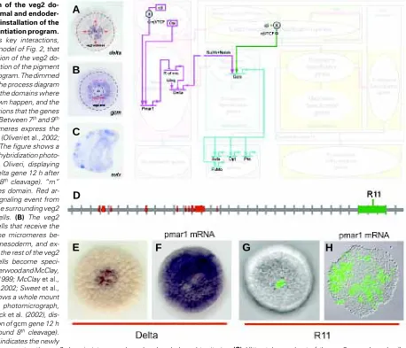
Refining the specification pattern: specification of the pig-ment cells
The portion of the network in the diagram of Fig. 3 tells us the mechanism by which the pigment cells are specified and ultimately differentiated, according to the network model. The pigment cells arise specifically from the mesodermal cells of the veg2 domain (Ruffins and Ettensohn, 1993, 1996). The Delta signaling ligand produced by the micromeres between 7th and 9th cleavage serves as the spatial cue that triggers the segregation of the mesodermal and endodermal fates of veg2 descendant cells (Fig. 3 A,B). Expression of the ligand Delta in the micromere descendants activates a Notch (N) receptor in the adjacent veg2 cells, which is required for normal
Fig. 2.Regulatory gene network model for endomesoderm specification from fertilization to just before gastrulation. This is a recent version of the model originally presented by Davidson et al. (2002a, 2002b). The cur-rent version of the model and the perturbation data on which it is based are available at www.its.caltech.edu/~mirsky/ endomes.htm (End-mes Gene Network Update) and www.its.caltech.edu/~mirsky/ qpcr.htm (End-mes Network QPCR Data), respectively. Short horizontal lines from which bent arrows extend represent cis-regu-latory elements responsible for expression of the genes named beneath the line. The arrows and barred lines indicate the inferred normal function of the input (acti-vation or repression), as deduced from changes in transcript levels due to the perturbations. Each input arrow constitutes a predic-tion of specific transcrippredic-tion factor target site sequence(s) in the cis-regulatory control element. Dot-ted lines indicate inferred but indi-rect relationships. Arrows inserted
A
B
D
C
E
F
G
H
specification of mesodermal fate in these cells (Sweet et al., 1999; McClay et al., 2000; Sweet et al., 2002). Localization of the Delta signal in the micromere descendants depends on the operation of the
pmar1 repression system, as explained above and illustrated in the diagram of Fig. 3. The response of Delta to the pmar1 repression system depends on the cis-regulatory element named R11 (Fig. 3 D-H) (R. Revilla-i-Domingo and E. Davidson, unpublished data). In normal embryos R11 drives expression of a reporter construct in the micromere descendants. When ‘r of mic’ is repressed everywhere in the embryo by ectopic expression of pmar1, the delta gene is activated in every cell (Fig. 3 E,F), and in the same embryos R11 also
drives expression of the reporter construct everywhere (Fig. 3 G,H) (R. Revilla-i-Domingo and E. Davidson, unpublished data).
Expression of the gcm gene begins in the single ring of mesoderm progenitor cells that directly receive the Delta micromere signal (Fig. 3B). As shown in the diagram of Fig. 3, activation of this gene depends on inputs from both the Notch signal transduction pathway, activated by the Delta signal, and (directly or indirectly) the nuclear β-catenin/TCF system (see diagram of Fig. 3), which is active in the whole of veg2 (Davidson et al., 2002a and A. Wikramanayake, unpublished data). The expression of gcm, therefore, reflects the creation of the new mesoderm-endoderm border, which did not exist formed border that segregates the veg2 domain into mesodermal and endodermal territories. (C) Ultimately, a subset of the veg2 mesodermal cells differentiate into pigment cells, and express the gene sutx (Calestani et al., 2003), among other pigment cell differentiation genes. The figure shows a whole mount in situ hybridization photomicrograph, modified from Calestani et al. (2003), displaying the expression of sutx gene in a gastrula stage embryo. (D-H) The cis-regulatory element R11 controls the localization of delta gene expression in the micromeres. (D) The R11 element consists of a sequence of genomic DNA near the coding sequence of the Delta gene. Each tic on the horizontal grey line representing genomic sequence demarcates 1 kb from the previous tic. 5' direction is to the left. Red blocks on the sequence indicate positions of the delta gene coding sequence. The green box on the sequence indicates the position of the R11 element. (E-F) pmar1 mRNA injection results in delta expression everywhere in the embryo. The figures show whole mount in situ hybridization photomicrographs, modified from Oliveri et al. (2002), comparing the expression of delta gene in normal blastula stage embryos (E), and embryos that have been injected with pmar1 mRNA (F). (G,H) The R11 element is responsible for localizing the expression of delta gene in the micromeres of normal embryos, and for driving the expression of the gene in every cell of embryos that have been injected with pmar1 mRNA (R. Revilla-i-Domingo and E. Davidson, unpublished data). The photomicrographs compare the expression of the GFP reporter gene in blastula stage embryos that have been injected with R11 reporter construct (G), and embryos that have been injected with pmar1 mRNA in addition to the R11 reporter construct (H).
Fig. 3.Segregation of the veg2 do-main into mesodermal and endoder-mal territories and installation of the pigment cell differentiation program. The diagram shows key interactions, extracted from the model of Fig. 2, that control the segregation of the veg2 do-main and the installation of the pigment cell differentiation program. The dimmed background shows the process diagram of Fig. 1E to indicate the domains where the interactions shown happen, and the developmental functions that the genes shown execute. (A) Between 7th and 9th
cleavage the micromeres express the signaling ligand Delta (Oliveri et al., 2002; Sweet et al., 2002). The figure shows a whole mount in situ hybridization photo-micrograph, from P. Oliveri, displaying the expression of delta gene 12 h after fertilization (around 8th cleavage). “m”
indicates micromeres domain. Red ar-rows indicate the signaling event from the micromeres to the surrounding veg2 endomesodermal cells. (B) The veg2 endomesodermal cells that receive the Delta signal from the micromeres be-come specified as mesoderm, and ex-press the gene gcm; the rest of the veg2 endomesodermal cells become speci-fied as endoderm (Sherwood and McClay, 1999; Sweet et al., 1999; McClay et al., 2000; Ransick et al., 2002; Sweet et al., 2002). The figure shows a whole mount in situ hybridization photomicrograph, modified from Ransick et al. (2002), dis-playing the expression of gcm gene 12 h after fertilization (around 8th cleavage).
before the Delta signal was received from the micromeres. The cis -regulatory element of gcm is responsible for integrating the spatial information provided by the inputs from the Notch transduction pathway, and the β-catenin/TCF system. In normal embryos this element drives the expression of a reporter construct in a localized region in the vegetal plate. But if a portion of this element, containing binding sites for the Notch transduction pathway, is eliminated, expression of the reporter construct is expanded to a broader region that includes the whole of the veg2 domain (A. Ransick and E. Davidson, unpublished data). In other words, now the cis -regula-tory element that controls gcm expression is ‘blind’ to the mesoderm-endoderm border established by the activation of the Notch transduc-tion pathway.
Ultimately, the gene gcm is expressed in the pigment cells (a prominent subset of the veg2 mesodermal cell types), where it activates a number of differentiation genes (see diagram of Fig. 3), the products of some of which are likely to be required for synthesis of the red quinone pigment that these cells produce (Davidson et al., 2002b; Ransick et al., 2002; Calestani et al., 2003). If translation of
gcm transcripts is blocked experimentally, the perturbed embryos show a perfectly normal morphology, except that they have no pigment cells (A. Ransick and E. Davidson, unpublished data).
The portion of the network depicted in Fig. 3 is a piece of the genetic program encoded in the cis-regulatory genomic sequence. It consists of a transcriptional apparatus, including R11 element, that localizes the Delta signal, and another transcriptional apparatus, including the Notch responsive element of the gcm gene, that interprets the signal. It explains why the cells in the inner ring of the veg2, and no others, give rise to pigment cells. And it also explains why elimination of expression of a single player in the program, gcm, results in the absence of the pigment cells. The overall function of this portion of the network is, first, to create a new domain of specification
in the embryo (the veg2 mesoderm), by setting a new border in the specification pattern; and then to install the program for pigment cell differentiation in the cells of the new domain. Other similar network subelements not yet resolved are undoubtedly responsible for differ-entiation of additional mesodermal cell types.
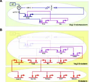
Stabilizing states of specification: the endoderm
Figure 4 illustrates the process by which the veg2 endoderm is specified. The veg2 lineage is born at 6th cleavage. By this time, the two spatial cues that trigger the specification of veg2 as endomesoderm are already operating. These initial cues consist of the autonomous nuclearization of β-catenin, which is a cofactor of the Tcf transcription regulator required for Tcf to function as a gene activator, and the early micromere signal (Ransick and Davidson, 1993, 1995; Logan et al., 1999). Two regulatory subcircuits execute the process by which the zygotic transcriptional apparatus interprets these initial cues, and by which it establishes an endomesodermal state of specification (Fig. 4A). The β-catenin/Tcf input activates the krox gene (Davidson
et al., 2002b). This gene stimulates expression of wnt8 gene and one of the transcription units of the otx gene. Wnt8 is a ligand which activates the β-catenin/Tcf system, and is itself a target of the β-catenin/Tcf input. This implies an autoreinforcing Tcf control loop, which is set up within the endomesodermal domain once this is defined (Davidson et al., 2002a). So, the result of the stimulation of
wnt8 expression, first by the β-catenin/Tcf system and later by krox, is to transfer control of the β-catenin/Tcf system from the autonomous cytoplasmic mechanism by which its activity was initiated to a zygotically controlled, intercellular signaling mechanism operating among the cells of the endomesoderm. The “community effect” (as defined by Gurdon, 1988; Gurdon et al., 1993) established by this regulatory subcircuit (dark blue connections in Fig. 4A) takes the cells out of a condition of alternative transcriptional possibility that is
Fig. 4.Stabilization of the endomesoderm specifica-tion state and installaspecifica-tion of the endoderm differentia-tion program. The diagram shows key interactions, ex-tracted from the model of Fig. 2, that control the stabiliza-tion of the endomesoderm state of specificastabiliza-tion and the installation of the endoderm differentiation program. (A) The box with green background shows the interactions that operate in the veg2 endomesoderm domain up to about 9th cleavage. Nuclearization of β-catenin is
autono-mous, and results in the activation of two regulatory subcircuits. Dark blue subcircuit: Wnt8 intercellular signal-ing among cells of the veg2 domain stimulates the nuclearization of β-catenin and establishes a “community effect,” which defines and locks the endomesodermal state of specification in the veg2 cells. Purple subcircuit: krox and otx cross-regulate, which results in a reinforcing loop that renders the endomesoderm state of specification independent of the initial inputs. (B) The box labeled “Veg2 Endoderm” shows the interactions that operate in the veg2 endoderm domain, from about 9th cleavage to
mesen-chyme blastula stage. The gatae gene is added to the krox-otx feedback loop (purple interactions), and together with β-catenin/TCF system, installs the endoderm specification program (red interactions). When β-catenin/TCF/Wnt8 in-puts disappear, the stabilization loop maintains the endo-dermal specification program active, which eventually re-sults in the activation of endodermal differentiation genes (lower box in the diagram labeled “Endoderm”).
A
A
B
their initial condition, and locks them into a stable state of gene expression.
The otx gene stimulates expression of the krox
gene. A regulatory subcircuit consisting of otx and krox
cross-regulation produces a transcription-level stabili-zation of the endomesodermal regulatory state (purple connections in Fig. 4A; see Davidson et al., 2002a). The otx gene also provides an input into the gatae
gene, which in turn has an input back into otx gene. This is a further positive feedback that links the gatae
gene into the stabilization circuitry (purple connections in Fig. 4B). The gatae gene plays an important role in endoderm specification (red connections in Fig. 4B), since, together with the β-catenin/Tcf system, it is responsible for the activation of many of the known endodermal regulators, including the bra, foxA and ui
genes (Davidson et al., 2002a and P.Y. Lee and E. Davidson, unpublished data). The FoxA transcrip-tion factor is a repressor that has multiple roles in the spatial control of gene expression patterns in the endoderm; Bra results in the activation of endodermal differentiation genes which are involved in cell motility and are needed for gastrulation and invagination to occur (Gross and McClay, 2001; Rast et al., 2002); the UI factor directly controls expression of endo-16 (Yuh
et al., 2001), which encodes a differentiation protein that is secreted in the lumen of the midgut. The crucial role that gatae plays in the specification of the endo-derm explains the phenotype shown by embryos in which translation of the gatae transcripts has been blocked. This treatment produces a severe interfer-ence with endoderm specification and gut develop-ment (P.Y. Lee and E. Davidson, unpublished data). During the late blastula stage, β-catenin disap-pears from the nuclei of the veg2 endodermal domain (Logan et al., 1999). But by this time, a network of stable intergenic interactions has been installed, so that the β-catenin inputs used earlier to set up tran-scriptional specification are no longer needed (Fig. 4B). We see here that the cis-regulatory interactions control the operation of at least three different regula-tory devices that are directly responsible for establish-ing at least part of the endoderm differentiation pro-gram. The first device consists of the “community effect,” which first defines and then locks on the endomesodermal specification state in the veg2 do-main (dark blue connections in Fig. 4A). The second device depends on a feedback loop, including krox
and otx (purple connections in Fig. 4A), which
gener-Understanding development and evolution
Developmental and evolutionary processes both have their root in the heritable genomic regulatory programs that determine how the body plan of each species is built (Davidson, 2001). It has been clear for a long time that the evolution of body plans has occurred by change in the genomic programs for the development of these body plans (Britten and Davidson, 1971), and it is now clear that we need to consider this in terms of change in the regulatory devices that execute these programs. The bilaterians all rely on essentially the same repertoire of regulatory genes to ates a robust and resilient regulatory structure in the already defined
endomesoderm domain. The third device consists of the addition of
gatae to the krox-otx feedback loop (purple connections in Fig. 4B), which ensures the operation of many endodermal regulatory genes in the endoderm. The result is a control system that drives the specification process forward as a progression of states, and it prevents it from reversing direction when the initial cues that trigger the specification process disappear. Progressivity and stability are fundamental properties of the developmental process. They derive from regulatory devices consisting of assemblages of cis-regulatory interactions.
control the developmental organization of their body plans. Analy-sis of cis-regulatory networks affords the means to focus on the significance of preserved uses of these genes, and on the exact consequences of differences in their use (Davidson, 2001).
Figure 5 compares the way certain genes are utilized in the specification of the endomesoderm of two different bilaterians, namely, the sea urchin and the starfish. All genes in Fig. 5, except for tbr, are central elements that control the specification of the endoderm in the sea urchin (see Figs. 2 and 4). The tbr gene, on the other hand, is activated exclusively in the micromere derived skeletogenic cells (see Fig. 2) (Croce et al., 2001; Fuchikami
et al., 2002; Oliveri et al., 2002). Its regulation depends on other genes specifically expressed in the micromere lineage (Oliveri
et al., 2002), and in turn, it drives expression of larval skeletogenic differentiation genes (Davidson et al., 2002a; Oliveri et al., 2002 and www.its.caltech.edu/~mirsky/endomes.htm). While the for-mation of the endoderm is at least superficially similar in the two species (Fig. 5A), starfish embryos do not have a micromere lineage, nor do they produce a larval skeleton (Fig. 5A).
Figure 5B shows that the cis-regulatory interactions that con-stitute the endodermal three-gene stabilizing loop in the sea urchin (see Fig. 4B), is found in identical form in the starfish (connections in bold in Fig. 5B) (Hinman et al., 2003). This set of identical cis-regulatory interactions must serve conserved evolu-tionary roles, since the possibility of convergence is ruled out by the number of similar functional starfish and sea urchin cis -regulatory interactions.
Sea urchins and starfish have diverged for at least 500 million years (Sprinkle and Kier, 1987; Smith, 1988; Bowring and Erwin, 1998). The reinforcing loop is therefore a regulatory device that was invented at least about 500 million years ago, and that has been conserved in at least two independently evolving lineages during all this time. 500 million years represents a very long genomic divergence, in the sense that comparisons of starfish and sea urchin DNA sequences around orthologous regions do not show any conservation distinguishable from random occur-rence between the cis-regulatory elements, even when the genes are similarly regulated (V. Hinman and E. Davidson, unpublished data). The preservation of this regulatory device suggests that the function it serves in the specification process must be essential. As we have already seen, in the sea urchin the regulatory feedback loop between krox and otx genes generates a robust regulatory structure in the endomesoderm domain, and the addi-tion of the gatae gene to this feedback loop ensures and maintains the operation of many endodermal regulatory genes after the initial transient inputs have disappeared (Davidson et al., 2002a and P.Y. Lee and E. Davidson, unpublished data). In the starfish,
gatae also drives the expression of many endodermal regulatory genes (Hinman et al., 2003), and in many other bilaterians, members of the Gata family of transcription regulatory genes are required for gut development (Reuter, 1994; Maduro et al., 2002; Patient and McGhee, 2002). What makes the reinforcing loop especially useful, and hence likely to be preserved during evolu-tion, may therefore be that it controls the installation and stabili-zation of the expression of the gatae gene in the endoderm (Hinman et al., 2003). Other intergenic feedback loops are used across the Bilateria to serve similar functions. For example a reinforcing feedback loop is found in the hox gene network that controls rhombomere specification in the mouse hindbrain (Nonchev et al., 1996; Barrow et al., 2000), in the regulatory
network for tracheal placode specification in Drosophila (Zelzer and Shilo, 2000), and in the specification of the oral ectoderm in sea urchin embryos (Amore et al., 2003), among others. It seems a general property of the developmental process to use feedback loops as a mechanism to achieve the progressivity of the process. The tbr gene, on the other hand, is used in completely different ways in the starfish and sea urchin embryos (Fig. 5B). It is required for the formation of the archenteron in the starfish embryo, and its expression is under the control of endodermal regulators (Otx, Gatae) (Hinman et al., 2003), whereas it is involved solely in skeletogenic functions in the sea urchin embryo (Croce et al., 2001; Oliveri et al., 2002 and www.its.caltech.edu/ ~mirsky/endomes.htm). The skeletogenic micromere lineage is a relatively recent echinoid invention (Wray and McClay, 1988; Tagawa et al., 2000). This suggests that in the sea urchin the skeletogenic use of tbr may have been coopted from an adult skeletogenic regulatory system, while an original embryonic endomesodermal regulatory element was lost (Hinman et al., 2003).
If indeed the larval skeletogenic lineage is the result of a cooption from the adult skeletogenic regulatory system, it repre-sents an example of how a regulatory subroutine can be “wired” into the specification system as the result of evolutionary change. How the intrinsic behavior of the subroutine is preserved in the new context, and how the rest of the developmental control system can cope with this change without disrupting its workabil-ity, speaks directly to the intrinsic robustness of the subroutine, and the robustness of the developmental process in general. Regulatory networks serve as the link between development and evolution. They provide a new means to address specific ques-tions about the robustness of the developmental process, and about the preservation of aspects of the process through evolu-tionary time. Questions such as these can only be answered by considering evolution and development together.
Conclusions
Gene network analysis identifies the mechanisms that control and operate the program for the developmental process. This will be true for all aspects of the developmental process that are required to generate the species-specific body plan. To address some of the general and fundamental questions about the pro-cess of development, though, will require understanding evolu-tion. Because gene regulatory networks underlie the processes of both development and evolution, unraveling their architecture in appropriately chosen species will be the key to understanding how genomes control development and how they evolve.
Acknowledgments
References
AMORE, G., YAVROUIAN, R.G., PETERSON, K.J., RANSICK, A., McCLAY, D.R. and DAVIDSON, E.H. (2003). Spdeadringer, a sea urchin embryo gene required separately in skeletogenic and oral ectoderm gene regulatory networks. Dev. Biol., in press.
BARROW, J., STADLER, H. and CAPECCHI, M. (2000). Roles of Hoxa1 and Hoxa2 in patterning the early hindbrain of the mouse. Development 127: 933-944. BOWRING, S. and ERWIN, D. (1998). A new look at evolutionary rates in deep time:
Uniting paleontology and high-precision geochronology. GSA Today 8: 2. BRITTEN, R.J. and DAVIDSON, E.H. (1971). Repetitive and non-repetitive DNA
sequences and a speculation on the origins of evolutionary novelty. Quart. Rev. Biol. 46: 111-138.
CALESTANI, C., RAST, J.P. and DAVIDSON, E.H. (2003). Isolation of mesoderm specific genes in the sea urchin embryo by differential macroarray screening. Development 130, in press.
CAMERON, R.A., HOUGH-EVANS, B.R., BRITTEN, R.J. and DAVIDSON, E.H. (1987). Lineage and fate of each blastomere of the eight-cell sea urchin embryo. Genes & Dev. 1: 75-85
CAMERON, R.A., FRASER, S.E., BRITTEN, R.J. and DAVIDSON, E. H. (1991). Macromere cell fates during sea urchin development. Development 113: 1085-1092
CROCE, J., LHOMOND, G., LOZANO., J.-C. and GACHE, C. (2001). Ske-T, a T-box gene expressed in the skeletogenic mesenchyme lineage of the sea urchin embryo. Mech. Dev. 107: 159-162.
DAVIDSON, E.H. (2001). Genomic Regulatory systems: Development and Evolution. San Diego, Academic Press.
DAVIDSON, E.H., CAMERON, R.A. and RANSICK, A. (1998). Specification of cell fate in the sea urchin embryo: Summary and some proposed mechanisms. Development 125: 3269-3290.
DAVIDSON, E.H. et al. (2002a). A provisional regulatory gene network for specifica-tion of endomesoderm and the sea urchin embryo. Dev. Biol. 246: 162-190. DAVIDSON, E.H. et al. (2002b). A genomic regulatory network for development.
Science 295: 1669-1678.
ETTENSOHN, C.A., ILLIES, M.R., OLIVERI. P. and DE JONG, D.L. (2003). Alx1, a member of the Cart1/Alx3/Alx4 subfamily of Paired-class homeodomain proteins, is an essential component of the gene network controlling skeletogenic fate specification in the sea urchin embryo. Development 130, 2917-2928. FUCHIKAMI, T. et al. (2002). T-brain homologue (HpTb) is involved in the
arch-enteron induction signals of micromere descendant cells in the sea urchin embryo. Development 129: 5205-5216.
GROSS, J.M. and McCLAY, D.R. (2001). The role of Brachyury (T) during gastrulation movements in the sea urchin Lytechinus variegates. Dev. Biol. 239: 132-147. GURDON, J. (1988). A community effect in animal development. Nature 336:
772-774.
GURDON, J., KATO, K. and LEMAIRE, P. (1993). The community effect, dorsalization and mesoderm induction. Curr. Opin. Genet. Dev. 3: 662-667.
HINMAN, V.F., NGUYEN, A., CAMERON, R.A. and DAVIDSON, E.H. (2003). Developmental gene regulatory network architecture across 500 MYA of echino-derm evolution. Proc. Natl. Acad. Sci. USA (In press).
HÖRSTADIUS, S. (1939). The mechanics of sea urchin development, studied by operative methods. Biol. Rev. Cambridge Philos. Soc. 14: 132-179.
KUROKAWA, D., KITAJIMA, T., MITSUNAGA-NAKATSUBO, K., AMEMIYA, S., SHIMADA, H. and AKASAKA, K. (1999). EpEts, an ets-related transcription factor implicated in primary mesenchyme cell differentiation in the sea urchin embryo. Mech. Dev. 80: 41-52.
LOGAN, C.Y. and McCLAY, D.R. (1997). The allocation of early blastomeres to the ectoderm and endoderm is variable in the sea urchin embryo. Development 124: 2213-2223.
LOGAN, C.Y., MILLER, J.R., FERKOWICZ, M. and McCLAY, D.R. (1999). Nuclear beta-catenin is required to specify vegetal cell fates in the sea urchin embryo. Development 126: 345-357.
MADURO, M.F., LIN, R. and ROTHMAN, J.H. (2002). Dynamics of developmental switch: Recursive intracellular and intranuclear redistribution of Caenorhabditis
elegans POP-1 parallels Wnt-inhibited transcriptional repression. Dev. Biol. 248: 128-142.
McCLAY, D.R., PETERSON, R., RANGE, R., WINTER-VANN, A. and FERKOWICZ, M. (2000). A micromere induction signal is activated by beta-catenin and acts through notch to initiate specification of secondary mesenchyme cells in the sea urchin embryo. Development 127: 5113-5122.
NONCHEV, S. et al. (1996). Segmental expression of Hoxa-2 in the hindbrain is directly regulated by Krox-20. Development 122: 543-554.
OLIVERI, P., CARRICK, D.M. and DAVIDSON, E.H. (2002). A regulatory gene network that directs micromere specification in the sea urchin embryo. Dev. Biol. 246: 209-228.
OLIVERI, P., DAVIDSON, E.H. and McCLAY, D.R. (2003). Activation of pmar1 controls specification of micromeres in the sea urchin embryo. Dev. Biol. 258: 32-43.
PATIENT, R.K. and McGHEE, J.D. (2002). The GATA family (vertebrates and invertebrates). Curr. Opin. Genet. Dev. 12: 416-422.
RANSICK, A. and DAVIDSON, E.H. (1993). A complete second gut induced by transplanted micromeres in the sea urchin embryo. Science 259: 1134-1138. RANSICK, A. and DAVIDSON, E.H. (1995). Micromeres are required for normal
vegetal plate specification in sea urchin embryos. Development 121: 3215-3222.
RANSICK, A. and DAVIDSON, E.H. (1998). Late specification of veg1 lineages to endodermal fate in the sea urchin embryo. Dev. Biol. 195: 38-48.
RANSICK, A., RAST, J.P., MINOKAWA, T., CALESTANI, C. and DAVIDSON, E.H. (2002). New early zygotic regulators expressed in endomesoderm of sea urchin embryos discovered by differential array hybridization. Dev. Biol. 246: 132-147.
RAST, J.P., AMORE, G., CALESTANI, C., LIVI, C.B., RANSICK, A. and DAVIDSON, E.H. (2000). Recovery of developmentally defined gene sets from high-density cDNA macroarrays. Dev. Biol. 228: 270-286.
RAST, J.P., CAMERON, R.A., POUSTKA, A.J. and DAVIDSON, E. H. (2002). brachyury target genes in the early sea urchin embryo isolated by differential macroarray screening. Dev. Biol. 246: 191-208.
REUTER, R. (1994). The gene serpent has homeotic properties and specifies endoderm versus ectoderm within the Drosophila gut. Development 120: 1123-1135.
RUFFINS, S.W. and ETTENSOHN, C.A. (1993). A clonal analysis of secondary mesenchyme cell fates in the sea urchin embryo. Dev. Biol. 160: 285-288. RUFFINS, S.W. and ETTENSOHN, C.A. (1996). A fate map of the vegetal plate of the
sea urchin (Lytechinus variegatus) mesenchyme blastula. Development 122: 253-263.
SHERWOOD, D. and McCLAY, D.R. (1999). LvNotch signaling mediates secondary mesenchyme specification in the sea urchin embryo. Development 126: 1703-1713.
SMITH, A. (1988). Echinoderm Phylogeny and Evolutionary Biology. Oxford, Clarendon Press.
SPRINKLE, J. and KIER, P. (1987). Fossil Invertebrates. Cambridge, MA, Blackwell Science.
SWEET, H., HODOR, P. and ETTENSOHN, C. (1999). The role of micromere signaling in Notch activation and mesoderm specification during sea urchin embryogenesis. Development 126: 5255-5265.
SWEET, H.C., GEHRING, M. and ETTENSOHN, C.A. (2002). LvDelta is a mesoderm-inducing signal in the sea urchin embryo and can endow blastomeres with organizer-like properties. Development 129: 1945-1955.
TAGAWA, K., HUMPHREYS, T. and SATOH, N. (2000). T-Brain expression in the apical organ of hemichordate tornaria larvae suggests its evolutionary link to the vertebrate forebrain. J. Exp. Zool. 288: 23-31.
WRAY, G. and McCLAY, D.R. (1988). The origin of spicule-forming cells in a ‘primitive’ sea urchin (Eucidaris tribuloides) which appears to lack primary mesenchyme cells. Development 103: 305-315.
YUH, C.-H., BOLOURI, H. and DAVIDSON, E.H. (2001). cis-Regulatory logic in the endo16 gene: Switching from a specification to a differentiation mode of control. Development 128: 617-629.