1
CLASSIFICATION OF FOETAL DISTRESS AND
HYPOXIA USING MACHINE LEARNING
ROUNAQ ABDALRAHMAN ABBAS
A thesis submitted in partial fulfilment of the requirements of Liverpool
John Moores University for the degree of Master of Philosophy
2
ACKNOWLEDGEMENTS
Firstly, I am grateful to Allah for the good health and wellbeing that were necessary to complete this dissertation.
I would like to express my sincere gratitude to my supervisor Dr.Abir Hussain for the continuous support of my M.PHIL study and related research, for her patience, motivation, and immense knowledge. Her guidance helped me in all the time of research and writing of this thesis. I could not have imagined having a better supervisor and mentor for my M.PHIL study.
I would like to take the opportunity to express my sincere appreciations to Prof.Dhiya Al-jumeily, for his continuous support and motivating words, which helped me through this entire journey.
My great gratitude to my family and everyone who supported me and believed in me especially my mother and my husband, I don't believe I can finish this dissertation without their support.
3
ABSTRACT
Foetal distress and hypoxia (oxygen deprivation) is considered a serious condition and one of the main factors for caesarean section in the obstetrics and gynaecology department. It is considered to be the third most common cause of death in new-born babies. Foetal distress occurs in about 1 in 20 pregnancies. Many foetuses that experience some sort of hypoxic effects can have series risks such as damage to the cells of the central nervous system that may lead to life-long disability (cerebral palsy) or even death. Continuous labour monitoring is essential to observe foetal wellbeing during labour. Many studies have used data from foetal surveillance by monitoring the foetal heart rate with a cardiotocography, which has succeeded traditional methods for foetal monitoring since 1960. Despite the indication of normal results, these results are not reassuring, and a small proportion of these foetuses are actually hypoxic. This study investigates the use of machine learning classifiers for classification of foetal hypoxic cases using a novel method, in which we are not only considering the classification performance only, but also investigating the worth of each participating parameter to the classification as seen by medical literature. The main parameters that are included in this study as indicators of metabolic acidosis are: pH level (which is a measure of the hydrogen ion concentration of blood to specify the acidity or alkalinity), as an indicator of respiratory acidosis; Base Deficit of extra-cellular fluid level and Base Excess (BE) (which is the measure of the total concentration of blood buffer base that indicates metabolic acidosis or compensated respiratory alkalosis). In addition to other parameters such as the PCO2 (partial pressure of carbon dioxide can reflect the hypoxic state of the foetus) and the Apgar scores (which shows the foetal physical activity within a specific time interval after birth).
4
The provided data was an open-source partum clinical data obtained by Physionet, including both hypoxic cases and normal cases. Six well known machine learning classifier are used for the classification; each model was presented with a set of selected features derived from the clinical data. Classifier evaluation is performed using the receiver operating characteristic curve analysis, area under the curve plots, as well as confusion matrix. The simulation results indicate that machine learning classifiers provide good results in diagnosis of foetal hypoxia, in addition to acceptable results of different combinations of parameters to differentiate the cases.
5
TABLE OF CONTENTS
Contents
List of Tables ... 7 List of Figures ... 8 ABBREVIATIONS ... 9CHAPTER ONE: INTRODUCTION ... 13
1.1 Overview ... 13
1.2 Problem statement ... 16
1.3 Aims and objectives ... 18
1.4 Research scope ... 19
1.5 Structure of the thesis ... 20
CHAPTER TWO: FOETAL DISTRESS CLASSIFICATION ... 23
2.1 Introduction ... 23
2.2 Foetal Distress ... 24
2.3 Foetal Hypoxia Causes ... 25
2.4 Foetal distress physiology ... 27
2.5 Evidence of Multi-organ Failure ... 29
2.6 Foetal hypoxia Diagnosis: ... 30
2. 6.1 Electronic foetal monitoring ... 32
2.6.2 Foetal blood sampling ... 34
2.7 Foetal non- reassuring status management ... 39
2.8 Summary of the chapter ... 41
CHAPTER THREE: MACHINE LEARNING TYPES AND APPLICATION 42 3.1 introduction ... 42
3.2 Machine Learning Development ... 42
3.4 Machine Learning: Algorithms Types ... 45
3.4.1 Supervised learning ... 45
3.4.2 Unsupervised learning... 46
3.4.3 Semi-supervised learning ... 46
3.4.4 Reinforcement learning ... 48
3.5 Machine Learning Classifiers ... 49
3.5.1 Artificial neural networks ... 50
3.5.2 Support vector machines (SVM) ... 55
3.5.3 k-nearest neighbours algorithm (KNN) ... 59
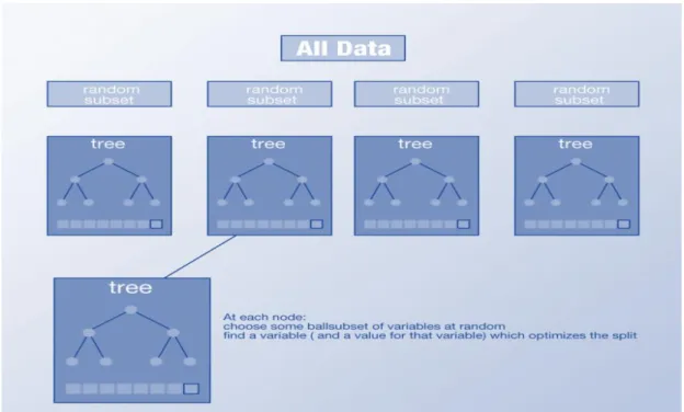
3.5.4 Decision Trees based classifiers ... 62
3.6 Summary of the Chapter ... 70
CHAPTER FOUR: METHODOLOGY ... 71
4.1 Introduction ... 71
4.2 Related research ... 72
4.3 Data collection and analysis ... 76
4.4 primary analysis of full Dataset ... 76
4.5 Data description ... 77
6
4.8 Class Distribution and visualization ... 81
4.9 Feature selection ... 83
4.10 Relationship between the various parameters and its effect on foetal hypoxia detection ... 86
4.11 Experimental classifiers of foetal data classification ... 89
4.12 General Approach of Classification process ... 91
4.13 Summary of the chapter... 93
CHAPTER FIVE: IMPLEMINTATION AND ALGORITHM EVALUATION ... 94
5.1 Introduction ... 94
5.2 Performance metrics ... 94
5.3 Data Mining Classification ... 97
5.5.1 General data classification process: ... 99
5.5.2 First round of classification ... 100
5.5.3 Second set of classification experiments ... 102
5.5.4 Third set of classification experiments ... 104
5.5 Chapter Summary ... 106
CHAPTER SIX: SIMULATION RESULTS AND ANALYSIS ... 107
6.1 Introduction ... 107
6.2 Classification results with full set of parameters ... 107
6.3 Classification results using five parameters ... 114
6.4 Classification using four parameters ... 116
6.5 Discussion ... 118
6.6 Summary of the chapter ... 126
CHAPTER SEVEN: CONCLUSION AND FUTURE WORK ... 128
7.1Introduction ... 128
7.2 Conclusion ... 128
7.3 Future work ... 132
7
List of Tables
Table 2.1 Foetal types and the related causes ... 26
Table 2.2 Thresholds for foetal health state ... 35
Table 4.1 Overview of various studies that presented classification results ... 75
Table 4.2 Patients and labour outcome statistics for the CT-UHB database ... 79
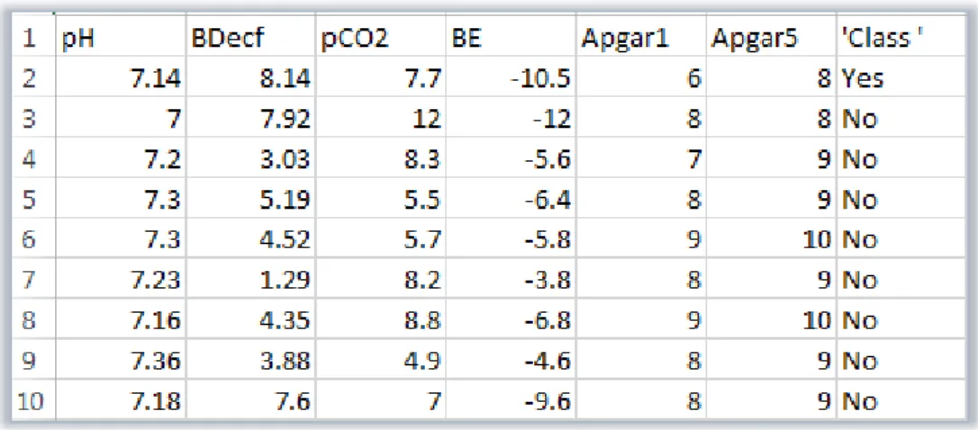
Table 4.3 labelled dataset including the main parameters. ... 80
Table 4.4 Output class distribution ... 81
Table 4.5 Experimental classifiers of foetal data classification ... 91
Table 5.1 Confusion Matrix ... 95
Table5.2 Performance Metrics ... 96
Table 5.3 Medical databases “Training dataset" ... 98
Table5.4 medical bases" predicting dataset" ... 98
Table6.1 performance results of all classifiers using six parameters ... 109
Table 6.2 ROC Summary (predictions) ... 110
Table 6.3 performance metrics for the second trial ... 115
Table6.4 ROC results for classifier comparison ... 116
Table6.5 performance metrics for the third trial ... 117
Table 6.6 variable importance of all classifiers using 5 parameters ... 123
8
List of Figures
Figure 1-1 foetal distress stages ... 15
Figure 2-1 Foetal distress pathophysiology ... 28
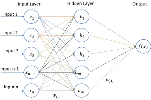
Figure 3.1 Typical ANN Classifier... 51
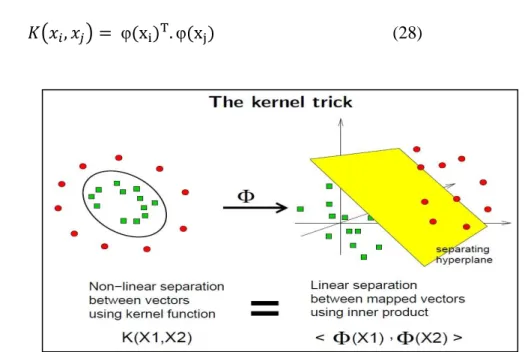
Figure 3.2 kernel trick demonstration ... 57
Figure 3.3 K-nearest neighbours algorithm (k-NN) example ... 60
Figure 3.4 Learning process of BNNP ... 68
Figure 4.1 Class distributions of the utilised data ... 82
Figure 4.2 Apgar scoring system ... 86
Figure 4.3 Linear (a) verse Non-linear (b) classifier classification problems ... 89
Figure 5.1 classification process map ... 91
Figure 5.2 (a) Learning stage: training data analysed by a classification algorithm ... 99
Figure 5.2 (b) Classification stage: test data are used to estimate the accuracy of the classification rule ... 99
Figure 5.3 (a) Learning stage: training data analysed by a classification algorithm ... 101
Figure 5.3 (b) Classification stage: test data are used to estimate the accuracy of the classification rules ... 101
Figure 5.4 (a) Learning stage: training data analysed by a classification algorithm ... 103
Figure 5.4 (b) Classification stage: test data are used to estimate the accuracy of the classification rules ... 103
Figure 5.5 (a) Learning stage: training data analysed by a classification algorithm ... 105
Figure 5.5 (b) Classification stage: test data are used to estimate the accuracy of the classification rules ... 105
Figure6.1 classifiers performance “using six parameters ... 109
... 110
Figure 6.2 Roc plot for all classifiers ... 110
Figure 6.3 variable importance plotted figures ... 113
Figure 6.4 classifiers performance using five parameters ... 114
... 115
Figure 6.5 ROC plots of all classifiers in the second trial ... 115
Figure 6.6 classifiers performance using four parameters ... 117
... 118
Figure 6.7 Roc plot of all classifiers in the third trial... 118
Figure 6.8 (a) SVM variable importance, (b) KNN variable importance ... 119
Figure 6.9 variable importance of 5 parameters ... 124
9
ABBREVIATIONS
ACOG
American college of Obstetricians and gynecologist criteria
AI Artificial intelligence
ANNs Artificial neural networks
AMIS Hospital information system
AS1 Apgar score at 1st
AS5 Apgar score at 5th
ACC Accuracy
AUC Area under curve
BDecf Base deficit
BE Base excess
BP Back-propagation
CART Classification and regression trees
CS Caesarean section
CTG Cardiotocography
CO2 Carbone dioxide
EFM Electronic Foetal monitoring
ECG Electric-cardiogram
FN False negative
FP False positive
FHR Foetal heart rate
FSS Foetal scalp blood sampling
10
GBDTs Gradient-boosted decision trees
HCO3 Bicarbonate
HIV Human immunosuppressive virus
HIE Hypoxic -ischemic encephalopathy
IA Intermittent auscultation
IUGR Intrauterine growth retardation
KDD Knowledge discovery in databases
KNN K-Nearest Neighbor
LSVM Linear Support vector machine
ML Machine learning
MLP Multilayer Perceptron
NNET Neural network
pH Power of hydrogen, expressing the blood acidity PCO2 Partial pressure of Carbone dioxide
PO2 Partial pressure of oxygen
PDA Patent dectus arteriosus
PUBS Percutaneous Umbilical Cord Blood Sampling
PCA Principle component analysis
PVL Periventricular leukomalacia
PE Processing element
RBF Radial basis function
ROC Receiver operating characteristic
RF Random forest
11
RCOG The Royal College of Obstetricians and Gynaecologists
RL Reinforcement learning
SOGC
The Society of Obstetricians and Gynaecologists of Canada
SSL Semi supervised learning
SVM Support vector machine
SVC Support vector classifier
SMOTE Synthetic Minority Over-sampling Technique
SE Sensitivity
SP Specificity
TPR True positive rate
TNR True negative rate
TN True negative
TP True positive
tSNE T-distributed stochastic neighbor embedding
UC Uterine contraction
12
PUPLICATION
2018 International Conference on Intelligent Computing August 15-18, 2018, Wuhan, China
Authors: Rounaq Abbas, Abir Hussain, Dhiya Al-Jumeily
Paper Title: Classification of Foetal Distress and Hypoxia using Machine Learning Approaches
The paper has been submitted and accepted to present by the 2018 International Conference on Intelligent Computing (ICIC2018).
13
CHAPTER ONE: INTRODUCTION
1.1 Overview
Foetal distress is a condition of foetal oxygen deprivation and accumulation of carbon dioxide, leading to hypoxia and acidosis during the ante-partum period (before labour) or intra-partum period (during the birth process) [1] as shown in Figure 1.1.
Foetal distress and hypoxia (oxygen deprivation) is considered as a serious conditions and one of the main reasons for caesarean section in the obstetrics and gynaecology department. Foetal distress occurs in about 1 in 20 pregnancies [2]. Foetal distress a severe condition that can lead to foetal death or brain damage. It is also considered to be the third most common cause of new-born death. Many of the foetuses have experienced hypoxia during different stages of the pregnancy period.
Foetal hypoxia can be classified as acute hypoxia or chronic hypoxia according to the stage of the intra-partum foetal life [2]. The former usually occurs during the labour process while the latter occurs during the first, second or the third trimester of the pregnancy. Various methods of intra-partum foetal surveillance have been employed to detect the signs of hypoxia as early as possible to minimize the risk of life-long disability such as cerebral palsy and to reduce the mortality rate among new-borns. Intra-partum monitoring of foetuses during labour has been commonly performed by monitoring the foetal heart with a technique such as the intermittent auscultation (IA), which is considered the most common method of foetal surveillance in labour [1]. However many of these methods have been subject to controversy as existing studies have found no benefit of its use in reducing the rates of cerebral palsy or peri-natal mortality [3].
Continuous labour monitoring is essential to observe the foetal wellbeing. There are many studies indicating foetal heart activity is the prominent source of information
14
about foetal health and especially the detection of foetal hypoxia. However, the physicians have found many challenges in identifying the perfect way of detecting foetal hypoxia by analysing foetal heart rate using the traditional and intermittent monitoring such as (IA) and Doppler machine [1]. Since 1960, cardiotocography (continuous electronic foetal heart rate monitoring) has been developed and replaced all other traditional methods to monitor foetal heart. Normal cardiotocography results in nearly half of all tracings indicating that enough oxygen is delivered to the foetus [4, 5]. However; these results are not encouraging [6], as a number of these foetuses are actually hypoxic, therefore a diagnostic test is necessary and compulsory. During the last decades, new methods have been used by antenatal care and during the labour process such as the cordocentesis (foetal blood sampling by ultrasound guided needle aspiration from the umbilical cord) [7], which can be used to detect hypoxia due to placental development abnormalities. This could cause many threats to the foetal health such as damage to the cells of the central nervous system that lead to life-long disability. Up to three quarters of infants with severe hypoxic-ischemic encephalopathy (HIE) die of multiple organ failure or lung infections caused by irregular breathing. Those who survive are commonly left with gross symptoms such as mental retardation, epilepsy, and cerebral palsy [8, 9]. Saling technique for detecting hypoxia has been the ideal method using a sample of blood from the foetus’s scalp during labour and analysing the pH value as an indicator [7]. Westgren et al. [10] used this technique in detecting the perinatal outcome, by using foetal scalp blood sampling to detect the pH value and lactate level, which may indicate hypoxia and identify which one will be the accurate indicator. They selected a “pH” value<7.20 as a cut-off value to recommend intervention, while the abnormal level of lactate in the foetal scalp blood was 3.08 mmol/L. The study findings suggest the measurement of the foetal scalp
15
lactate levels is more useful than pH analysis due to the complexity of pH analysis, demanding of a relatively large amount of blood (30-50 μl), while 5 μl of blood was enough to detect the lactate level. Another reason for the undesirability of using pH as an indicator was the sampling failure rates of 11% that have been reported. The study of Westgren and his colleagues [10]concluded that using lactate level was more successful and accurate in predicting perinatal outcome. James et al. [11]concluded that umbilical cord blood gas analysis can give an indication of preceding foetal hypoxic stress. In this research work, Machine-learning (ML) classifiers have been used for the classification of foetal hypoxia. The relationships between the various parameters and their effect on hypoxic state detection were studied.
16
1.2 Problem statement
The early stages of Embryogenesis, foetal growth, and survival of the perinatal period all depend on optimal maternal health and normal placental development. Maternal exposure to a persistently hypoxic situation could lead to critical injury to vital organs. Failure of the normal placental function could have profound acute and chronic effects on the developing foetus and lead to many complications such as; intrauterine growth restriction (IUGR), asphyxia, multi-organ failure, premature delivery, and perinatal demise. In the United States, IUGR and prematurity complicate about 12% of the deliveries and represent the leading cause of perinatal mortality and morbidity to this day, accounting for up to 75% of perinatal deaths [12]. In addition to long-term disabilities such as cerebral palsy, hearing loss, retinopathies, and chronic lung disease, there are also the associated substantial emotional burdens for affected families and health care costs to the society as well machine learning
[12].
Many procedures have been used in the management and prevention of foetal hypoxia, however most of the diagnostic decisions that affect a foetus’s health are conducted after the delivery and dependent widely on the physician’s experience. Extensive researches indicate that there are high risks of wrong diagnosis due to poor investigation and irregular antenatal follow up for the mother during the pregnancy especially in the third world countries. The diagnosis of foetal asphyxia can be tricky as there are many abnormal signs and symptoms that will be developed by new-born babies but will not necessarily cause foetal complication or serious outcome.
17
Diagnosis of foetal hypoxia is still considered to be difficult because the consequences of hypoxia/acidosis are very different, depending on whether this is acute or chronic. The normal human foetus is adapted to survive labour and has compensatory mechanisms that allow it to withstand even severe hypoxia and acidosis for short periods of time. Several studies have looked at the neurological outcome of neonates who were severely asphyxiated at delivery and showed that the pH level can be highly sensitive to the results of the final diagnosis. Findings indicated that the predictive value of acidosis at birth for neurological sequelae, especially in term neonates, depending on the pH values alone is poor, while the long morbidity rate was much higher with the chronic type hypoxia [13].
Finding other parameters that can help in diagnosis and classification of neonatal cases using artificial intelligence (AI) methods was another challenge in this research. ML classifiers are used for this purpose, it has been trained using medical data sets of infants with both normal and hypoxic states, labelled by using the pH Level and BDecf (which are the measure of the total concentration of blood buffer base that indicates the metabolic acidosis or compensated respiratory alkalosis) as a threshold for both the respiratory and metabolic acidosis which are the main types of foetal hypoxia [14].
An alternative pathway to diagnose and manage foetuses delivered with hypoxic state is necessary to improve the neonatal care as well as to conquer the challenges facing the medical staff.
18
1.3 Aims and objectives
The aim of this study is to provide a robust and effective diagnostic support classifier to improve the diagnostic accuracy of foetal hypoxic infants using ML methods. Determine the parameters that affect the diagnostic decisions using computer science approaches. This could assist physicians in the clinical management and follows up of hypoxic neonates after delivery and prevents many serious medical complications.
The objectives of this thesis are as follows.
1. Review and comprehend foetal hypoxia (asphyxia), in accordance with the clinical guidelines.
2. Review and evaluate various research studies that are aimed at improving the classification or diagnosis of foetal hypoxia.
3. Develop and evaluate various diagnostic procedure or classification methods using a machine learning classifiers trained with data records of cases with foetal hypoxia.
4. Examine various medical parameters using data science approaches to find relations between those parameters for the diagnosis of foetal hypoxia.
19
1.4 Research scope
This study focuses on providing a simple yet powerful method to enable the monitoring and follow-up of new-borns with foetal hypoxia as early as possible, even before real signs and symptoms appear, this can be achieved by monitoring the infant’s clinical tests at delivery and finding a final classification of the cases through an ML-based diagnostic classifier. This study also focuses on defining the exact parameters of the new-born clinical tests that can provide a better way of following-up the foetal state and give a better decision whether the infant has a sign of respiratory acidosis or any other symptoms of metabolic acidosis.
The following is a list of the main types of foetal hypoxia that this study will investigate:
Respiratory acidosis: occurs when the arterial partial pressure of carbon dioxide (PCO2) is elevated above the normal range (>44 mm Hg) leading to a blood pH lower than 7.35 [15]. By definition, the diagnosis of respiratory acidosis requires measurement of PCO2 and pH.
Metabolic acidosis, defined as the accumulation of non-carbonic acid equivalents, arises from excessive production or inadequate excretion of hydrogen ions or from an increased loss of bicarbonate. In practice metabolic acidosis may result from birth asphyxia, cold stress, hypovolaemia, sepsis, congenital heart disease (particularly hypo plastic left heart syndrome, coarctation and interruption of the aortic arch) and renal disease. The majority of conditions in which foetal acidosis is present are associated with foetal hypoxemia and the accumulation of lactic acid in the foetal tissues and blood. When the cells are not receiving adequate oxygen, they revert to anaerobic metabolism as a compensatory
20
mechanism, which produces acidic by-products, such as lactic acid; when too many acid by-products are in the blood, acidosis occurs. BDecf levels and the base excess can be used as the best threshold in detecting the metabolic acidosis. While respiratory acidosis means the acidosis is due to impaired gas exchange (elevated carbon dioxide), metabolic acidosis is acidosis caused by metabolic reasons, such as a low HCO3- or the occurrence of anaerobic metabolism. In simplest terms, HCO3- represents the metabolic component and PCO2 represents the respiratory component of acid base status [20].
Mixed acidaemia: Mixed acidaemia is metabolic acidosis that develops when respiratory acidosis is prolonged. The baby is not getting rid of enough carbon dioxide, which causes acidosis (respiratory acidosis). Then, the prolonged oxygen deprivation causes anaerobic metabolism, which produces a metabolic acidosis. This is the most common pattern seen after prolonged end-stage bradycardia [20].
1.5 Structure of the thesis
The remainder of this thesis is organised as follows:
Chapter Two will discuss the main concept of foetal distress, including the definition, types and the main causes of foetal hypoxia. The next section will give a detailed explanation of the pathophysiological changes of the foetus during and after a hypoxic state, with the main adaptive mechanism of the foetus that helps to overcome the early stages of oxygen deprivation. All the diagnostic procedures and different methods of diagnosis are also included in this section, in addition to the main criteria of acute intrapartum hypoxia, which will be used as a parameter during the classification process. The parameters that have been used in this study as an evidence of foetal
21
hypoxia will be according to the guidelines of both the Society of Obstetricians and Gynaecologists of Canada (SOGC) and The American College of Obstetricians and Gynaecologists (ACOG).
Chapter Three will discuss machine learning technology as an AI discipline, resulting in huge development of human knowledge. Different types of ML problems, as well as types of classifiers, will be provided with a brief description of each type. The importance of the machine learning concept and how it influences human knowledge in different aspects has also been included in this chapter. In addition to the main ML classifiers that will be used for data classification and detecting the hypoxic cases will be discussed at the end of Chapter Three.
Chapter Four will discuss the project’s methodology, depending on related research and studies, which give us a better idea of how we can deal with the medical data and the classification process in general. The following section of this chapter will include the main steps of the primary data analysis. Understanding the data we use is considered an important step in analysis and will give us a better idea about how we can deal with it. Preparing the dataset by eliminating missing data, observing the classes’ distribution and features selection will be included in this chapter as important steps for the classification process. The last section of Chapter Four will discuss the relationships between different parameters that will be used for the classification. Although we depended on the SOGC and ACOG guidelines in detecting hypoxic state, in this study we will examine different combinations of these parameters and observe any differences in the final decision of the classification.
Chapter Five will explain the classifiers implementation on the preprocessed data as well as the main evaluation methods that will be used for each classifier. This chapter will have an explanation of the classification as one of the ML problems, in addition
22
to the general approach of three different experiments of the classification process using a different combination of parameters.
Chapter Six will include the simulation results of the three classification experiments using six different classifiers in each experiment, then will discuss and analyse these results to determine the performance of each classifier in detecting the hypoxic cases. Chapter Seven will have the final conclusion of our study, providing the best classifier/s and will determine if different combination of parameters will be useful to detect the hypoxic cases. Finally, the main ideas of possible future work will be discussed in the last section of Chapter Seven.
23
CHAPTER TWO: FOETAL DISTRESS
CLASSIFICATION
2.1 Introduction
This chapter will introduce foetal hypoxia as a serious condition that could be considered the third most common cause of new-born death. In addition to its serious effect on the mother, it is also responsible for about1 in 20 of pregnancy terminations by caesarean section to save the foetus before developing serious complication such as life-long disabilities, or even death. The main causes of each type of this disorder will be discussed and how it can be developed during the antenatal and postnatal period. The main diagnostic tests are discussed with the main clinical criteria and variable threshold values of foetal hypoxic cases according to different national committee guidelines such as The American College of Obstetricians and Gynaecologists criteria (ACOG) [16], the Society of Obstetricians and Gynaecologists of Canada (SOGC) [17] and the Royal College of Obstetricians and Gynaecologists (RCOG) [18].The main management lines that could prevent or minimise the risk of serious complication will be discussed.
24
2.2 Foetal Distress
Foetal distress is a condition of foetal oxygen deprivation and accumulation of carbon dioxide, leading to hypoxia and acidosis during the antepartum period or intrapartum period. Foetal distress and hypoxia (oxygen deprivation) is considered a serious condition and one of the main reasons for caesarean section in the obstetrics and gynaecology department. Foetal distress occurs in about 1 in 20 pregnancies. Many of the foetuses have experienced hypoxia during different stages of the pregnancy period. It is common among physicians to use the term foetal distress to explain foetal hypoxia, or birth asphyxia, however each term has a different meaning and explains a distinct process of foetal health.
According to the SOGC task force report on cerebral palsy and asphyxia, there are different definitions of each hypoxic state of the newly born infant. The following definitions explain the accurate meaning of each term:
Hypoxemia: decreased oxygen content in blood Hypoxia: decreased oxygen content in tissues Acidaemia: increased H+ content in blood Acidosis: increased H+ content in tissues Asphyxia: hypoxia with metabolic acidosis
25
2.3 Foetal Hypoxia Causes
Foetal hypoxia can be classified according to the stage of the intra-partum foetal life, which can be acute or chronic hypoxia. The former usually occurs during the labour process while the latter occurs during the first, second or third trimester of the pregnancy [2].
Each type has many factors contributing to a reduction in normal oxygen levels as shown in Table 2.1.
26
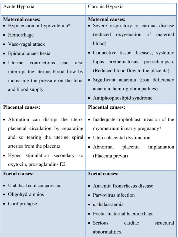
Table 2.1 Foetal types and the related causes
Acute Hypoxia Chronic Hypoxia
Maternal causes:
Hypotension or hypovolemia* Hemorrhage
Vaso-vagal attack Epidural anaesthesia
Uterine contractions can also interrupt the uterine blood flow by increasing the pressure on the fetus and blood supply
Maternal causes:
Severe respiratory or cardiac disease (reduced oxygenation of maternal blood)
Connective tissue diseases; systemic lupus erythematosus, pre-eclampsia. (Reduced blood flow to the placenta) Significant anaemia (iron deficiency
anaemia, hemo-globinopathies). Antiphosphoslipid syndrome Placental causes:
Abruption can disrupt the utero-placental circulation by separating and so tearing the uterine spiral arteries from the placenta.
Hyper stimulation secondary to oxytocin, prostaglandins E2
Placental causes:
Inadequate trophoblast invasion of the myometrium in early pregnancy* Utero-placental dysfunction
Abnormal placenta implantation (Placenta previa)
Foetal causes:
Umbilical cord compression
Oligohydramnios Cord prolapse
Foetal causes:
Anaemia from rhesus disease Parvovirus infection
α-thalassaemia
Foetal-maternal haemorrhage
Serious cardiac structural abnormalities.
*Any causes of haemorrhage or hypotension and hypovolaemia can reduce the maternal blood supply and so oxygen delivery to the uterus.
27
2.4 Foetal distress physiology
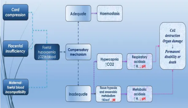
Foetal oxygen concentration (PO2) is lower than maternal (40mm Hg in umbilical vein compared to 95 mmHg in maternal artery). Oxygen saturation in the umbilical vein is almost the same as the maternal arterial blood and this could be explained by the fact that there is a higher haemoglobin concentration with higher affinity for oxygen in the foetal blood. This considers the first compensatory mechanism helping the foetus to release more oxygen to the tissues during the intra-partum foetal life.
Another compensatory mechanism for low oxygen concentration (PO2) provided by the maternal blood is redistribution of the blood to the most vital organs secondary to high cardiac output and increasing the extraction of the tissue oxygen [19] [25]. Figure 2.1 shows the patho-physiological process of foetal hypoxic disorder.
At the early stage of the foetal distress or during short-term episodes of acute hypoxia (lasting a few minutes) caused by uterine contractions and/or compressions of the umbilical cord [20], the foetus may tolerate the oxygen flow reduction with no signs of hypoxic complications. The foetus may be able to adapt the hypoxic effects by causing the blood flow to be redistributed and increasing the blood supply containing the nutrients and oxygen to the most vital organs such as the brain, myocardium, and upper body and reducing the perfusion of the kidneys, gastrointestinal tract, and lower extremities [21].
28
Figure 2-1 Foetal distress pathophysiology
In the case of chronic hypoxia, the long-time redistribution of oxygen has adverse consequences for the foetal heart. Further reduction of the foetal oxygenation will be overcome by the deterioration in cardiac output and the development of arterial hypertension. Barker et al. [22] indicated the “foetal origins of adult disease hypothesis”, “Surviving babies seem to be susceptible to the development of serious postnatal complication such as cardiovascular disease later in life”. Barker’s theory states “physiologic adaptation enables the foetus to survive a period of intrauterine deprivation resulting in permanent reprogramming of the development of key organs that may have pathological consequences in postnatal life”. Furthermore, reduction in oxygen perfusion will cause carbon dioxide accumulations in the foetal blood causing an increase in the partial pressure of carbon dioxide (PCO 2) and a concomitant
29
decrease in pH producing respiratory acidosis which is similar to the adult respiratory acidosis [22].
Continuous hypoxia deprives the foetus of the required oxygen to perform the aerobic reactions, resulting in accumulation of organic acids with the accumulation of pyruvic and lactic acids as a result of anaerobic metabolism and subsequently developing metabolic acidosis [23].
Compensatory foetal response to prolonged or profound reductions of oxygen can be summarised as foetal tachycardia (FHR >160 bpm), decreased movement, tone and breathing due to the decrease in oxygen consumption and finally decreasing the blood flow to some organs such as Kidney, Lungs, Gut, Liver and the Peripheral tissue (increase the anaerobic metabolism) compared to the increased blood flow to the Brain, Heart and Adrenals [24].
Thus, any damage that has happened to the foetal brain is correlated to the damage of other organs. Severe respiratory or metabolic acidosis indicates damage to several organs such as Lungs, Heart and Kidneys. Accordingly, evidence of multi organ failure, as well as metabolic acidosis, is required to diagnose foetal birth asphyxia [25].
2.5 Evidence of Multi-organ Failure
The following is a list of evidence for Multi-organ failure [25]:
Apgar score <3 at 5 minutes, means the neonatal vital signs have weakened with or without neonatal neurological squeals (hypotonia, seizure, irritability). Kidney-oliguria, anuria.
Lung-respiratory distress syndrome (RDS), pulmonary hypertension. Heart-cardiomyopathy, patent dectus arteriosus (PDA).
30
Liver–hypoglycaemia (low blood sugar) elevated liver enzymes. Biochemical profile (pH <7.0, Base deficit 8-16).
2.6 Foetal hypoxia Diagnosis:
Various methods of intra-partum foetal surveillance have been employed to detect the signs of hypoxia as early as possible to minimize the risk of life-long disability such as cerebral palsy and to reduce the mortality rate among the new-borns.
During hypoxia, numbers of cardio-respiratory system modifications we be made as hypoxia progresses in foetal life. These responses have been used as a compensatory mechanism to preserve the oxygenation of vital organs such as the brain and heart. As the foetal hypoxia progresses, Foetal heart pattern can be changed as a response to hypoxia. These pattern changes can be considered a useful way to detect the correlation between the foetal distress and the FHR changes [26]. Foetal heart rate monitoring, typically by use of the electronic assessment methods such as cardiotocography (CTG) and intermittent auscultation, are accepted methods of antenatal screening. However, the precise relationship between hypoxia and foetal heart rate changes is not well understood. This situation is likely to explain some of the inaccuracy inherent in diagnoses of asphyxia based on foetal heart rate observations [26].
Another way of foetal monitoring was summarised by Manning et al. [27]. Manning assessed the general foetal activity by evaluating five biophysical variables (foetal breathing movements, foetal movements, foetal tone, qualitative amniotic fluid volume, and the non-stress test) to form a biophysical profile of the foetus. Although Manning et al. had determined the foetal distress condition and perinatal mortality rate by studying each single variable, however the false negative rate was low and was similar between tests, while the false positive rate was high (>50%) and varied
31
significantly between tests. On the other hand, studying all variables together shows significant changes in both the false negative and false positive rates as compared to any single test. According to Thacker et al.’s study [28], it has been shown that foetal condition is diagnosed more accurately using an ensample of measurements of all the biophysical variables profile instead of studying each variable
Many other methods have been used through the antenatal care and during the labour process such as the cordocentesis. Cordocentesis is a foetal blood sampling by ultrasound guided needle aspiration from the umbilical cord, usually used to detect hypoxia due to placental development abnormalities [29]. The main challenge to the physicians is still the detection of foetal hypoxia during labour/birth process because foetuses usually are subjected to maximum stress due to the increased duration and frequency of the uterine contraction [29].
This may cause many threats to the foetal health such as damage to the cells of the central nervous system that leads to life-long disability. Up to three quarters of infants with severe hypoxic-ischaemic encephalopathy (HIE) die of multiple organ failure or lung infections caused by irregular breathing. Those who survive are commonly left with gross symptoms such as mental retardation, epilepsy, and cerebral palsy [30]. Continuous labour monitoring is essential to observe the foetal well-being. There are many studies indicating that foetal heart activity is the prominent source of information about foetal health and especially for detection of foetal hypoxia [31].
32
2. 6.1 Electronic foetal monitoring
Intra-partum monitoring of foetuses during labour has been commonly performed by monitoring the foetal heart such as the intermittent auscultation, which is main method of foetal surveillance in labour [32].
Significant changes can be detected using IA, if the time period was immediately after the contraction. Repetitive decelerations of FHR detected by IA may give the first clue of the Asphyxia presence and may be further clarified by the EFM changes. It was a challenging attempt to use the correlation between the FHR pattern and the foetal health state as an outcome predictor. However, the ability of the EFM to predict the outcome was poor, particularly in low-risk pregnancies. An evaluation study comparing the EFM and IA role in foetal hypoxia detection showed that, although the operative delivery rate by caesarean section was significantly increased, however, the foetal outcome in preventing serious complication was not improved. Many of these electronic methods have been the subject of recent controversy, as existing studies have found no benefit from using such methods in reducing rates of cerebral palsy or peri-natal mortality. Therefore, physicians have found many challenges to identify the best way of detecting foetal hypoxia by analysing the foetal heart rate [3]. In 1960, cardiotocography (continuous electronic foetal heart rate monitoring) was introduced to replace all the other traditional methods in monitoring the foetal heart. Since then, this method has been routinely used by obstetricians to assess the foetal heart rate and uterine contraction (UC) [33].
Examining Cardiotocography trace patterns by healthcare professionals to interpret the intra-partum FHR patterns and provide suitable management decisions is a big
33
challenge [34]. CTG records need high proficiency of the medical staff to identify the suspicious and pathological changes of the FHR that may correlate with the maternal uterine contraction. According to ACOG, SOGC, RCOG and FIGO (International Federation of Gynaecology and Obstetrics
)
guidelines, any deceleration in the FHR ≥15 bpm for ≥ 15 sec or isolated prolonged deceleration ≥ 3 min will indicate the foetal distress condition [35].
Their decision will identify the appropriate course of action (such as performing a Caesarean section). However, there is great variability among physicians in terms of how they perform this task. Furthermore, because significant hypoxia is usually rare, false alarms are common, leading physicians to disregard truly abnormal signals. Approximately 50 percent of birth-related brain injuries are deemed preventable, with incorrect CTG interpretation leading the list of causes [36, 37]. On the other hand, it is possible that some physicians miss some abnormalities of the FHR which are considered life-threatening changes that lead to life-long handicaps in the babies as well as the emotional distress of the parents and the financial compensation for the families who suffer from a medical catastrophe during labour.
Many techniques and methods have been used ante-natally to assess the foetal well-being and help understanding some of the life events before birth such as ‘ultrasound
imaging’ which, provide some ideas about the foetal size, skeleton abnormalities and vital organs development and Doppler test, which studies the foetal circulation and detects the vessels (arteries, veins) abnormalities before the birth [38]. Although, these methods can provide the physicians with a good assessment of the foetal wellbeing, unfortunately, none of the discussed methods can give an accurate diagnosis of the foetal hypoxia or asphyxia.
34
2.6.2 Foetal blood sampling
Further diagnostic tests are important in case of a typical (non-reassuring) foetal heart rate FHR signs. Foetal scalp sampling or umbilical cord sampling has been considered an essential test to confirm the foetal asphyxia diagnosis. In early 1980s, cordocentesis technique was used to identify the acid base state of the foetus and identify the early sign of respiratory acidosis (when the umbilical cord pH< 7.0) and metabolic acidosis (BDecf 8-16). Cordocentesis or Percutaneous Umbilical Cord Blood Sampling (PUBS) is a diagnostic test firstly performed by Daffos under an ultrasound guidance using a fixed needle guide attached to the base of the ultrasound transducer to study the biochemical profile of the umbilical blood sample [39]. According to Okamura et al. [40],Cordocentesis has considered “the precise evaluation of foetal condition to determine the timing of the delivery and to prevent the neurological sequelae caused by hypoxia” [40].
2.6.2.1 Foetal scalp blood sampling (FSS)
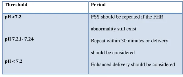
Foetal scalp blood sampling (FSS) should be performed when there is a typical (non-reassuring) FHR pattern detected by either IA or the EFM. Scalp blood sampling can be used in detecting fetal hypoxia by providing the pH level as an indicator. Table 2.2 presents the thresholds of the pH that relate to the clinical health state of the foetus and required management by the health staff.
35
Table 2.2 Thresholds for foetal health state
Threshold Period
pH >7.2
pH 7.21- 7.24
pH < 7.2
FSS should be repeated if the FHR abnormality still exist
Repeat within 30 minutes or delivery should be considered
Enhanced delivery should be considered
Foetal blood sampling can be considering an ideal method in detecting foetal hypoxia. Many researchers depended on this technique in detecting and predicting foetal hypoxia depending on the pH level, such as Saling et al. [7] and Westgren et al. [10]. However, this technique has some limitations as it gives instant, not continuous results making repeated sampling mandatory to follow the foetal status. In addition, there are difficult technical restrictions such as the operator skills, risk of sample contamination with the amniotic fluid, risk of foetal infection that may be transported from an active maternal infection (HIV, Genital herpes). Other limitations are the large amount of the blood that is required for the analysis (at least 30-50 ml) and the requirement of the cervix dilation as it should be at least 2cm dilated for the sampling to be done. Furthermore, FSS can give a false negative result in the case of metabolic acidosis, as it takes a long time for the H+ ions to cross the peripheral tissue in to the blood stream [10].
36
2.6.2.2 Umbilical cord blood sampling
According to the SOGC, it is important to do the cord blood gas sampling after all deliveries and it has been recommended to be done routinely for all neonates. Many international organizations have different recommendations about the use of umbilical artery or the vein blood sample. SOGC recommends measuring both umbilical artery and vein samples assessment. However, if only one is possible, it should be the arterial sample. On the other hand, ACOG recommends sampling the umbilical artery only for selective measurement of the cord gases (ACOG, 2006) [16]. While the Royal College of Obstetricians and Gynecologist recommends selective measurements of acid base status in the umbilical artery as a minimum (RCOG, 2001) [18].
Physicians usually use Umbilical cord blood sampling to detect five important thresholds to diagnose foetal asphyxia (pH, PCO2, PO2, BDecf and BE) [41]. In addition to the Apgar score, which is an expression of the infant’s physiological condition at one point in time, which includes subjective components (colour, heart rate, reflex irritability, muscle tone and respiration) [42].
PCO2 level is usually increased in the case of respiratory acidosis, which can be developed rapidly following the first neonatal breaths as a normal response to the umbilical cord clamping after delivery. The cord clamping or in the case of cord compression will cause interruption of the blood flow from the foetus to the placenta, which will cause accumulation of CO2 in the foetal blood vessels causing a decrease in the pH level as a result of the reaction between the CO2 and water producing bicarbonate and hydrogen ions, which will accumulate in the blood vessels causing the decrease in the pH level.
37
On the other hand, PCO2 will not change in the case of metabolic acidosis as in this case the pathogenesis will be different from the respiratory acidosis. Metabolic acidosis takes longer to develop as a result of decreasing the PO2 level that causes the foetus to shift to anaerobic metabolism to maintain the energy balance. As a result of the anaerobic metabolism, lactic acid will accumulate in the tissue and dissociate into lactate and hydrogen ions causing a decrease in the pH level when some of the hydrogen ions move from the tissue to the blood vessels [24].
Using pH in detecting the foetal hypoxia is considered an important indicator to quantify perinatal asphyxia [43]. However, pH alone will not differentiate between the respiratory and metabolic acidosis as it increases in both conditions as shown in Figure 2.1. The last threshold of the umbilical cord blood sampling is the base deficit/base excess which can provide a better differentiation between the respiratory and metabolic acidosis as they are both normal in the respiratory acidosis while in case of metabolic acidosis the base deficit will increase and the base excess will decrease [44]. In the case of a normally delivered infant with no sign of hypoxia or any other health state that could be related to oxygen level deprivation, the umbilical cord blood sampling should have the following thresholds:
Normal values in an umbilical arterial sample in a term newborn [44]:
pH: 7.18 – 7.38
PCO2: 32 – 66 (mmHg) HCO3: 17 – 27 (mmol/L) PO2: 6 – 31 (mmHg)
Base excess: -8 – 0 (mmol/L) Base deficit: 0 – 12
38
In this case the infant will not need any further intervention by the medical staff. However any changing of these levels could be an early sign of developing foetal hypoxia disorder. Although the hypoxic state can be varied in its severity, we will depend on two of the main guidelines that are followed by many physicians; the first guideline for acute intra-partum hypoxia is from the society of Obstetricians and Gynaecologists of Canada
Criteria of acute intra-partum hypoxia according to the Society of Obstetricians and Gynaecologists of Canada (SOGC):
Apgar score 0 – 3 for >15 minutes
Neonatal neurological signs (hypotonia, coma, seizure) Evidence of multi-organ failure
Umbilical cord pH < 7.0
Umbilical cord arterial base deficit < 16 mmol/L
It cannot conclude the existence of the hypoxic acidaemia without any of these criteria. All these conditions should be present for definite diagnosis of foetal acidaemia. The American College of Obstetricians and Gynaecologists (ACOG) have another guideline to classify the health state of the foetus according to the stage of the foetal life. The ante-partum birth asphyxia could happen when the foetus is still inside the womb, while the intra-partum birth asphyxia could happen during or after delivery. Therefore, there are two different sets of criteria to diagnose the foetal hypoxia.
39
A. The American College of Obstetricians and Gynaecologists criteria of intra-partum birth asphyxia:
Sudden or sustained bradycardia or the absence of FHR variability in the presence of persistent, late or variable decelerations.
Apgar score 0-3 beyond 5 minutes
Onset of multi organ failure within 72 hours of birth.
Early imaging study showing evidence of acute non focal abnormality
B. Ante-partum criteria of Asphyxia:
Evidence of metabolic acidosis (Umbilical cord pH< 7.0, base deficit 8-12)
Early onset of sever or moderate neonatal encephalopathy in infants born at 34 or more weeks of gestation
Cerebral palsy of the spastic quadriplegic or dyskinetic type.
Exclusion of other identifiable etiologies such as trauma, coagulation disorders, infectious conditions, or genetic disorders.
2.7 Foetal non- reassuring status management
The initial treatment used for non-reassuring foetal status is the intrauterine resuscitation. This will prevent any unnecessary intervention.
The main intrauterine resuscitation techniques that can be done by the physicians during the intra-partum stage are:
Maintain continuous oxygen supply to the foetus is the first priority during foetal distress management. Physicians usually start by changing the mother’s position to decrease the unnecessary pressure on the uterus or the placenta.
40
Ensuring the mother is well hydrated and has adequate oxygen.
Amnio-infusion: refers to the instillation of fluid into the amniotic cavity. This procedure is typically performed during labour through an intrauterine pressure catheter introduced trans-cervically after rupture of the foetal membranes. Alternatively, fluid can be infused through a needle trans-abdominally (the reverse process of amniocentesis). The rationale for amnio-infusion is that augmenting amniotic fluid volume may decrease or eliminate problems associated with a severe reduction or absence of amniotic fluid [45].
Tocolysis (using some medication or techniques to suppress the uterine contraction temporarily and delay the preterm delivery).
Nonetheless, there are some conditions in which emergency cesarean section is mandatory. However, due to the over-diagnosis of foetal distress and potential misinterpretation of the foetal heart rate, it is recommended to confirm a potential foetal distress diagnosis with a foetal blood acid base study. Overall, this condition points to the importance of prenatal care and proper monitoring of the mother and foetus throughout pregnancy.
41
2.8 Summary of the chapter
In this chapter, foetal asphyxia disorders are discussed. Main types of foetal hypoxia classified according to the main reason of oxygen level deprivation have been shown. Foetal hypoxia general causes can be summarized according to the time of hypoxic state development during foetal life into acute and chronic hypoxia. Discussion on the foetal hypoxia path physiological process to understand the main relationships that may affect the diagnostic variable combination was provided. Other information has been included in this chapter on the diagnostic procedures of the foetal hypoxia including, the electronic foetal monitoring and foetal blood sampling. Each of them is presented with the main clinical features and diagnostic criteria based on latest clinical guidelines and references. In general, this deep investigation of the different types of foetal hypoxia and the variable ways of diagnosis showed that foetal blood sampling during the ante partum stage of the foetal life as a diagnostic tool, can be depended upon by the physicians to start the management process according to the ACOG guideline. There is no big deference between the American College (ACOG) and the SOGC in identifying the hypoxic foetus criteria apart from the base deficit threshold and the timing of the asphyxia. The next chapter will discuss machine learning classifiers as well as the types that could help in analysing foetal data for classification purposes.
42
CHAPTER THREE: MACHINE LEARNING TYPES
AND APPLICATION
3.1 introduction
In this chapter, Machine learning classifiers will be identified as an artificial intelligence discipline. ML classifiers allow the computers to imitate the way by which humans can deal with large data automatically via various methods including analysis, self-training, observation and experience. Different classifiers that show advantage in data analysis especially medical data and various applications of real world problems, such as the decision tree classifiers, neural network and the support vector classifiers will be discussed.
3.2 Machine Learning Development
Machine learning is an artificial intelligence discipline geared toward the technological development of human knowledge. It is considered the fundamental sub-area of artificial intelligence and one of the fastest growing fields in computer science [46]. As digital technology increasingly infiltrates our daily life, more data is continuously getting bigger, either generated or collected. Machine learning technology plays a significant role in data analysis in variant fields such as astronomy and biology. The collection of datasets is getting larger every year and also it is represented in different ways, not just numbers or character strings anymore but images, video, audio, documents web pages, graphs and more.
43
Machine learning has the ability to improve its own performance through the use of software and algorithms that use artificial intelligence techniques to imitate the ways humans seem to learn, such as repetition and experience through continuous exposure to new scenarios, testing and adaptation. Machine learning classifiers are allows computers to perform new learning situations automatically without human intervention or assistance, which can be done via analysis, self-training, observation and experience.
Jaime et al. [47] showed that the computer systems do have the ability to perform any task through examples or previously solved tasks; in addition computers cannot improve the task performance on the basis of past mistakes or obtain new abilities by observing and imitating experts.
Machine learning research has been extended to identify the possibility of instructing the computer system in a new way to match the human abilities. Alpaydin et al. [48] stated that
“With advances in computer technology, we currently have the ability to store and process a large amount of data, as well as access it from physically distant locations over a computer network. Most data acquisition devices are digital now and record reliable data”.
Programming the computer systems through machine learning methods can optimise the standard performance by using example data or past experience. Machine learning usually uses statistical theories in building a mathematical classifier, and then refines up this classifier to some parameters using the training data or past experience. The developed classifier may have predictive characteristics to make future predictions, or descriptive features to gain knowledge from data, or both.
44
The learning process can be achieved by training the classifier through an efficient algorithm to solve the optimised problem. The representation of the learned classifier and algorithmic solution for interpretation needs to be efficient as well.
The major advantage of machine learning is the fact that it can extract patterns from massive amounts of data which humans cannot do because humans cannot retain everything in memory and they cannot perform obvious/redundant computations for hours and days to come up with interesting patterns. Machine learning has found major applications in finance, healthcare, entertainment, robotics, and many more. Once a Machine Learning classifier with good generalization capabilities is learned, it can handle previously unseen scenarios and take decisions accordingly.
Some tasks cannot be defined well except by example; that is it is possible to specify input and output pairs but not a concise relationship between inputs and desired outputs. Machines are able to adjust their internal structure to produce correct outputs for a large number of sample inputs.
Machine learning methods can often be used to extract any relationships or correlations that are hidden among large piles of data [50]. Machines can adapt to the changing environment over time, which would reduce the need for constant redesign.
45
3.4 Machine Learning: Algorithms Types
Machine learning algorithms are organized in specific assembling, based on the desired outcome of the algorithm. Common algorithm types include, Supervised learning, unsupervised learning, Semi-supervised learning, Reinforcement learning [52]. We will use the supervised learning as the main concept for data classification in this research.
3.4.1 Supervised learning
It is considered the most important methodology in machine learning, which means the ability of systems to infer a function of variant tasks depending on supervised training data. The training data usually consist of a set of training samples. Each instance has a pair consisting of input data (typically a vector) and a desired result as an output value (also called the supervisory signal) [53]. Supervised learning involves learning a mapping between a set of input variables X and an output variable Y and applying this mapping to predict the outputs for unseen data. An optimal consequence will allow for the algorithm to correctly determine the class labels for unseen instances. The main characteristic of the supervised classifiers is the generalization ability, which means having the ability to produce reasonable outputs for inputs not encountered during the training [52].
The standard formulation of the supervised learning task is the classification, when the learner needs to learn (approximate the behavior of) a function that maps a vector into one of several classes by observing several input-output examples of the function [52]. In the building phase, the training procedure continues until the algorithm is able to achieve the best accuracy on the given data.
46
3.4.2 Unsupervised learning
Unsupervised learning is also one type of machine learning model that can be applied to drive implication from training datasets involving input data without output (labelled responses). In unsupervised learning, the system will learn by a set of particular input patterns that reflect the statistical structure without specific target output or environmental evaluation for each input [54]. Unsupervised learning has more similarity to the human brain compared to supervised learning. For instance there are about 10 million photoreceptors in the human eye with constantly changing activities in visualization of different objects, providing all the information that indicates what the objects are. The structural and physiological properties of neocortex synapses are known to be influenced by the sensory neurons’ pattern of activity. However, none of the information is available, making the unsupervised learning more essential in computational models for synaptic variation. Although the unsupervised learning shows no benefit in data analysis for prediction of a response as the supervised learning does, it shows a precise importance in identifying unknown subgroups among the variables.
3.4.3 Semi-supervised learning
Semi-supervised learning is a learning paradigm concerned with the study of how computers and natural systems such as humans learn in the presence of both labelled and unlabelled data. Availability of large amounts of unlabelled data compared to the
47
small amounts of labelled data, in addition to the expensive labelling or data annotating has motivated machine learners to study new methods that can use information of the input distribution.
Semi-supervised learning (SSL) can be considered a midway between supervised and unsupervised learning. The standard semi-supervised learning has data sets divided into two parts, one points with labels while for the other points, the labels are not known [54].
Semi-Supervised Learning in Practice
Semi-supervised learning is useful whenever there are far more unlabelled data than labelled. This type of algorithm is a significant tool in machine learning due to the ability to use unlabelled data to enhance supervised learning tasks and can yield considerable improvement in accuracy especially when the data is expensive and scarce [55]. This is the case in many application areas of machine learning, for example: In speech recognition, it costs almost nothing to record huge amounts of speech, but labelling it requires some human to listen to it and type a transcript. Billions of Webpages are directly available for automated processing, but to classify them reliably, humans have to read them.
Another example is the study of the protein sequences. Protein sequences are nowadays acquired at industrial speed (by genome sequencing, computational gene finding, and automatic translation), to resolve a three-dimensional (3D) structure or to determine the functions of a single protein may require years of scientific work.
48
3.4.4 Reinforcement learning
In the machine learning domain, Reinforcement learning (RL) is usually identified as techniques whereby an algorithm learns from the regular consequences of its actions instead of being explicitly taught based on previous experiences (exploitation). The machine is exposed to an environment where it trains itself continually using trial and error. This machine learns from past experience and tries to capture the best possible knowledge to make accurate business decisions [56].
The reinforcement machine learning instructs the algorithm about the policy of how to act, given an observation of the world. Every action has some impact in the environment, and the environment provides feedback that guides the learning algorithm [57].This kind of machine learning would not emphasise which action should be taken under consideration, but instead would have to discover which action produced such an excellent reward. RL is usually utilised in different applications to solve a number of complex tasks. For instance, RL has performed in medical diagnosis, speech recognition, bioinformatics, computational vision, spell recognition, and robots Locomotion [52].
49
3.5 Machine Learning Classifiers
Machine learning algorithms are used to optimize the performance criterion using example data or past experience [58]. .
Machine learning uses the theory of statistics in building mathematical classifiers, because the core task is to make inference from sample data. In training, an efficient classifier is needed to solve the optimization problem, as well as to store and process the massive amount of data generated. Once the classifier is learned, its representation and algorithmic solution for inference needs to be efficient. For the classification and regression problem, there are various choices of Machine Learning classifiers each of them can be viewed as a black box that solves the same problem [59].
In this study, we chose different types of classifiers, based on their ability to classify a real world medical data. For instance, Jezewski et al. 2010 [80] and Spilka et al.2012 [84] used the NNET and SVM to classify their data and the results showed an acceptable accuracy outcomes. In addition to use the cross-validation and test harness to determine which classifier performs best on test data [125]. Studying our data characteristics (real world data, simple row data containing numerical variables and categorical classes) encouraged us to choose the decision tree based classifiers as well. Its methodology considered very easy to understand, as it doesn’t require analytical background to interpret the data. It has been considered one of the quickest ways to classify variables and find the relationship between them to predict the target variable, and it can control both numerical and categorical variables.