R A P I D C O M M U N I C A T I O N
Open Access
Incorporation of a hinge domain improves
the expansion of chimeric antigen receptor
T cells
Le Qin
1,2,3†, Yunxin Lai
1,2,3†, Ruocong Zhao
1,2,3, Xinru Wei
1,2,3, Jianyu Weng
4, Peilong Lai
4, Baiheng Li
1,2,3,
Simiao Lin
1,2,3, Suna Wang
1,2,3, Qiting Wu
1,2,3, Qiubin Liang
5, Yangqiu Li
6, Xuchao Zhang
7, Yilong Wu
7, Pentao Liu
8,
Yao Yao
1,2,3, Duanqing Pei
1,2, Xin Du
4and Peng Li
1,2,3*Abstract
Background:
Multiple iterations of chimeric antigen receptors (CARs) have been developed, mainly focusing on
intracellular signaling modules. However, the effect of non-signaling extracellular modules on the expansion and
therapeutic efficacy of CARs remains largely undefined.
Methods:
We generated two versions of CAR vectors, with or without a hinge domain, targeting CD19, mesothelin,
PSCA, MUC1, and HER2, respectively. Then, we systematically compared the effect of the hinge domains on the
growth kinetics, cytokine production, and cytotoxicity of CAR T cells in vitro and in vivo.
Results:
During in vitro culture period, the percentages and absolute numbers of T cells expressing the CARs
containing a hinge domain continuously increased, mainly through the promotion of CD4+ CAR T cell expansion,
regardless of the single-chain variable fragment (scFv). In vitro migration assay showed that the hinges enhanced
CAR T cells migratory capacity. The T cells expressing anti-CD19 CARs with or without a hinge had similar antitumor
capacities in vivo, whereas the T cells expressing anti-mesothelin CARs containing a hinge domain showed
enhanced antitumor activities.
Conclusions:
Hence, our results demonstrate that a hinge contributes to CAR T cell expansion and is capable of
increasing the antitumor efficacy of some specific CAR T cells. Our results suggest potential novel strategies in CAR
vector design.
Keywords:
CAR T cell, Hinge domain, Expansion, CD4+ T cell, CD19, Mesothelin
Background
In the last 5 years, chimeric antigen receptor (CAR) T cells
have emerged from bench to bedside and made headlines
in clinical trials at a number of academic institutions [1
–
4].
CARs are recombinant receptors that specifically target
tumor-surface antigens. Once the CARs are transfected
into T cells, the cells acquire supraphysiologic properties
and act as
“
living drugs
”
[5, 6]. Multiple iterations of CARs
have been developed, mainly focusing on intracellular
sig-naling modules, which are deemed crucial for CAR design
[7, 8]. To achieve appropriate costimulatory signals so as to
activate effector T cells, improve response, and prolong
per-sistence, many different kinds of costimulatory receptors
can be incorporated (e.g., CD28 [9, 10], 4-1BB [11, 12],
OX40 [13], ICOS [14], and CD27 [15]), alone or in tandem
[16]. However, the effect of non-signaling extracellular
modules, such as hinge and TM domains, on the
prolifera-tion of the transduced T cells and therapeutic efficacy of
CARs remains largely unclear [17].
A hinge domain is a structure between the targeting
moiety and the T cell plasma membrane [18]; these
sequences are generally derived from IgG subclasses
* Correspondence:[email protected] †Equal contributors
1Key Laboratory of Regenerative Biology, South China Institute for Stem Cell
Biology and Regenerative Medicine, Guangzhou Institutes of Biomedicine and Health, Chinese Academy of Sciences, Guangzhou 510530, China
2
Guangdong Provincial Key Laboratory of Stem Cell and Regenerative Medicine, South China Institute for Stem Cell Biology and Regenerative Medicine, Guangzhou Institutes of Biomedicine and Health, Chinese Academy of Sciences, Guangzhou 510530, China
Full list of author information is available at the end of the article
(such as IgG1 and IgG4), IgD and CD8 domains, of which
IgG1 has been most extensively used [19–21]. Currently,
studies of the hinge domain mainly focus on the following
four aspects: (1) reducing binding affinity to the Fc
γ
re-ceptor, thereby eliminating off-target activation [19, 21];
(2) enhancing the single-chain variable fragment (scFv)
flexibility, thereby relieving the spatial constraints between
tumor antigens and CARs, in turn promoting synapse
formation between the CAR T cells and target cells; for
example, to overcome steric hindrance in MUC1-specific
CAR, a flexible and elongated hinge of the IgD isotype
can be inserted [20]; (3) reducing the distance between an
scFv and the target epitope, for example, anti-CD22 CAR
needs a hinge domain to exert optimal cytotoxicity [22];
and (4) facilitating the detection of CAR expression using
anti-Fc reagents. Nevertheless, the influences of the hinge
domain on CAR T cell physiology are not well
understood.
To better understand the effect of the hinge domain
on CAR T cells, we generated two versions of CARs,
with or without a hinge domain, targeting CD19,
mesothelin, PSCA (prostate stem cell antigen), MUC1,
and HER2 (human epidermal growth factor receptor 2),
respectively [23–31]. We systematically compared the
effect of the hinge domains on the growth kinetics,
cyto-kine production, and cytotoxicity of CAR T cells in vitro
and in vivo. We revealed that the incorporation of a
hinge into CAR constructs can substantially increase the
CAR T cell percentage during the in vitro culture period,
enhance the invasiveness of CAR T cells. In addition, we
found that anti-CD19 CAR T cells with or without a
hinge domain have similar abilities to eliminate leukemia
cells, whereas a hinge domain can enhance the in vivo
antitumor activity of anti-mesothelin CAR T cells.
Methods
Cells and culture conditions
NALM6-GL (acute lymphoblastic leukemia line, stably
transfected with GFP and luciferase) and A549-GL (human
lung cancer cell line, stably transfected with GFP and
lucif-erase) cell lines were cultured in RPMI-1640 (Gibco, Life
Technologies). HEK293T cells used for lentivirus
produc-tion were cultured with DMEM (Gibco, Life Technologies).
DMEM and RPMI-1640 media were supplemented with
10% heat-inactivated FBS (Gibco, Life Technologies),
10 mM of HEPES, 100 U/ml of penicillin, 100
μ
g/ml of
streptomycin, and 2 mM of
L-glutamine (Gibco, Life
Technologies).
Human peripheral blood mononuclear cells (PBMCs)
from healthy donors were obtained from Guangdong
General Hospital, after obtaining informed consent and
for research use only. Pan T cells were enriched from
the PBMCs using a
“Pan T cell isolation kit”
(Miltenyi
Biotec, Germany). CD8+ T cells were positively isolated
from the Pan T cells using
“CD8 microbeads”
(Miltenyi
Biotec, Germany); the unlabeled cells that passed
through the column were collected, representing the
CD4+ T cells.
Construction of chimeric antigen receptors (CARs)
CD19.28z, Meso.28z, HER2.28z, and PSCA.28z CARs
were constructed by linking sequences from a signal
peptide derived from GM-CSF (GenBank; AAA58735.1,
aa 1–19) to the corresponding antigen-specific
single-chain variable fragment (scFv); these CARs do not have
a spacer domain. Anti-CD19 scFv derived from FMC63
monoclonal antibody, anti-Mesothelin scFv derived from
SS1 monoclonal antibody, anti-HER2 scFv derived from
FRP5 monoclonal antibody, and PSCA scFv derived
from humanized 1G8 monoclonal antibody. The CARs
were codon-optimized and chemically synthesized using
Genscript. A spacer containing a hinge domain and a
CH3 domain derived from human IgG4 (GenBank;
AAC82527.1, aa 98–329) was included in PSCA-H.28z
CAR; an additional IgD hinge spacer was included in
MUC1-H.28z CAR. The scFv of anti-MUC1 CAR
de-rived from HMFG2 monoclonal antibody. The resulting
products were sub-cloned into a Pwpxld-based lentiviral
backbone plasmid encoding the transmembrane and
intracellular domains of CD28 (aa 153–220) and the
intracellular domain of CD3-
ζ
(aa 52–163). CD19-H.28z
CAR and Meso-H.28z CAR were constructed by
over-lapping PCR, adding the hinge domain and the CH3 of
IgG4 to the 3′
end of the scFv.
Lentiviral production and transduction
removed on day 5. Fresh medium was added every
2 days to maintain an appropriate cell density ranging
from 5 × 10
5to 1 × 10
6cells/ml.
Flow cytometry
All of the samples were analyzed with an LSR Fortessa
or C6 (BD Bioscience), and the data were analyzed using
FlowJo software. CAR T cells were detected by GFP, and
T cell phenotypes were evaluated via CD3 PE-cy7 (clone
OKT3, eBioscience), CD3 BV421 (clone UCHT1, BD),
CD8a PE (clone HT8a, eBioscience), CD8a PE-cf594
(clone RPA-T8, BD), and CD4 APC (clone OKT4,
eBioscience). All FACS plots representing the CAR T cell
phenotypes were gated on CD3 and GFP double positive
cells.
In vitro tumor-killing assays and cytokine-release assays
The target leukemia cells, NALM6-GL, were co-cultured
with GFP T, 19.28z, or 19-H.28z T cells at the indicated
E:T ratios in triplicate in U-bottomed 96-well plates for
18 h; A549-GL cells were co-cultured with GFP T,
Meso.28z T, or Meso-H.28z T cells, each well of 96-well
plates contained 200
μ
l supernatants. One hundred
microliter supernatants from the wells with E:T ratios of
1:1 were collected and used for detecting the
concentra-tions
of
IL-2
and
IFN-
γ
using
an
ELISA
kit
(eBioscience). The luciferase substrate
D-luciferin
(potas-sium salt, 150
μ
g/ml, Cayman Chemical, USA) was
added to a 100
μ
l/well, and the viability of the target
cells was monitored by a microplate reader at a 450-nm
excitation wavelength. The background luminescence
was negligible (<1% of the signal from the wells with
only target cells); therefore, the target cell viability (%)
was calculated as (experimental signal/maximal signal) ×
100%, and the killing percentage was calculated as 100%
−
viability percentage.
Transwell cell migration assay
For the 19.28z T and 19-H.28z T cells, Nalm6 cell lysates
were used as a chemoattractant in the lower chamber,
while for the Meso.28z T and Meso-H.28z T cells, A549
cell lysates were used as a chemoattractant in the lower
chamber. The T cells were cultured in an insert coated
with Matrigel for 24 h, and the cells that transmigrated
to the lower chamber were counted by flow cytometry.
The percentage of migration was calculated as follows:
(CAR T cells migrating through the Matrigel chamber
membrane/total CAR T cells in insert membrane before
assay begin) × 100.
In vivo studies
NSI mice (NOD-scid-IL2Rg
−/
−mice, Guangzhou Institutes
of Biomedicine and Health (GIBH)), aged 6–12 weeks
were used to construct the xenograft tumor mouse
models. All of the animal studies were carried out in
ac-cordance with instructional guidelines from the China
Council on Animal Care and under protocols approved by
the guidelines of the Ethics Committee of Animal
Ex-periments at GIBH. Equivalent numbers of male and
female mice were used. The NALM6-GL leukemia
lines were intravenously inoculated into 2 × 10
5cells,
and the A549-GL carcinoma lines were inoculated
into 5 × 10
5cells subcutaneously (flank). The mice
then received an adoptive transfer of CAR T cells
intravenously 3–14 days later, as indicated in the
indi-vidual experiments. To control the differences in
transduction efficiency, non-transduced T cells were
supplemented to ensure that both the number of
CAR+ T cells, and the total number of T cells
remained constant across all CAR T cell groups. The
leukemia burden was evaluated using a cooled CCD
camera system (IVIS 100 Series Imaging System,
Xenogen,
Alameda,
CA,
USA).
The
mice
were
injected intraperitoneally with
D-luciferin firefly
potas-sium salt at 75 mg/kg and then imaged 5 min later
with an exposure time of 30 s. Quantification of the
total and average emissions was performed using
Living Image software (Xenogen).
Statistics
All graphs report the mean ± SEM. All statistical
ana-lyses were performed with Prism software version 6.0
(GraphPad). The statistical significance of all data was
calculated using an unpaired Student’s
t
test with the
Bonferroni correction for multiple comparisons, where
applicable.
P
< 0.05 was considered significant and is
designated with an asterisk in all figures.
Results
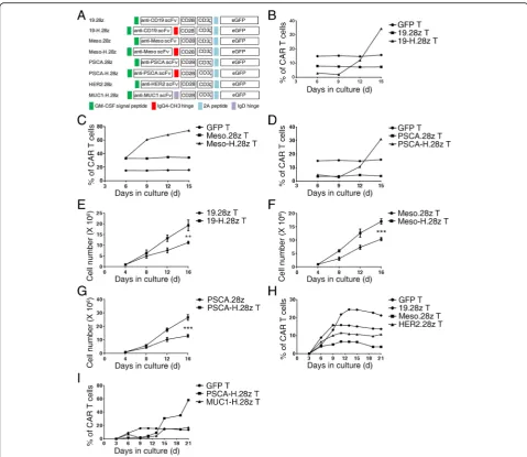
MUC1-H.28z T cells also increased from 6.9 to 17.3%,
al-though the MUC1-H.28z CAR was designed with an IgD
hinge, which indicates that the ability to increase the CAR
T cell percentage is not restricted to IgG4-CH3 hinges
(Fig. 1i). The absolute number of CAR T cells also
signifi-cantly increased (Fig. 1e–g). In contrast, the percentages of
CAR T cells without a hinge domain tended to be stable
throughout the in vitro expansion process (Fig. 1b–d;
Additional file 1: Figure S1), and their total cell numbers
were less than those of CAR T cells containing a hinge
domain (Fig. 1e–g). Taken together, these results
demon-strate that the incorporation of a hinge domain can
promote the growth of CAR T cells in vitro.
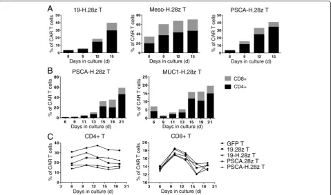
Hinge incorporation mainly promotes CD4+ CAR T cell
expansion
To identify which T cell subsets were most responsive to
the incorporation of a hinge domain, we monitored the
ratios of CD4+ and CD8+ CAR T cells in culture and
found that the percentages of CD4+ 19-H.28z T and
Fig. 1Hinge incorporation can promote the expansion of CAR T cells.aSchematic representation of CAR constructs specific for different
CD4+ Meso-H.28z T cells increased from 3.13 to 29.6%
(Fig. 2a, left; Additional file 2: Figure S2) and from 19.9
to 46.5% (Fig. 2a, middle), respectively. In addition, the
percentages of CD4+ PSCA-H.28z T cells increased
from 3.06 to 34.3% (Fig. 2a, right) and from 0.92 to
46% (Fig. 2b, left). The CD4+ MUC1-H.28z T cells
also increased from 4.14 to 14.9% (Fig. 2b, right).
However, the influence of the hinge domain on the
percentages of CD8+ CAR T cells was less
pro-nounced, as the percentages of CD8+ 19-H.28z T and
CD8+ Meso-H.28z T cells increased from 0.4 to
10.5% (Fig. 2a, left; Additional file 2: Figure S2) and
from 14.1 to 23.8% (Fig. 2a, middle), respectively. In
addition, the CD8+ PSCA-H.28z T cells increased
from 0.92 to 6.34% (Fig. 2a, right) and from 0.67 to
12.6% (Fig. 2b, left). The CD8+ MUC1-H.28z T cells
also increased from 2.85 to 4.61% (Fig. 2b, right). The
percentage of CAR T cells without a hinge domain
tended to be stable throughout the in vitro culture
period (Additional file 3: Figure S3). Interestingly,
when we isolated CD4+ T and CD8+ T cells and
cultured them separately in vitro, the percentages of
CAR T cells with or without a hinge domain all
tended to be stable in both CD4+ T and CD8+ T
cells, consistent with the GFP control T cells (Fig. 2c).
This suggests that the use of a hinge to enhance CD4
+ CAR T cell expansion requires the co-participation
of CD8+ CAR T cells. In summary, hinge
incorpo-ration mainly promotes CD4+ CAR T cell expansion
during the in vitro culture period.
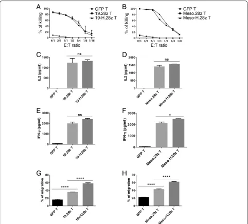
Hinge incorporation can enhances migratory capacity of
CAR T cells
To study whether the incorporation of a hinge domain
af-fects the cytotoxicity of CAR T cells, we compared the
killing capacities of anti-CD19 and anti-mesothelin CARs
with and without a hinge. Both 19.28z T and 19-H.28z T
cells efficiently lysed the NALM6-GL (Fig. 3a), indicating
that the killing capacities of these two CARs were similar.
Similarly, there were no significant differences between
the lysis capacities of Meso.28z T and Meso-H.28z CAR T
cells (Fig. 3b). For cytokine production, both 19-H.28z T
and Meso-H.28z T cells produced similar levels of IL2 and
IFN-
γ
compared with their hinge-free counterparts
Fig. 2Hinge incorporation mainly promotes CD4+ CAR T cell expansion.aFlow cytometric analysis of the percentage of CD4+ and CD8+ 19-H.28z T
(Fig. 3c–f ). Next, we compared the migratory capacity of
GFP T, 19.28z T, and 19-H.28z T cells, using NALM6 cell
lysate as a chemoattractant in the lower chamber of the
transwell plate. Interestingly, we found that the 19-H.28z
T cells transmigrated the Matrigel more efficiently than
the 19.28z T cells (Fig. 3g). Similar results were also
ob-tained in the Meso-H.28z T cells (Fig. 3h), suggesting that
hinge incorporation enhanced the migratory and invasion
capabilities of CAR T cells.
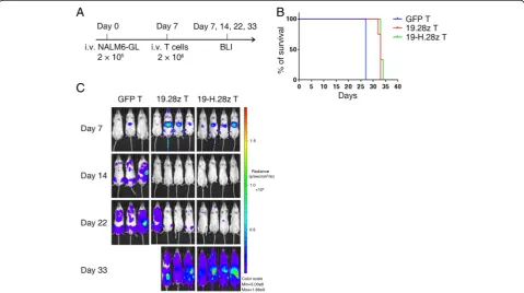
19-H.28z T and 19.28z T cells have comparable anti-tumor
efficacy in vivo
Subsequently, we evaluated the in vivo antitumor
capaci-ties of CAR T cells with or without the hinge domain in
cell-line-derived xenograft-bearing mice. Immune
defi-cient NSI mice were intravenously injected with 2 × 10
5NALM6-GL cells, followed by the infusion of a single
dose of 2 × 10
6GFP, 19.28z, or 19-H.28z T cells on day 7
(Fig.
4a)
[32–36].
Bioluminescence
imaging
(BLI)
showed a reduced tumor burden in the mice infused
with 19.28z T and 19-H.28z T cells compared with those
infused with GFP T cells on day 14. However, both the
19.28z T and 19-H.28z T-cell-infused mice relapsed on
day 22 (Fig. 4c). The survival times of the 19.28z T and
19-H.28z T cell groups also showed no significant
differ-ence (Fig. 4b). In summary, the introduction of a hinge
into anti-CD19 specific CARs did not enhance their in
vivo antitumor capacities.
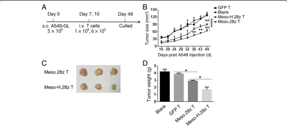
Meso-H.28z T cells exhibit enhanced antitumor capacities
in vivo
To study whether a hinge can affect the antitumor
capacities of CARs that are specific for solid tumors, a
solid-tumor mouse model was established, in which
A549-GL cells were subcutaneously injected into NSI
mice. Mice bearing an established tumor were treated
I.V. with two doses of Meso.28z, Meso-H.28z, or GFP T
cells, with the first dose on day 7 and the second on day
10. The tumor diameters were measured every 6 days.
On day 49, the mice were sacrificed (Fig. 5a).
Interest-ingly, tumor growth in both the Meso.28z T and
Meso-H.28z T cell groups was delayed compared with the
blank and GFP T cell group, but the delay was greater in
the Meso-H.28z T group (Fig. 5b). Results consistent
with these were also obtained from weight measurement
and photographic inspection of the tumors (Fig. 5c–d).
Fig. 419-H.28z T and 19.28z T cells have comparable antitumor efficacy in vivo.aTimeline and events of the experiment with intravenous
These results demonstrated that the incorporation of a
hinge domain enhanced the antitumor capacities of
anti-mesothelin CARs.
Discussion
Despite the remarkable progress in CAR T cell-based
immune therapy, several obstacles remain [37, 38]. For
example, the efficiency of CAR T cell expansion requires
improvement. Recently, some groups have reported that
an optimal CD4/CD8 ratio is important for the in vivo
antitumor activity of CAR T cells, and the percentage of
CD4+ CAR T cells is positively correlated with patient
recovery rates [39–42]. Because CD8+ T cells tend to be
preferentially expanded in current T cell in vitro culture
systems [43], a method to promote the expansion of
CD4+ T cells is urgently needed. Herein, we found that
both IgG4-CH3 and IgD hinges were able to
continu-ously increase the CAR T cell percentages and absolute
cell numbers during the in vitro culture period [20, 44],
and even more importantly, the additional CAR T cells
were mainly CD4+. However, when we isolated the CD4
+ T and CD8+ T cells and cultured them separately in
vitro, the increased-growth effect disappeared. The
mechanism of the increased growth will be studied by us
in the future.
To date, many different versions of anti-CD19 CARs
have been used in clinical trials [38]. The scFvs of these
CARs are almost all derived from FMC63 mAb [45],
while various different hinge domains and costimulatory
molecules have been used. For example, CTL019, which
is the most widely used CAR in CD19+ leukemia and
lymphoma treatment, has a CD8
α
hinge [46, 47], while
CD19RCD28 CAR uses a modified IgG4 hinge and Fc
region [48], and also has a hinge-deleted version [19].
The functional differences between CD28 and 41-BB
costimulatory molecules have already been well
charac-terized [49]; however, the influence of the hinge domain
on anti-CD19 CARs has not been studied. Our results
showed that the killing activities of 19.28z T and 19-H.28z
T cells were similar whether in vitro or in vivo. There are
several possible explanations of this result: the location of
the CD19 epitope recognized by the FMC63 mAb is not
membrane-proximal, there are no steric inhibitory effects
between FMC63 mAb and its epitope, or the density of
the CD19 molecule on tumor cells is high [50]. These
factors may also partially explain the greater popularity of
CD19 in clinical trials compared with CD20 and CD22.
Mesothelin is a glycosyl–phosphatidyl inositol-linked
cell surface glycoprotein, which is highly expressed in
mesothelioma, and lung, pancreas, breast, ovarian, and
other cancers, and has been used as a tumor antigen of
CAR T cells in several trials [51]. We have shown that
Meso-H.28z T cells have a better tumor-eradication
capacity than Meso.28z T cells in tumor-bearing mice,
suggesting that the mesothelin epitope recognized by the
Fig. 5Meso-H.28z T cells exhibit enhanced antitumor capacities in vivo.aTimeline of events of the xenograft experiment with the subcutaneous
scFv may be membrane-proximal, or that there exist
steric inhibitory effects between scFv and its epitope, like
in SM3 mAb (a MUC1-specific mAb) [20]. Thus, a hinge
is necessary to reduce the distance or ameliorate the
steric inhibitory effects between the scFv and its epitope.
Furthermore, a hinge can also improve the flexibility of
the scFv, which may also be one of the reasons why
Meso-H.28z T cells exhibit better anti-tumor activities
than Meso.28z T cells. The importance of Ab flexibility
has also been demonstrated in naive B cells which
co-express cell surface IgM, which lacks a hinge, and IgD,
whose elongated monomeric hinge is the longest of all
Ab isotypes [52]. As a result, IgD can assume a
“T
shape”
in which Fab regions can engage Ag in virtually
any orientation, making the scFv omni-directional.
Conclusions
To summarize, our data demonstrate that the
incorpo-ration of a hinge domain can enhance CAR T cell
expan-sion during the in vitro culture period, mainly by
promoting CD4+ CAR T cell proliferation, and a hinge
domain can also enhance the antitumor efficacy of some
specific CARs. Our results suggest potential novel
strategies in CAR vector design.
Additional files
Additional file 1: Figure S1.Hinge incorporation can promote the
expansion of CAR T cells. Flow cytometric analysis of the percentage of 19.28z, 19-H.28z T cells, Meso.28z, Meso-H.28z T cells, PSCA.28z, PSCA-H.28z T cells, and GFP control T cells from day 6 to 15 during the in vitro culture period. The data are representative of independent experiments verified with cells from over three individual healthy human donors. (JPG 2876 kb)
Additional file 2: Figure S2.Hinge incorporation promotes CD4+
anti-CD19 CAR T cell expansion. Flow cytometric analysis of the percentage of CD4+ and CD8+ GFP T, 19.28z T, and 19-H.28z T during the in vitro culture period. The data are representative of independent experiments verified with cells from over three individual healthy human donors. (JPG 2320 kb)
Additional file 3: Figure S3.Percentages of CD4+ and CD8+ CAR T
cells without a hinge domain both tended to be stable throughout the in vitro culture period. Flow cytometric analysis of the percentage of CD4 + and CD8+ (A) 19.28z T, (B) Meso.28z T, (C) PSCA.28z T, (D) HER2.28z T, and (E) GFP control T cells during the in vitro culture period. The data are representative of independent experiments verified with cells from over three individual healthy human donors. (JPG 924 kb)
Abbreviations
BLI:Bioluminescence imaging; CAR: Chimeric antigen receptor; HER2: Human epidermal growth factor receptor 2; Meso: Mesothelin; PSCA: Prostate stem cell antigen; scFv: Single-chain fragment variable
Acknowledgements
Dr. Guohua Huang and Dr. Qiuhua Deng helped him to collect some data and gave several suggestions for the revision. So I added Dr. Huang and Dr. Deng into author list of this manuscript in the revision. However, I think I should thank them in the acknowledgment instead of giving them the authorship after carefully reading the BioMed Central authorship policy.
Funding
This study was supported by the National Natural Science Foundation of China (NSFC)—81272329, 81522002, 81570156, and 81327801, Strategic Priority Research Program of the Chinese Academy of Sciences (XDA01020310), the Natural Science Fund for Distinguished Young Scholars of Guangdong Province (2014A030306028), the Guangdong Provincial Applied Science and Technology Research & Development Program (2016B020237006), the Guangdong Provincial Outstanding Young Scholars Award (2014TQ01R068), the Guangdong Provincial Basic Research Program (2015B020227003), the Guangdong Provincial Research and Commercialization Program (2014B090901044), the Guangdong Province and Chinese Academy of Sciences Joint Program for Research and Commercialization Program (2013B091000010), the Guangzhou Basic Research Program (201510010186), the MOST funding of the State Key Laboratory of Respiratory Disease, and the National Basic Research Program of China (973 Program) (2011CB504004 and 2010CB945500), the Frontier and key technology innovation special grant from the Department of Science and Technology of Guangdong province, (2014B020225005), and the Guangzhou Science Technology and Innovation Commission Project (201504010016).
Availability of data and materials
All relevant data and materials within this work are made available in this manuscript. Any additional information can be made freely available to any scientist on reasonable request.
Authors’contributions
LQ, YL, YL, XZ, YW, PL, YY, DP, XD, and PL conceived and designed the study. LQ, YL, RZ, XW, and YY analyzed and interpreted the data. JW and PL provisioned study the material. BL, SL, SW, QW, and QL provided an administrative support. LQ and PL wrote the manuscript. LQ, YL, and RZ performed the experiments. PL provided the financial support. All authors read and approved the final manuscript.
Competing interests
The authors declare that they have no competing interests.
Consent for publication
Consent to publish has been obtained from the participants.
Ethics approval and consent to participate
All animal experimental protocols were performed in accordance with instruction guidelines from the China Council on Animal Care and approved by the guidelines of the Ethics Committee of Animal Experiments at Guangzhou Institutes of Biomedicine and Health (GIBH). Human PBMCs from healthy donors were obtained with informed consent for research purposes, and the procedures were approved by the Research Ethics Board of GIBH. Consent to publish has been obtained from the participant to report individual patient data.
Publisher’s Note
Springer Nature remains neutral with regard to jurisdictional claims in published maps and institutional affiliations.
Author details
1
Key Laboratory of Regenerative Biology, South China Institute for Stem Cell Biology and Regenerative Medicine, Guangzhou Institutes of Biomedicine and Health, Chinese Academy of Sciences, Guangzhou 510530, China.
2Guangdong Provincial Key Laboratory of Stem Cell and Regenerative
Medicine, South China Institute for Stem Cell Biology and Regenerative Medicine, Guangzhou Institutes of Biomedicine and Health, Chinese Academy of Sciences, Guangzhou 510530, China.3State Key Laboratory of
Respiratory Disease, Guangzhou Institutes of Biomedicine and Health, Chinese Academy of Sciences, Guangzhou 510530, China.4Department of Hematology, Guangdong General Hospital/Guangdong Academy of Medical Sciences, Guangzhou 510080, Guangdong, China.5InVivo Biomedicine Co.
Ltd, Guangzhou 510000, China.6Institute of Hematology, Medical College,
Jinan University, Guangzhou 510632, China.7Guangdong Lung Cancer Institute, Medical Research Center, Guangdong General Hospital, Guangdong Academy of Medical Sciences, Guangzhou, China.8Wellcome Trust Sanger
Received: 20 December 2016 Accepted: 3 March 2017
References
1. Park JH, Geyer MB, Brentjens RJ. CD19-targeted CAR T-cell therapeutics for hematologic malignancies: interpreting clinical outcomes to date. Blood. 2016;127(26):3312–20.
2. Jackson HJ, Rafiq S, Brentjens RJ. Driving CAR T-cells forward. Nat Rev Clin Oncol. 2016;13(6):370–83.
3. Cheadle EJ, Gornall H, Baldan V, Hanson V, Hawkins RE, Gilham DE. CAR T cells: driving the road from the laboratory to the clinic. Immunol Rev. 2014; 257(1):91–106.
4. Cai B, Guo M, Wang Y, Zhang Y, Yang J, Guo Y, Dai H, Yu C, Sun Q, Qiao J, et al. Co-infusion of haplo-identical CD19-chimeric antigen receptor T cells and stem cells achieved full donor engraftment in refractory acute lymphoblastic leukemia. J Hematol Oncol. 2016;9(1):131.
5. Barrett DM, Grupp SA, June CH. Chimeric antigen receptor- and TCR-modified T cells enter main street and wall street. J Immunol. 2015;195(3):755–61. 6. Heiblig M, Elhamri M, Michallet M, Thomas X. Adoptive immunotherapy for
acute leukemia: new insights in chimeric antigen receptors. World J Stem Cells. 2015;7(7):1022–38.
7. Dotti G, Gottschalk S, Savoldo B, Brenner MK. Design and development of therapies using chimeric antigen receptor-expressing T cells. Immunol Rev. 2014;257(1):107–26.
8. Sadelain M, Brentjens R, Riviere I. The basic principles of chimeric antigen receptor design. Cancer Discov. 2013;3(4):388–98.
9. Savoldo B, Ramos CA, Liu E, Mims MP, Keating MJ, Carrum G, Kamble RT, Bollard CM, Gee AP, Mei Z, et al. CD28 costimulation improves expansion and persistence of chimeric antigen receptor-modified T cells in lymphoma patients. J Clin Invest. 2011;121(5):1822–6.
10. Kowolik CM, Topp MS, Gonzalez S, Pfeiffer T, Olivares S, Gonzalez N, Smith DD, Forman SJ, Jensen MC, Cooper LJ. CD28 costimulation provided through a CD19-specific chimeric antigen receptor enhances in vivo persistence and antitumor efficacy of adoptively transferred T cells. Cancer Res. 2006;66(22):10995–1004.
11. Long AH, Haso WM, Shern JF, Wanhainen KM, Murgai M, Ingaramo M, Smith JP, Walker AJ, Kohler ME, Venkateshwara VR, et al. 4-1BB costimulation ameliorates T cell exhaustion induced by tonic signaling of chimeric antigen receptors. Nat Med. 2015;21(6):581–90.
12. Milone MC, Fish JD, Carpenito C, Carroll RG, Binder GK, Teachey D, Samanta M, Lakhal M, Gloss B, Danet-Desnoyers G, et al. Chimeric receptors containing CD137 signal transduction domains mediate enhanced survival of T cells and increased antileukemic efficacy in vivo. Mol Ther. 2009;17(8):1453–64.
13. Hombach AA, Heiders J, Foppe M, Chmielewski M, Abken H. OX40 costimulation by a chimeric antigen receptor abrogates CD28 and IL-2 induced IL-10 secretion by redirected CD4(+) T cells. Oncoimmunology. 2012;1(4):458–66.
14. Guedan S, Chen X, Madar A, Carpenito C, McGettigan SE, Frigault MJ, Lee J, Posey Jr AD, Scholler J, Scholler N, et al. ICOS-based chimeric antigen receptors program bipolar TH17/TH1 cells. Blood. 2014;124(7):1070–80. 15. Song DG, Ye Q, Poussin M, Harms GM, Figini M, Powell Jr DJ. CD27
costimulation augments the survival and antitumor activity of redirected human T cells in vivo. Blood. 2012;119(3):696–706.
16. Zhong XS, Matsushita M, Plotkin J, Riviere I, Sadelain M. Chimeric antigen receptors combining 4-1BB and CD28 signaling domains augment PI3kinase/AKT/Bcl-XL activation and CD8+ T cell-mediated tumor eradication. Mol Ther. 2010;18(2):413–20.
17. Guest RD, Hawkins RE, Kirillova N, Cheadle EJ, Arnold J, O'Neill A, Irlam J, Chester KA, Kemshead JT, Shaw DM, et al. The role of extracellular spacer regions in the optimal design of chimeric immune receptors: evaluation of four different scFvs and antigens. J Immunother. 2005;28(3):203–11. 18. Moritz D, Groner B. A spacer region between the single chain
antibody- and the CD3 zeta-chain domain of chimeric T cell receptor components is required for efficient ligand binding and signaling activity. Gene Ther. 1995;2(8):539–46.
19. Almasbak H, Walseng E, Kristian A, Myhre MR, Suso EM, Munthe LA, Andersen JT, Wang MY, Kvalheim G, Gaudernack G, et al. Inclusion of an IgG1-Fc spacer abrogates efficacy of CD19 CAR T cells in a xenograft mouse model. Gene Ther. 2015;22(5):391–403.
20. Wilkie S, Picco G, Foster J, Davies DM, Julien S, Cooper L, Arif S, Mather SJ, Taylor-Papadimitriou J, Burchell JM, et al. Retargeting of human T cells to tumor-associated MUC1: the evolution of a chimeric antigen receptor. J Immunol. 2008;180(7):4901–9.
21. Hombach A, Hombach AA, Abken H. Adoptive immunotherapy with genetically engineered T cells: modification of the IgG1 Fc‘spacer’domain in the extracellular moiety of chimeric antigen receptors avoids‘off-target’ activation and unintended initiation of an innate immune response. Gene Ther. 2010;17(10):1206–13.
22. James SE, Greenberg PD, Jensen MC, Lin Y, Wang J, Till BG, Raubitschek AA, Forman SJ, Press OW. Antigen sensitivity of CD22-specific chimeric TCR is modulated by target epitope distance from the cell membrane. J Immunol. 2008;180(10):7028–38.
23. Hu Y, Sun J, Wu Z, Yu J, Cui Q, Pu C, Liang B, Luo Y, Shi J, Jin A, et al. Predominant cerebral cytokine release syndrome in CD19-directed chimeric antigen receptor-modified T cell therapy. J Hematol Oncol. 2016;9(1):70. 24. Morello A, Sadelain M, Adusumilli PS. Mesothelin-targeted CARs: driving T
cells to solid tumors. Cancer Discov. 2016;6(2):133–46.
25. Yuan J, Kashiwagi S, Reeves P, Nezivar J, Yang Y, Arrifin NH, Nguyen M, Jean-Mary G, Tong X, Uppal P, et al. A novel mycobacterial
Hsp70-containing fusion protein targeting mesothelin augments antitumor immunity and prolongs survival in murine models of ovarian cancer and mesothelioma. J Hematol Oncol. 2014;7:15.
26. Morgenroth A, Cartellieri M, Schmitz M, Gunes S, Weigle B, Bachmann M, Abken H, Rieber EP, Temme A. Targeting of tumor cells expressing the prostate stem cell antigen (PSCA) using genetically engineered T-cells. Prostate. 2007;67(10):1121–31.
27. Hillerdal V, Ramachandran M, Leja J, Essand M. Systemic treatment with CAR-engineered T cells against PSCA delays subcutaneous tumor growth and prolongs survival of mice. BMC Cancer. 2014;14:30.
28. Liu J, Pan C, Guo L, Wu M, Guo J, Peng S, Wu Q, Zuo Q. A new mechanism of trastuzumab resistance in gastric cancer: MACC1 promotes the Warburg effect via activation of the PI3K/AKT signaling pathway. J Hematol Oncol. 2016;9(1):76.
29. You F, Jiang L, Zhang B, Lu Q, Zhou Q, Liao X, Wu H, Du K, Zhu Y, Meng H, et al. Phase 1 clinical trial demonstrated that MUC1 positive metastatic seminal vesicle cancer can be effectively eradicated by modified Anti-MUC1 chimeric antigen receptor transduced T cells. Sci China Life Sci. 2016;59(4):386–97. 30. Javle M, Churi C, Kang HC, Shroff R, Janku F, Surapaneni R, Zuo M, Barrera C,
Alshamsi H, Krishnan S, et al. HER2/neu-directed therapy for biliary tract cancer. J Hematol Oncol. 2015;8:58.
31. Hegde M, Mukherjee M, Grada Z, Pignata A, Landi D, Navai SA, Wakefield A, Fousek K, Bielamowicz K, Chow KK, et al. Tandem CAR T cells targeting HER2 and IL13Ralpha2 mitigate tumor antigen escape. J Clin Invest. 2016; 126(8):3036–52.
32. Ye W, Jiang Z, Li GX, Xiao Y, Lin S, Lai Y, Wang S, Li B, Jia B, Li Y, et al. Quantitative evaluation of the immunodeficiency of a mouse strain by tumor engraftments. J Hematol Oncol. 2015;8:59.
33. Ye W, Jiang Z, Lu X, Ren X, Deng M, Lin S, Xiao Y, Lin S, Wang S, Li B et al. GZD824 suppresses the growth of human B cell precursor acute lymphoblastic leukemia cells by inhibiting the SRC kinase and PI3K/AKT pathways. Oncotarget. 2016. doi:10.18632/oncotarget.10881. 34. Xiao Y, Jiang Z, Li Y, Ye W, Jia B, Zhang M, Xu Y, Wu D, Lai L, Chen Y, et al.
ANGPTL7 regulates the expansion and repopulation of human hematopoietic stem and progenitor cells. Haematologica. 2015;100(5):585–94.
35. Xiao Y, Wei X, Jiang Z, Wang X, Ye W, Liu X, Zhang M, Xu Y, Wu D, Lai L, et al. Loss of Angiopoietin-like 7 diminishes the regeneration capacity of hematopoietic stem and progenitor cells. J Hematol Oncol. 2015;8:7. 36. Jiang Z, Deng M, Wei X, Ye W, Xiao Y, Lin S, Wang S, Li B, Liu X, Zhang G, et al.
Heterogeneity of CD34 and CD38 expression in acute B lymphoblastic leukemia cells is reversible and not hierarchically organized. J Hematol Oncol. 2016;9(1):94. 37. Dai H, Wang Y, Lu X, Han W. Chimeric Antigen Receptors Modified T-Cells
for Cancer Therapy. J Natl Cancer Inst. 2016;108(7):djv439.
38. Geyer MB, Brentjens RJ. Review: Current clinical applications of chimeric antigen receptor (CAR) modified T cells. Cytotherapy. 2016;18(11):1393-409. 39. Turtle CJ, Hanafi LA, Berger C, Gooley TA, Cherian S, Hudecek M,
Sommermeyer D, Melville K, Pender B, Budiarto TM, et al. CD19 CAR-T cells of defined CD4+:CD8+ composition in adult B cell ALL patients. J Clin Invest. 2016;126(6):2123–38.
lymphoma with a defined ratio of CD8+ and CD4+ CD19-specific chimeric antigen receptor-modified T cells. Sci Transl Med. 2016;8(355):355ra116. 41. Adusumilli PS, Cherkassky L, Villena-Vargas J, Colovos C, Servais E, Plotkin J,
Jones DR, Sadelain M. Regional delivery of mesothelin-targeted CAR T cell therapy generates potent and long-lasting CD4-dependent tumor immunity. Sci Transl Med. 2014;6(261):261ra151.
42. Zhu Z, Cuss SM, Singh V, Gurusamy D, Shoe JL, Leighty R, Bronte V, Hurwitz AA. CD4+ T cell help selectively enhances high-avidity tumor antigen-specific CD8+ T cells. J Immunol. 2015;195(7):3482–9. 43. Golubovskaya V, Wu L. Different Subsets of T Cells, Memory, Effector
Functions, and CAR-T Immunotherapy. Cancers. 2016;8(3):36.
44. Hudecek M, Sommermeyer D, Kosasih PL, Silva-Benedict A, Liu L, Rader C, Jensen MC, Riddell SR. The nonsignaling extracellular spacer domain of chimeric antigen receptors is decisive for in vivo antitumor activity. Cancer Immunol Res. 2015;3(2):125–35.
45. Zola H, MacArdle PJ, Bradford T, Weedon H, Yasui H, Kurosawa Y. Preparation and characterization of a chimeric CD19 monoclonal antibody. Immunol Cell Biol. 1991;69(Pt 6):411–22.
46. Maude SL, Frey N, Shaw PA, Aplenc R, Barrett DM, Bunin NJ, Chew A, Gonzalez VE, Zheng Z, Lacey SF, et al. Chimeric antigen receptor T cells for sustained remissions in leukemia. N Engl J Med. 2014;371(16):1507–17. 47. Porter DL, Hwang WT, Frey NV, Lacey SF, Shaw PA, Loren AW, Bagg A, Marcucci KT, Shen A, Gonzalez V, et al. Chimeric antigen receptor T cells persist and induce sustained remissions in relapsed refractory chronic lymphocytic leukemia. Sci Transl Med. 2015;7(303):303ra139. 48. Huls MH, Figliola MJ, Dawson MJ, Olivares S, Kebriaei P, Shpall EJ,
Champlin RE, Singh H, Cooper LJ. Clinical application of Sleeping Beauty and artificial antigen presenting cells to genetically modify T cells from peripheral and umbilical cord blood. J Vis Exp. 2013;72:e50070.
49. Kawalekar OU, O’Connor RS, Fraietta JA, Guo L, McGettigan SE, Posey Jr AD, Patel PR, Guedan S, Scholler J, Keith B, et al. Distinct signaling of coreceptors regulates specific metabolism pathways and impacts memory development in CAR T cells. Immunity. 2016;44(2):380–90.
50. Du X, Beers R, Fitzgerald DJ, Pastan I. Differential cellular internalization of anti-CD19 and -CD22 immunotoxins results in different cytotoxic activity. Cancer Res. 2008;68(15):6300–5.
51. O’Hara M, Stashwick C, Haas AR, Tanyi JL. Mesothelin as a target for chimeric antigen receptor-modified T cells as anticancer therapy. Immunotherapy. 2016;8(4):449–60.
52. Loset GA, Roux KH, Zhu P, Michaelsen TE, Sandlie I. Differential segmental flexibility and reach dictate the antigen binding mode of chimeric IgD and IgM: implications for the function of the B cell receptor. J Immunol. 2004; 172(5):2925–34.
• We accept pre-submission inquiries
• Our selector tool helps you to find the most relevant journal • We provide round the clock customer support
• Convenient online submission • Thorough peer review
• Inclusion in PubMed and all major indexing services • Maximum visibility for your research
Submit your manuscript at www.biomedcentral.com/submit