R E S E A R C H
Open Access
Long non-coding RNAs defining major
subtypes of B cell precursor acute
lymphoblastic leukemia
Alva Rani James
1,2,3, Michael P. Schroeder
1, Martin Neumann
1,2,3, Lorenz Bastian
1,2,3, Cornelia Eckert
2,3,4,
Nicola Gökbuget
2,3,5, Jutta Ortiz Tanchez
1, Cornelia Schlee
1, Konstandina Isaakidis
1, Stefan Schwartz
1,
Thomas Burmeister
6, Arend von Stackelberg
2,3,4, Michael A. Rieger
2,3,5, Stefanie Göllner
7, Martin Horstman
8,
Martin Schrappe
9, Renate Kirschner-Schwabe
2,3,4, Monika Brüggemann
10, Carsten Müller-Tidow
7, Hubert Serve
2,3,5,
Altuna Akalin
11and Claudia D. Baldus
1,2,3,10*Abstract
Background:Long non-coding RNAs (lncRNAs) have emerged as a novel class of RNA due to its diverse mechanism in cancer development and progression. However, the role and expression pattern of lncRNAs in molecular subtypes of B cell acute lymphoblastic leukemia (BCP-ALL) have not yet been investigated. Here, we assess to what extent lncRNA expression and DNA methylation is driving the progression of relapsed BCP-ALL subtypes and we determine if the expression and DNA methylation profile of lncRNAs correlates with established BCP-ALL subtypes.
Methods:We performed RNA sequencing and DNA methylation (Illumina Infinium microarray) of 40 diagnosis and 42 relapse samples from 45 BCP-ALL patients in a German cohort and quantified lncRNA expression. Unsupervised clustering was applied to ascertain and confirm that the lncRNA-based classification of the BCP-ALL molecular subtypes is present in both our cohort and an independent validation cohort of 47 patients. A differential
expression and differential methylation analysis was applied to determine the subtype-specific, relapse-specific, and differentially methylated lncRNAs. Potential functions of subtype-specific lncRNAs were determined by using co-expression-based analysis on nearby (cis) and distally (trans) located protein-coding genes.
Results:Using an integrative Bioinformatics analysis, we developed a comprehensive catalog of 1235 aberrantly dysregulated BCP-ALL subtype-specific and 942 relapse-specific lncRNAs and the methylation profile of three subtypes of BCP-ALL. The 1235 subtype-specific lncRNA signature represented a similar classification of the molecular subtypes of BCP-ALL in the independent validation cohort. We identified a strong correlation between the DUX4-specific lncRNAs and genes involved in the activation of TGF-βand Hippo signaling pathways. Similarly, Ph-like-specific lncRNAs were correlated with genes involved in the activation of PI3K-AKT, mTOR, and JAK-STAT signaling pathways. Interestingly, the relapse-specific lncRNAs correlated with the activation of metabolic and signaling pathways. Finally, we found 23 promoter methylated lncRNAs epigenetically facilitating their expression levels.
(Continued on next page)
* Correspondence:[email protected]
1Department of Hematology and Oncology, Charité, University Hospital Berlin, Campus Benjamin Franklin, 12203 Berlin, Germany
2German Cancer Research Center (DKFZ), 69120 Heidelberg, Germany Full list of author information is available at the end of the article
(Continued from previous page)
Conclusion:Here, we describe a set of subtype-specific and relapse-specific lncRNAs from three major BCP-ALL subtypes and define their potential functions and epigenetic regulation. The subtype-specific lncRNAs are reproducible and can effectively stratify ALL subtypes. Our data uncover the diverse mechanism of action of lncRNAs in BCP-ALL subtypes defining which lncRNAs are involved in the pathogenesis of disease and are relevant for the stratification of BCP-ALL subtypes.
Keywords:BCP-ALL subtypes, DUX4, Ph-like, NH-HeH, Subtype-specific lncRNAs, Key signaling pathways, Relapse-specific lncRNAs, Epigenetically altered lncRNAs,
Background
B cell precursor acute lymphoblastic leukemia
(BCP-ALL) is the most prevalent disease in children but affects also adults. Despite improvements in treatment
regimens such as chemotherapy and allogeneic
hematopoietic stem cell transplantation, the prognosis remains poor for patients in high-risk groups and at re-lapse [1]. Various risk subtypes have been established based on the cytogenetic analysis and molecular genetics studies. These subtypes are classified based on the pres-ence of high hyperdiploidy (51–65 chromosomes) [2], hypodiploidy (less than 44 chromosomes) [3], and fusion
genes, such as BCR-ABL, ETV6-RUNX, and MLL [4].
About 70–80% of both adults and pediatric cases of
BCP-ALL constitute these subtypes, although the fre-quency may differ [5].
Recent efforts taking advantage of whole transcriptome sequencing (RNA-seq) have refined this classification by identifying novel BCP-ALL subtypes [6]. RNA-seq ana-lyses identified cytogenetically non-detectable recurrent
rearrangements and gene fusions, which allowed
characterization of additional subtypes based on distinct gene expression profiles [7]. For example, the DUX4 sub-type is defined mainly by the IGH-DUX4 [8] gene fusions; the Ph-like subtype is a high-risk subtype with a gene ex-pression profile similar to Ph-positive ALL, but lacking
BCR-ABL1 fusion gene [9]; and the near haploid/high
hyperdiploid (NH-HeH) (51–67 chromosomes) subtype is
a common subtype [10, 11] comprising 30% of all
pediatric BCP-ALL. These subtypes are clinically relevant with distinct gene expression profile and have been widely studied in the recent past.
Nevertheless, we are far from complete understanding of BCP-ALL subtypes and their heterogeneity and its as-sociated molecular mechanisms, which pose a major challenge for improving diagnosis and therapy. Recent studies have suggested that long non-coding RNAs (lncRNAs) and small non-coding RNAs (e.g., micro-RNAs) play a key role in development and progression of leukemia [11] and thus constitute as new biomarkers and potential targets for novel therapies [12].
lncRNAs are arbitrarily defined as transcripts longer than 200 bp and lacking an extended protein-coding
open reading frame (ORF). It has become apparent that lncRNAs are frequently spliced and polyadenylated and
are mainly transcribed by RNA polymerase II [13].
lncRNA expression has been reported as highly
tissue-specific even though the expression abundance is generally lower compared to protein-coding genes [14]. The expression specificity has been extended to a wide variety of physiological and pathological mechanisms like cancer development and pluripotency [15]. lncRNAs can act either proximally (in the cis region) or distally (in the trans region) interfering in the transcriptional regulation of protein-coding genes [16]. Like proteins,
various lncRNAs are attributed to oncogenic or
tumor-suppressive activities exerting various cellular functions [17, 18]. In addition, lncRNAs regulate gene expression at the epigenetic [19] and post-transcription levels [20]. Genome-wide association studies in cancer
have disclosed that 80% of cancer-associated
single-nucleotide polymorphisms (SNPs) [21] are in
non-coding regions [22], including lncRNAs, suggesting that a significant portion of the genetic etiology of can-cer can be related to lncRNAs. Moreover, lncRNAs are reported to be useful for disease prognosis, exemplified
by the lncRNA HOTAIR [23] (HOX transcript antisense
RNA), which is upregulated in acute myeloid leukemia (AML) patients.
So far, the majority of profiling studies explored the role of single lncRNAs in leukemia including AML [24], chronic lymphocytic leukemia (CLL) [25], and pediatric ALL [21,26]. Yet a comprehensive genomic and epigen-etic delineation of lncRNA deregulations in BCP-ALL subtypes and their molecular and functional insights during the evolution of the disease are lacking.
In the present study, we explored lncRNA landscapes in DUX4, Ph-like, and NH-HeH BCP-ALL subtypes and extracted novel biological and functional insights of BCP-ALL subtype-specific lncRNAs and their epigenetic activity. On the basis of RNA-seq transcriptional and DNA methylation survey of lncRNAs, we have
deter-mined 1235 subtype-specific and relapse-specific
lncRNAs. Overall, this work provides a most compre-hensive and integrative insight that highlight the impact of lncRNAs on relevant pathways that are dysregulated in the molecular subgroups of BCP-ALL and may pro-vide new approaches for prognosis and treatment.
Method Patient samples
The patients (n= 45, Table1) included in this study were selected based on the lack of fusion genes detectable by routine diagnostic workup (BCR-ABL, MLL transloca-tions, ETV6-RUNX1) from 25 pediatric and 20 adult pa-tients. From these patients, we had collected 40 bone marrow samples at initial diagnosis (ID) and 42 bone marrow samples at relapse (REL). All patients were
treated in population-based German study trials
(GMALL for adult and BFM for pediatric patients) [26]. The study was designed to include relapsed BCP-ALL patients with paired samples from diagnosis and relapse. Due to poor RNA quality, selected samples had to be ex-cluded from further downstream analysis: these inex-cluded 5 samples from ID and 3 samples from REL with insuffi-cient quality of RNA-seq data. Out of 45 patients, 37 pa-tients had paired samples. A written informed consent to participate in these trails according to the Declaration of Helsinki was obtained from all patients. The studies were approved by the ethics board of Charité, Berlin.
Overview of RNA-seq and DNA methylation array data To generate transcriptome profiles of patient samples, mRNA was isolated by using Trizol reagent (Life Tech-nologies, Grand Island, NY) procedure from the bone marrow mononuclear cells (MNCs) of the ID and REL samples. The paired-end RNA sequencing was performed on an Illumina HiSeq4000 platform (multiplexing) in the
high-throughput sequencing core facility, German Cancer Research Center, Heidelberg, Germany. The RNA-seq was performed by using six samples per lane, which resulted in an average of 64 million mapped paired reads per sam-ple. For methylation, genomic DNA was isolated using unstranded Allprep extraction (Qiagen, Hilden, Germany) from the bone marrow of same patients (ID and REL sam-ples) was then hybridized onto an Illumina Infinium HumanMethylation450K. From the DNA methylation chip, we identified 60,021 probes annotated to 7190 lncRNAs.
RNA-seq read alignment and quantitative extraction The STAR aligner (version 2.4.0.1) [27] (2-pass align-ment parameters) was used to align paired-end reads to the human genome reference. The human genome refer-ence files used for processing RNA-seq samples were the hg19 (GRCh37) genome version for alignment and tran-script annotation from GENCODE version 19 (equiva-lent Ensembl GRCh37). The transcriptome construction and gene-level counts for each sample were obtained using StringTie. The read count information from the files generated by StringTie was extracted using the
“prepDE.py” python script provided by the StringTie
[28]. We detected 84% of 13,860 lncRNAs (including
23,898 transcripts) annotated by GENCODE (V19) from our samples (FPKM > 0 for multi-exon lncRNAs and FPKM > 0 for single exonic lncRNAs) showing that our sequencing depth was good.
Sample clustering and differential expression analysis for subtype-specific and relapse-specific lncRNAs
We performed PCA using the prcomp R function on
13,860 lncRNAs from RNA-seq and 60,021 CpGs on 7190 lncRNAs from DNA methylation datasets. The PCA plots were plotted using python matplotlib axes3D
Table 1The patient information of the discovery cohort
Patient and sample information
Patients (n= 45) Paired (ID/REL):n= 37 Unpaired (ID or REL only):n= 8
Samples (n= 82) ID: 40
REL: 42
Patients in Subtypes Samples ID/REL (paired/unpaired)
DUX4 (n= 12) n= 23 ID (n= 12)
REL (n= 11)
Paired (n= 11) Unpaired (n= 1; ID only)
Ph-like (n= 11) n= 21 ID (n= 10)
REL (n= 11)
Paired (n= 10)
Unpaired (n= 1; REL only)
NH-HeH (n= 9) n= 16 ID (n= 7)
REL (n= 9)
Paired (n= 7)
Unpaired (n= 2; REL only)
LH (n= 3) n= 6 ID (n= 3)
REL (n= 3)
Paired (n= 3) Unpaired (n= 0)
Unassigned (n= 10) n= 16 ID (n= 8) REL (n= 8)
Paired (n= 6)
Unpaired (n= 4; 2 ID/2 REL only)
function. The R bioconductor package Linear Models for Microarray (LIMMA)Voom [29] was used on gene-level expression data for identifying the subtype-specific and relapse-specific differentially expressed (DE) lncRNAs. The subtype-specific DE lncRNAs were identified by implementing separate design matrix for the three sub-types (DUX4, Ph-like, and NH-HeH). For example, for DUX4 subtype, we used 23 samples from DUX4 subtype as treatment group and 59 samples from the rest of cohort as control group to perform the subtype-specific analysis. Within our cohort (82 samples from 45 patients), some patients had imbalances in matching ID or REL samples. For example, 2 pediatric and 6 adult patients had no matching ID or REL samples (Additional file1: Table S1). LIMMA voom leveraged the sample imbalances and confounders (patient and samples) with its duplicatecorre-lation function. We implemented duplicatecorrelation
function, which addressed all patient effects by estimating correlations of multiple samples from the same patient while allowing us to compare across the subtypes. Add-itionally, we included the ID and REL time factors into the design (makeContrasts) to avoid the inflation of the vari-ance due to time factor for each subtype.
The relapse-specific DE lncRNAs within each subtype were identified by analyzing DE lncRNAs ID versus REL samples within each subtype separately. The significant DE genes were assigned based on thepvalue < 0.01 and
fold change of ≥±1.5. The lncRNAs from GENCODE
version 19 (equivalent Ensembl GRCh37) were used as reference annotation. The heatmaps and correlation-based (Spearman method) hierarchical clustering of DE
lncRNAs were performed on z-score transformed
LIMMA-normalized gene expression values using the R Bioconductor package ComplexHeatmap. The validation of 1235 subtype-specific was performed by unsupervised hierarchical clustering on an independent BCP-ALL co-hort (validation coco-hort) of 47 BCP-ALL patients.
Differential methylation data analysis
The ID and REL samples from the same patients were assayed with the Illumina 450k methylation array. All the beta values have been normalized using the Subset-quantile Within Array Normalization (SWAN) method using Partek® Genomics Suite®. In order to de-tect differentially methylated regions, we used the R package bumphunter [30] using the most variant quar-tile of the CpG probes. Bumphunter searches for differ-entially methylated regions in an annotation-unbiased manner. Separate bumphunter runs have been per-formed for ID and REL samples for every three subtypes (DUX4, Ph-like, and NH-HeH), comparing each subtype versus the rest of the cohort on theM value. The cutoff was chosen individually at 0.95, the quantile used for picking the cutoff using the permutation distribution. In
addition to that, 1000 resamples were performed for computing the null distribution. We associated the dif-ferentially methylated regions from three BCP-ALL sub-types using HOMER (hypergeometric optimization of motif enrichment) suite of tool with the reference file
GRCh37.74, using the-geneparameter. The HOMER tool
provided us with annotation of each probe; we separated lncRNAs from the output. The genomic regions were di-vided into promoter (± 2 kb from transcription start site, transcription termination site, TSS) and gene body. The gene body was defined if the CpGs were annotated in exonic, intronic, or TTS. The regions mapped to lncRNAs were then used for analysis. The significantly differentially hyper-methylated (methylation difference value≥0.2;Pvalue≤0.05) and hypo-methylated (methy-lation difference value≤0; P value ≤0.05) regions were used for further analysis. The intronic and intergenic dif-ferentially methylated (DM) lncRNAs were then mapped using“BedTools”with the B lymphocyte cell line“
wgEn-codeBroadHmmGm12878HMM.bed” in order to find
the epigenetic markers. The significance of enrichment was calculated using Fisher’s exact test. The epigeneti-cally altered lncRNAs were assigned if promoter methyl-ated lncRNAs were differentially expressed and their DNA methylation values (log-transformed beta values) and expression values (log-transformed FPKM values) are correlated. The most significant correlations (Pear-son correlations coefficient, two-tailed P value ≤0.05) were classified, later called as epigenetically altered lncRNAs.
Functional predictions using guilt-by-association approach
In our study, we used the“guilt-by-association” [31] ap-proach by establishing the pairwise expression
correla-tions between DE lncRNAs (from all BCP-ALL
subtypes) and itscisandtransprotein-coding (PC) genes in order to predict the functions of subtype-specific
lncRNAs. We determined the cisand trans PC genes of
DE lncRNAs using the GREAT tool (version v3.0.0) [32].
All PC genes from GENCODE v19 annotation (n =
20,698) were used in the analysis. The individualcisand
trans genes for each DE lncRNAs were located within a genomic window of 100 kb and greater than 100 kb, re-spectively. From each dataset, we then computed the pairwise expression correlation using Pearson
correl-ation method between each lncRNAs and its cis and
trans coding gene. The significantly co-expressed PC
genes (Pearson correlation coefficient ≥0.55 and
terms were considered significant only if it is enriched withPvalue≤0.05.
Results
Unique lncRNA expression profiles characterize BCP-ALL subtypes
To identify BCP-ALL subtype-specific lncRNAs, we ana-lyzed transcriptome profiles from paired initial diagnosis (ID) and relapse (REL) samples of 25 pediatric and 20 adult BCP-ALL patients lacking known chromosomal translocations like BCR-ABL, KMT2A, and ETV6-RUNX1. Based on expression signatures of PC gene and fusion gene detection by RNA expression and DNA methylation profiles, the samples (n= 82) were classified into different molecular subtypes (Additional file 1: Table S1), namely, double homeobox, 4 (DUX4) (n= 23),
Ph-like (n = 21), near haploid or high hyperdiploid
(NH-HeH; n= 16), and low-hypodiploid (LH;n = 6) and
others (n= 16).
The unsupervised clustering using principle compo-nent analysis (PCA) on the expression (FPKM value) of 13,860 GENCODE lncRNAs revealed a distinct separation into three major BCP-ALL subtypes
corre-sponding to DUX4, Ph-like, and NH-HeH (Fig. 1a).
There was no change in subgroup classification from initial diagnosis to relapse, with all samples clustering consistently to one subgroup and no samples changed subgroup from ID to REL. This observation is in con-cordance to the predefined molecular classification based on PC expression signatures. In particular, sam-ples of the DUX4 subtype showed robust separation
to the remaining samples highlighting a
subtype-specific lncRNA signature.
When the level of lncRNA gene expression profile across all BCP-ALL samples was compared with that of protein-coding genes, the former generally showed lower expression levels to the latter [37] (Additional file2:
Fig-ure S1a, Additional file 1: Table S1). To unveil DE
lncRNAs across these three major molecular BCP-ALL
subtypes, we performed a DE analysis between the sub-types. We obtained 1235 significant DE subtype-specific
lncRNAs (P value ≤0.01 and absolute fold change ≥±
1.5) defining signatures of three subtypes (Fig. 1b, Add-itional file 2: Figure S2a-c, Additional file 1: Table S1). Of these, 24 lncRNAs were commonly detected in all 3 BCP-ALL subtypes. Out of the 1235 subtype-specific
lncRNAs, 23 overlapped with previously defined
cancer-related lncRNAs in the lnc2cancer database [35] (Additional file1: Table S1). For example, the oncogenic lncRNAs PVT1 [36] and GAS5 [37] were differentially
upregulated in the DUX4 subgroup, andEGOT[38] was
upregulated DE in Ph-like subgroup.
Validation of the subtype-specific lncRNAs using an independent BCP-ALL cohort
To ascertain if this subtype-specific lncRNAs (n= 1235) could stratify the molecular subtypes of BCP-ALL sam-ples beyond our discovery cohort (n= 45), we performed an unsupervised hierarchical clustering on an independ-ent validation cohort (n = 47; Additional file 1: Table S1). Patients from the validation cohort included only adult patients with samples collected at ID. The result was a robust separation of DUX4, Ph-like, and NH-HeH (Fig.1c) subtypes, which is in concordance with previous observations. The lncRNA signature classified correctly on our validation cohort with 100% sensitivity and speci-ficity. This validation indicates the ability of our subtype-specific lncRNAs in stratification of subtypes in BCP-ALL.
Identification and inferred functions of lncRNAs associated with molecular subtypes of BCP-ALL
As lncRNAs can exert their function by regulating protein-coding genes located at their incis and/ortrans [39–42] regions, we performed functional enrichment analyses using guilt-by-association approach based on the correlation between neighboring (cis, within ±100 kb) and distally (trans, > ± 100 kb window) located protein-coding (PC) genes of the subtype-specific lncRNAs (see the“Method”section). Expression of both
cis and trans PC genes showed a higher tendency to-wards positive correlation with the expression of the corresponding lncRNAs (Table2).
Out of these significantly co-expressed (Pearson cor-relation coefficient ≥0.55, two-tailed P value ≤0.05) cis
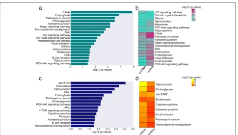
protein-coding genes we identified, 58 DUX4- and 24 Ph-like-specific lncRNAs demonstrated activation of key signaling pathways involved in proliferation, apoptosis, and differentiation in leukemia (Additional file 3: Table S2). For example, in thecis-based co-expression analysis, we identified a strong correlation between DUX4-specific lncRNAs and genes involved in the TGF-beta,
Hippo, and P53 signaling pathways (Fig. 2a,
Add-itional file 3: Table S2). Ph-like-specific lncRNAs were correlated with genes involved in JAK-STAT, mTOR,
and PIK3-AKT signaling pathways (Fig. 2c,
Add-itional file 3: Table S2). The trans-based co-expression analysis revealed same vital signaling pathways in DUX4 subtype (Additional file2: Figure S3a-b, Additional file3: Table S2), whereas in Ph-like subtype, we identified
add-itional signaling pathways, including P53 and
mitogen-activated protein kinase (MAPK) pathways (Additional file 2: Figure S3c, Additional file 3: Table
S2). The strongly co-expressed cis PC genes with DE
lncRNAs (n= 32) include oncogenes such asIL2RA[43],
TGFB2[44], andCDK6 [45] activated in signaling
path-ways from DUX4 and Ph-like subgroups
(Add-itional file2: Figure S4a-d, Table3).
However, there were no significant pathways identified within NH-HeH subtype. We next related the functions of DUX4 and Ph-like-specific DE lncRNAs obtained fromcis-based analysis to those functions identified with
DE PC genes. We observed an overlap of 100% (n= 18,
Additional file 3: Table S2) of pathways from the DUX4 subtype between lncRNA-based and PC-based func-tional enrichment analysis (Fig. 2b). In the Ph-like sub-type, we identified 60% (9 out of 15) equal pathways between DE PC-based and DE lncRNA-based functional
enrichment analysis (Additional file 3: Table S2 and
Fig. 2d). However, we identified Ph-like-specific
lncRNAs to be strongly correlated with genes involved in key signaling pathways than Ph-like-specific protein-coding genes. For example, we identified mTOR and PI3K-AKT exclusively in the Ph-like-specific lncRNA-based pathway analysis. Together, our analyses highlight
important functions of BCP-ALL subtype-specific
lncRNAs whose expression correlates tightly with that of cancer-related oncogenes.
Table 2Number of BCP-ALL subtype-specific lncRNAs co-expressed with itscisandtransPC genes
Subtype CisPC genes (n= 929)
lncRNAs co-expressed withcisPC genes (n= 621)
TransPC genes (n= 753)
lncRNAs co-expressed withtransPC genes (n= 552)
DUX4 669 451 (736) 492 379 (736)
Ph-like 260 170 (383) 261 173 (383)
The table represents the number of significantly (Pearson correlation coefficient≥0.55, two-tailedPvalue≤0.05) co-expressed subtype-specific lncRNAs with their
Relapse-specific lncRNAs driving BCP-ALL progression To gain insights into the possible role of lncRNAs driving BCP-ALL progression, we investigated dysreg-ulation of lncRNAs at relapse. For each molecular BCP-ALL subtype, we performed a differential expres-sion analysis of lncRNAs between ID and REL
sam-ples (Fig. 3). Nine hundred forty-seven lncRNAs
(Additional file 4: Table S3) emerged as significantly
DE (absolute fold change ≥± 1.5; P value ≤0.01)
be-tween ID and REL from the three subtypes. Around
20% (n = 186) of those DE lncRNAs were
upregu-lated, and 80% were downregulated at relapse. The hierarchical clustering on relapse-specific lncRNAs within each subtype (DUX4, Ph-like, NH-HeH) identi-fied clear separation between ID and REL (Fig. 3a–c). While majority of relapse-specific lncRNAs are novel, we identified a few previously reported onco-lncRNAs (Table 4) within our set.
The putative molecular functions of relapse-specific lncRNAs were identified using the previously described guilt-by-association approach. Relapse-specific lncRNAs within Ph-like and NH-HeH subtypes did not reveal any
significant correlation with activation of pathways. In contrast, in the DUX4 subtype, we identified 56% (n =
321) relapse-specific lncRNAs correlated with cis PC
genes (Additional file 4: Table S3). These DUX4
relapse-specific lncRNAs showed correlation with the PC genes involved in vital signaling pathways and meta-bolic pathways, including NF-kappa B-signaling pathway, cell adhesion molecule (CAMS) and metabolic pathways
(number of genes involved ≥3 and P value ≤0.05)
(Fig. 3d, Additional file 4: Table S3). These results indi-cate that relapse-specific markers from DUX4 subtype may be functionally engaged in metabolic and signaling pathways.
Subtype-specific BCP-ALL lncRNAs show epigenetic alterations
For the analysis of the methylation status of loci located at the lncRNAs genomic position in the BCP-ALL sub-types, we used DNA methylation array data (collected from Illumina 450k methylation array) from the same patients (n= 45) including matched ID and REL samples (n= 82). The distribution of DNA methylation levels of Fig. 2The molecular pathways of lncRNAs involved in the DUX4 and Ph-like BCP-ALL subgroups.aThe barplot plot depicts the molecular pathway analysis from the functional enrichment analysis for nearby (≤100 kb proximity)cisprotein-coding genes correlated (Pearson correlation coefficient≥0.55 and two-tailedPvalue≤0.05) with DE lncRNAs in the DUX4 subtype.bThe heatmap depicts the concordance between the protein-coding and lncRNA-based predictions for DUX4 subtypes.cThe barplot depicts the molecular pathway analysis from the functional enrichment analysis for nearby (≤100 kb proximity)cisprotein-coding genes correlated (Pearson correlation coefficient≥0.55 and two-tailedP
CpG sites (n= 60,021, Additional file5: Table S4) associ-ated with 7160 lncRNAs was compared with CpG sites associated with PC genes across all BCP-ALL samples. Unlike the expression levels, the distribution of DNA methylation (hypo-methylation or hyper-methylation) between lncRNAs and PC genes was similar (Add-itional file2: Figure S1b). Given the robust separation of BCP-ALL subtypes on DNA methylation profile of CpGs associated with lncRNAs on the PCA analysis (Fig. 4a), we next studied the differential hypo-methylated
(methy-lation difference value < 0; P value ≤0.05) and
hyper-methylated (methylation difference value > 0.2; P
value ≤0.05) CpGs associated with lncRNAs in each
subtype (see the “Method” section). The hierarchical clustering of differentially methylated (DM) lncRNAs showed distinct methylation patterns for each subtype, concordant with the DE lncRNA signature (Fig. 4b–d,
Additional file5: Table S4). In the DUX4 and NH-HeH
subtypes, the number of hypo-methylated lncRNAs (dif-ferential methylation value < 0, P value ≤0.05) was higher compared to the number of hyper-methylated lncRNAs. We classified the DM lncRNAs based on their
genomic regions as gene body methylated and
promoter-TSS methylated. In the promoter methylated Table 3Subtype-specific lncRNAs and oncogenes
Subtype-specific lncRNAs Pearson correlation coefficient Pvalue Oncogene
RP11-347C18.3 0.56 3.25E−008 CDK6
RP11-461F16.3 0.62 5.21E−010
RP11-96H19.1 0.62 3.89E−010
RP11-228B15.4 0.64 7.68E−011
MME-AS1 0.56 3.68E−008
CTB-39G8.3 0.57 1.78E−008
AC002454.1 0.72 2.21E−014
RP11-582 J16.4 0.55 8.08E−008
AC009970.1 0.64 6.23E−011
RP11-229P13.20 0.66 1.44E−011
LINC00114 0.57 3.06E−008
CTB-118N6.3 0.61 9.70E−010
SOCS2-AS1 0.62 4.94E−010
CTD-2561B21.10 0.61 9.91E−010
RP11-413E1.4 0.56 4.36E−008
KB-1460A1.1 0.55 7.77E−008
AC012309.5 0.59 4.10E−009
RP11-37B2.1 0.59 4.76E−009
ASB16-AS1 0.65 3.86E−011
LINC00426 0.62 6.32E−010
LINC01071 0.57 2.46E−008
RP11-536K7.5 0.74 5.11E−15 IL2RA
RP11-224O19.2 0.98 1.08E−061 TGFB2
AC004837.5 0.83 6.11E−023
RP11-251M1.1 0.79 7.39E−019
CTD-2571L23.8 0.75 2.94E−016
RP11-35O15.1 0.65 3.36E−011
AC139100.3 0.58 1.00E−008
RP11-158M2.3 0.58 1.50E−008
RP11-672A2.5 0.56 4.68E−008
CTD-2357A8.3 0.55 7.46E−008
RP11-677M14.3 0.55 6.68E−008
Fig. 3Relapse-specific DE lncRNAs from BCP-ALL subtypes.a–cHeatmap depicting the hierarchical clustering on relapse-specific DE lncRNA signature onz-score transformed LIMMA normalized expression values from DUX4, Ph-like, and NH-HeH subtypes. Each heatmap shows the up- and downregulated lncRNAs specific to ID and REL samples.dMolecular pathway analysis with the number of genes involved in each pathway from the enrichment analysis of the nearby (< 100 kb proximity)cisprotein-coding genes correlated (Pearson correlation > 0.55 and
Pvalue≤0.05) with relapse-specific DE lncRNAs in the DUX4 subtype. The legend box indicates the number of ID and REL samples within each group. Abbr.: CAMs; cell adhesion molecules
Table 4Previously reported lncRNAs identified as relapse-specific lncRNAs in BCP-ALL subtypes
Relapse-specific lncRNAs Disease association
TCL6 (DUX4) Chromosomal translocation T cell leukemia/lymphoma [49]
LINC00312 (DUX4, Ph-like, NH-HeH) Proliferation, invasion, and migration of thyroid cancer, nasopharyngeal carcinoma [50]
miR-17-92a-1 (DUX4, Ph-like, NH-HeH) Development, progression, and aggressiveness of colorectal cancer [51]
lncRNAs, we identified the same trend with high
degree of hypo-methylated and lower number
hyper-methylated lncRNAs in DUX4 and NH-HeH subtypes. However, the Ph-like subtype has shown a higher degree of hyper-methylated DM lncRNAs than
hypo-methylated DM lncRNAs. The list of
subtype-specific DM lncRNAs from the three subtypes contained previously defined epigenetically altered lncRNAs from other cancer types, for example, we
ob-served the oncogenic lncRNAsLINC00312 [46], PVT1,
and TCL6 [47], which are differentially methylated in at least one of the three subtypes. Together, these data
illustrate epigenetically altered list of lncRNAs in three BCP-ALL subtypes.
Correlation between subtype-specific differentially expressed and differentially methylated lncRNAs
23 lncRNAs harbored significant hypo-methylation and
hyper-methylation pattern (Pearson correlation,
two-tailed P value ≤0.05) at the promoter region
(Table5) across the three BCP-ALL subtypes.
Of these 23 putative epigenetically facilitated lncRNAs, 15 were related to the DUX4 subgroup (Fig.5a) including
the novel lncRNAs R11-138M12.1 and RP11-624M8.1.
These were significantly hypo-methylated at their promoter region and transcriptionally upregulated in the DUX4 sub-group (Pearson correlation coefficient =−0.69; P value = 5.1E−13 forR11-138 M12.1; Pearson correlation coefficient =−0.50;Pvalue = 1.36E−06 forRP11-624M8.1; Fig.5b, c). In the Ph-like subtype, we observed 7 lncRNAs with pro-moter methylation (Fig.5d); interestingly, the same lncRNA
R11-138 M12.1showed significant hyper-methylation at the promoter region and a concordant downregulation in the Ph-like subgroup (Fig. 5e). Besides these novel lncRNAs, we identified lncRNAs previously reported in the context of different cancers from our epigenetically altered results.
The lncRNAs PVT1 (Pearson correlation coefficient =−
0.40, two-tailedPvalue≤0.001) andDIO3OS[48] (Pearson
correlation coefficient =−0.31, two-tailedPvalue = 0.0037) are examples, which we observed in the DUX4 and NH-HeH subtype with significant anti-correlation (two--tailedPvalue≤0.01) to its expression level.
Around 46% (n= 512) of DM subtype-specific lncRNAs were localized in the intronic and intergenic genomic re-gions. We next aimed to investigate whether these lncRNA regions have chromatin markers encoded within their genomic location. A recent human genome-wide chromatin marker study [49] has provided a rich resource to identify chromatin markers. Genome-wide mapping of B lymphocyte cell line by searching for epigenetic markers within our DM subtype-specific intronic and intergenic regions revealed a significant number of lncRNAs (n= 53; Additional file 5: Table S4, Fisher extract test P value = 2.2E−16) with enchancer and in-sulator markers (Additional file 5: Table S4). Out of
these, lncRNAs, RP11-134O21.1, RP11-398B16.2,
RP11-689B22.2, CTC-458I2.2, and LINC00880 were DE expressed, with a significant negative correlation between DNA methylation and the expression levels Table 5The list of significantly correlated DNA methylation and the expression of the promoter methylated lncRNAs (n= 23) from BCP-ALL subtypes
DM lncRNAs Pearson correlation coefficient Pvalue Methylation Absolute fold change Subtypes
AC003075.4 −0.31 0.004 1.43 −1.26 DUX4
AC099754.1 −0.32 0.002 −1.74 3.2
AC104655.3 −0.26 0.017 −2.27 2.07
CACNA1C-AS1 −0.45 2.03E−05 1.97 −1.62
CTB-25B13.9 −0.26 0.016 −1.73 1.46
IGF2-AS −0.24 0.028 −1.33 4.95
LINC01006 −0.39 0.001 −2.06 2.53
PVT1 −0.40 0.001 −2.13 1.15
RGMB-AS1 −0.26 0.0193 −1.48 5.96
RP11-125B21.2 −0.35 0.001 −1.75 4.11
RP11-138M12.1 −0.70 5.21E−13 −5.98 3.77
RP11-367G6.3 −0.30 0.004 1.98 −1.63
RP11-624M8.1 −0.50 1.34E−06 −3.34 4.13
RP11-789C17.3 −0.36 0.001 −2.27 3.2
SERTAD4-AS1 −0.25 0.0232 −1.98 1.79
LINC01006 −0.38 0.0003 1.44 −1.56 Ph-like
RP11-138M12.1 −0.70 5.21E−13 2.06 −1.44
RP11-305F18.1 −0.64 5.36E−11 1.76 −2.08
AC099754.1 −0.33 0.002 1.21 −1.36
ACVR2B-AS1 −0.36 0.0009 2.18 −1.75
LINC00996 −0.39 0.0003 −1.56 2.11
ERICH1-AS1 −0.40 0.0006 −1.82 2.21
DIO3OS −0.31 0.0037 −1.76 4.05 NH-HeH
U3 −0.83 1.346E-22 −2.01 2.43
in the DUX4 subtype (Table 6). These findings suggest that epigenetic silencing and activation of promoter lncRNAs may be a mechanism that contributes to the dys-regulation of expression of lncRNAs.
Discussion
Although previous studies have demonstrated the in-volvement of lncRNAs in acute leukemias [21,25],
com-prehensive characterization of the transcriptome,
epigenetic regulation and functional contribution of lncRNAs in distinct BCP-ALL subtypes are lacking.
LncRNAs, as the novel class of functional molecules, are involved in cancer biology and have previously been reported in different molecular subtypes in vari-ous cancers. However, their role in BCP-ALL subtypes has not been investigated. Here, we unravel the lncRNA landscape using transcriptome and methy-lome data from 45 (adult and pediatric) relapsed BCP-ALL patients focusing on the three molecular subtypes namely DUX4, Ph-like, and NH-HeH.
Our integrated transcriptomic analyses using RNA-seq and DNA methylation bring significant novel insights Fig. 5The epigenetically altered promoter methylated lncRNAs and their expression.aThe promoter methylated lncRNAs with significant negative correlation with DE expression profile from the DUX4 subtypes.b,cTwo representative examples of hypo-methylated lncRNAs with increased expression profile from DUX4 subtype. lncRNAs,RP11-138M12.1(Pearson correlation coefficient =−0.69, two-tailedPvalue = 5.21E−13),
RP11-624MB.1(Pearson correlation coefficient =−0.50,Pvalue = 1.36E−06) are examples with hypo-methylation and upregulated expression pattern with significant inverse correlation between DNA methylation and expression levels.dThe promoter methylated lncRNAs with significant negative correlation with DE expression profile from the Ph-like subtypes.eA representative example of the promoter hyper-methylated lncRNA,
RP11-138M12.1(Pearson correlation coefficient =−0.69, two-tailedPvalue = 5.21E−13) with downregulated expression pattern, and with inverse correlation within the Ph-like subtype
Table 6The list of significantly correlated DNA methylation and the expression of the intronic and intergenic methylated lncRNAs (n= 5) from DUX4 BCP-ALL subtypes
DM lncRNAs Absolute fold change Methylation value Pearson correlation rate Pvalue Epi-markers Biotype
RP11-134O21.1 2.54 −1.56 −0.63 1.9E−010 Enhancer Intron
RP11-398B16.2 2.08 −1.85 −0.47 0.0007 Insulator
RP11-689B22.2 1.52 −3.37 −0.47 0.008 Enhancer
CTC-458I2.2 −1.16 3.38 −0.42 0.0001 Enhancer
LINC00880 −1.45 2.23 −0.25 0.02 Enhancer Intergenic
and advances: they provide the most comprehensive novel datasets so far for BCP-ALL subtypes. We provide a resource of subtype-specific and relapse-specific lncRNAs and potential lncRNA functions and uncover their epigenetic alterations within the BCP-ALL sub-types. We identified 1235 DE subtype-specific lncRNAs dysregulated in at least one of the three subtypes. These 1235 DE subtype-specific lncRNAs successfully stratified subtypes in our discovery cohort, an independent valid-ation cohort.
Another important aspect of our study is the identifi-cation of relapse-specific dysregulated lncRNAs across three BCP-ALL subtypes. A closer look into the relapse-specific lncRNA signature identified lncRNAs
previously described as oncogenic including
RP11-701P16.5[50],SLC38A3[51], andLINC00312, which are upregulated in relapsed samples within DUX4 subtype.
Importantly, apoptosis suppressor lncRNA in
Myc-driven lymphomas miR-17/92 cluster host gene
(MIR17HG) [52] is upregulated in relapsed samples within the Ph-like subtype and downregulated in re-lapsed samples within DUX4 and NH-HeH subtypes. Overall, the relapse-specific lncRNAs highlight the onco-genic relevance in BCP-ALL subtypes: near haploid or
high hyperdiploid (NH-HeH; n = 16) and low
hypodip-loid. Besides the oncogenic properties, lncRNAs can act as prognostic markers [53,54] and aid at disease diagno-sis and treatment. A subset of our relapse-specific lncRNAs (n= 61, Additional file 4: Table S3) overlaps
with the prognostic markers identified from 14
Pan-Cancer datasets [42], including lung
cancer-associated transcript 1 (LUCAT1), which is previously reported for its drug resistance in solid cancer [55]. Within the DUX4 subtype, we identified the upregulated expression ofLUCAT1at relapse, potentially providing a novel insight into treatment resistance for BCP-ALL subtypes. Together, this illustrates the catalog of relevant
lncRNAs in different subtypes of BCP-ALL as
subtype-specific and relapse-specific markers with the potential of RNA-based approaches in the treatment of BCP-ALL subtypes.
The dissection of the regulatory pathways mediated by
the molecular subtype-specific and relapse-specific
lncRNAs revealed the activation of pivotal signaling pathways across three BCP-ALL subtypes. The func-tional analysis by means of the guilt-by-association approach highlights the subtype-specific and relapse-specific lncRNAs associated with activation of signaling pathways and metabolic pathways that are associated with leukemogenesis including TGF-Beta, hippo, P53,
JAK-STAT, cytokine-cytokine receptor, endocytosis,
mTOR, and metabolic pathways. Characterization of the lncRNAs involved in these pathways may potentially re-veal novel targets in molecular therapies.
The functional insights of relapse-specific and
subtype-specific lncRNAs revealed biological relevance to BCP-ALL subtypes including cell cycle functions, sig-nal transduction, cell migration, and metabolic pro-cesses. Some of the functions predicted here corroborate previous studies emphasizing the strengths of the employed guilt-by-association. For example, lncRNA
AC002454.1, which we associated to the PIK3-AKT pathway in Ph-like subtype, was recently reported to regulate cyclin-dependent kinase (CDK6) to participate in cell cycle disorder [56]. TheCDK6gene appears to be frequently dysregulated in hematopoietic malignancies [45] and is hence attributed a critical role in tumorigen-esis, also shown in ALL driven by mixed lineage leukemia (MLL) fusion proteins [57]. In Ph-like subtype, both CDK6and AC002454.1 are correlated and upregu-lated specifically in Ph-like samples, suggesting they displayed enhancer-like functions. We identified 8 relapse-specific lncRNAs (Additional file4: Table S3) as-sociated with metabolic pathways in the DUX4 subtype overlapping with lncRNAs [58] reported to synergistic-ally dysregulate metabolic pathways in multiple tumor contexts.
Besides known lncRNAs, we also identified novel lncRNAs associated with activation of key signaling pathways. For instance, in the DUX4 subtype, we identi-fied a set of novel lncRNAs associated with TGF-beta pathway, including the antisense RP11-224019.2, with a significant positive correlation to the TGFB gene. Re-cently, a number of lncRNAs were documented to be as-sociated with TGFß signaling pathway, including MEG3
regulating the TGFB2 pathway in breast cancer [40].
However, lncRNAs associated with the TGFß pathway in BCP-ALL subtypes have not been reported. The
co-expression of RP11-224019.2 and TGFB in DUX4
subtype may indicate their functional relatedness or regulatory relationships. In addition to that, a notable observation was a strong correlation between relapse-specific lncRNAs with genes involved in the activation of metabolic pathways in the DUX4 subtype. We identified 112 relapse-specific lncRNAs co-expressed with 29 (Additional file4: Table S3) PC genes activated in metabolic pathways, including previously reported 8 biomarker lncRNAs. For example, we identified
onco-genic lncRNA LUCAT1 reported to be associated with
poor prognosis in lung cancer [59]. However, the
Although we observed that subtype-specific lncRNAs and subtype-specific protein-coding genes were pre-dicted to activate or inhibit the same pathways, some important exclusivity was observed. For instance, the signaling pathways such as the PI3K and mTOR in Ph-like subtype was enriched only in the lncRNA-based enrichment analysis, whereas these pathways did not appear to be enriched/dysregulated in the mRNA-based analysis. The PI3K and mTOR signaling pathways control proliferation, differentiation, and survival of hematopoietic cells [54]. Consistent with our results, other studies indicated the potency of lncRNAs facili-tating the cancer cell growth through mTOR and PI3K signaling pathways [33, 44, 55], yet reports on BCP-ALL subtypes have been lacking so far. Consid-ering the functional nexus between Ph-like-specific lncRNAs and the activation of pathways such as mTOR and PI3K signaling pathways, targeting those lncRNAs may be a promising novel therapeutic target for BCP-ALL subtypes.
Our work also underscores the importance of epi-genetic alterations in modulating lncRNA transcrip-tional activities. Although previous studies [60–62] have demonstrated cross-talk between DNA methyla-tion and transcripmethyla-tional activities of lncRNAs, their role in the etiology of BCP-ALL subtypes has not
been investigated. DNA methylation analyses of
lncRNAs revealed that DNA methylation might
underlie the differential expression of BCP-ALL
subtype-specific lncRNAs. Some subtype-specific
lncRNAs identified here have been reported by
previ-ous studies. For example, SOX2-OT (67, [63]),
LINC00312 [46], TCL6, and PVT1 are onco-lncRNAs, which are promoter methylated in one of the three
subtypes. The lncRNA, PVT1, was reported for its
MYC activity [64, 65] and as oncogenic lncRNA with
multiple roles in cell growth, dysfunction, and
differ-entiation in AML [66]. Both lncRNAs, LINC00312
and TCL6, have been extensively investigated on expres-sion levels but not on the epigenetic level. The promoters
of both TCL6 and LINC00312 were observed to be
hyper-methylated with corresponding diminished expres-sion in the DUX4 and NH-HeH samples. Notably, the DNA methylation analysis of lncRNAs revealed that DNA methylation might underlie the differential expression of subtype-specific lncRNAs.
Conclusions
Overall, our study provides an in-depth analysis of the lncRNA transcriptome and epigenome in BCP-ALL sub-types and provides novel lncRNA markers associated with subtype and relapse specificity and with epigen-etic alterations in BCP-ALL subtypes. Additionally, we also demonstrated these lncRNAs might contribute to
the regulation of key signaling pathways involved in BCP-ALL. In summary, our study provides a compre hensive set of dysregulated lncRNAs from BCP-ALL
subtypes derived using different integrative
ap-proaches. This subtype-specific lncRNAs and their mechanisms of action in detail might provide promis-ing avenues for future studies to investigate key bio-markers and potential therapeutic targets in BCP-ALL subtypes.
Additional files
Additional file 1:Table S1.RNA-seq data used for analysis and subtype-specific lncRNAs from three subtypes (XLSX 13450 kb)
Additional file 2:Figure S1.The distribution of lncRNAs and PC gene expression and DNA methylation levels across samples.Figure S2. BCP-ALL subtype-specific differentially expressed lncRNAs. Figure S3. Comparison of molecular pathways from cis and trans based analysis on subtype-specific DE lncRNAs.Figure S4.The subtype-specific lncRNAs co-expressed with oncogenes involved in key signaling pathways in DUX4 and Ph-like subtypes. (PDF 1510 kb)
Additional file 3:Table S2.The functionally involved subtype-specific lncRNAs from DUX4 and Ph-like subtypes. Thetransandcisco-expressed
subtype-specific lncRNAs (XLSX 143 kb)
Additional file 4:Table S3.The relapse-specific lncRNAs from three sub-types. The lncRNAs involved in functions from DUX4 subtype (XLSX 371 kb)
Additional file 5:Table S4.DNA methylation array dataset. The differentially methylated lncRNAs from three subtypes. List of cis-acting epigenetically active lncRNAs. (XLSX 59994 kb)
Abbreviations
BCP-ALL:B cell precursor acute lymphoblastic leukemia; DE: Differential expression; DM: Differential methylation; DUX4: Double homeobox, 4; JAK-STAT2: Janus kinase/signal transducers and activators of transcription; LH: Low-hypodiploid; MTOR: Mammalian target of rapamycin; NH-HeH: Near haploid or high hyperdiploid; Ph-like: Philadelphia like;
PIK3-AKT: Phosphatidylinositol 3-kinase; PVT1: Plasmacytoma variant translocation 1; SOX2-OT: SOX2 overlapping transcript; TCL6: T cell leukemia/lymphoma 6; TGFß: Transforming growth factor beta
Acknowledgements
We are grateful to Johanna Angermaier for discussions and Ulf Leser for the suggestions in data analysis.
Funding
This work was supported by the German Cancer Aid (Deutsche Krebshilfe) [grant number 111533].
Availability of data and materials
Microarray data used in this study are available at the European Genome-Phenome Archive through the accession number EGAS00001002856.
Authors’contributions
Ethics approval and consent to participate
Ethical approval for this study was given by the ethics board of Charité University of Medicine, Berlin, Germany.
Consent for publication Not applicable
Competing interests
The authors declare that they have no competing interests.
Publisher’s Note
Springer Nature remains neutral with regard to jurisdictional claims in published maps and institutional affiliations.
Author details 1
Department of Hematology and Oncology, Charité, University Hospital Berlin, Campus Benjamin Franklin, 12203 Berlin, Germany.2German Cancer Research Center (DKFZ), 69120 Heidelberg, Germany.3German Cancer Consortium (DKTK), 69120 Heidelberg, Germany.4Department of Pediatric Hematology/Oncology, Charité, University Hospital Berlin, Campus Rudolf Virchow, 13353 Berlin, Germany.5Department of Medicine II, Department of Hematology/Oncology, Goethe University Hospital, 60590 Frankfurt/M, Germany.6Department of Hematology, Oncology and Tumor Immunology, Charité University Hospital Berlin, Campus Virchow-Klinikum, 13353 Berlin, Germany.7Department of Hematology, Oncology & Rheumatology, University Clinic Heidelberg, 69120 Heidelberg, Germany.8Department of Pediatric Hematology and Oncology, Research Institute Children’s Cancer Center, University Medical Center Hamburg, 20251 Hamburg, Germany. 9Department of Pediatrics, University Hospital Schleswig-Holstein, Campus Kiel, 24105 Kiel, Germany.10Department of Hematology and Oncology, University Hospital Schleswig-Holstein, Campus Kiel, 24105 Kiel, Germany. 11
Bioinformatics Platform, Berlin Institute for Medical Systems Biology (BIMSB), Max Delbrück Center (MDC), 13125 Berlin, Germany.
Received: 10 September 2018 Accepted: 25 December 2018
References
1. Portell CA, Wenzell CM, Advani AS. Clinical and pharmacologic aspects of blinatumomab in the treatment of B-cell acute lymphoblastic leukemia. Clin Pharmacol. 2013;5:5–11.
2. Ito C, Kumagai M, Manabe A, Coustan-Smith E, Raimondi SC, Behm FG, et al. Hyperdiploid acute lymphoblastic leukemia with 51 to 65 chromosomes: a distinct biological entity with a marked propensity to undergo apoptosis. Blood. 1999;93(1):315-20.
3. Heerema NA, Nachman JB, Sather HN, Sensel MG, Lee MK, Hutchinson R, et al. Hypodiploidy with less than 45 chromosomes confers adverse risk in childhood acute lymphoblastic leukemia: a report from the children’s cancer group. Blood. 1999;94(12):4036–45.
4. Jones D, Luthra R, Cortes J, Thomas D, O’Brien S, Bueso-Ramos C, et al. BCR-ABL fusion transcript types and levels and their interaction with secondary genetic changes in determining the phenotype of Philadelphia chromosome positive leukemias. Blood. 2008;112(13):5190–192. 5. Lilljebjörn H, Fioretos T. New oncogenic subtypes in pediatric B-cell
precursor acute lymphoblastic leukemia. Blood. 2017;130:1395–401. 6. Holmfeldt L, Wei L, Diaz-Flores E, Walsh M, Zhang J, Ding L, et al. The
genomic landscape of hypodiploid acute lymphoblastic leukemia. Nat Genet. 2013;45(3):242–52.
7. Mullighan CG. Genomic profiling of B-progenitor acute lymphoblastic leukemia. Best Pract Res Clin Haematol. 2011;24(4):489-503.
8. Lilljebjörn H, Henningsson R, Hyrenius-Wittsten A, Olsson L, Orsmark-Pietras C, Von Palffy S, et al. Identification of ETV6-RUNX1-like and DUX4-rearranged subtypes in paediatric B-cell precursor acute lymphoblastic leukaemia. Nat Commun. 2016;7:11790.https://doi.org/10.1038/ncomms11790.
9. Tran TH, Loh ML. Ph-like acute lymphoblastic Leukemia. Hematology Am Soc Hematol Educ Program. 2016;2016(1):561-66.
10. Paulsson K, Lilljebjörn H, Biloglav A, Olsson L, Rissler M, Castor A, et al. The genomic landscape of high hyperdiploid childhood acute lymphoblastic leukemia. Nat Genet. 2015;47(6):672–6.
11. Garzon R, Volinia S, Papaioannou D, Nicolet D, Kohlschmidt J, Yan PS, et al. Expression and prognostic impact of lncRNAs in acute myeloid leukemia.
Proc Natl Acad Sci U S A. 2014;111(52):18679–84 Available from:http:// www.pubmedcentral.nih.gov/articlerender.fcgi?artid=4284555&tool= pmcentrez&rendertype=abstract.
12. Chen S, Liang H, Yang H, Zhou K, Xu L, Liu J, et al. Long non-coding RNAs: the novel diagnostic biomarkers for leukemia. Environ Toxicol Pharmacol. 2017;55:81–6. 13. Kung JTY, Colognori D, Lee JT. Long noncoding RNAs: past, present, and
future. Genetics. 2013;193(3):651–69.
14. Ward M, McEwan C, Mills JD, Janitz M. Conservation and tissue-specific transcription patterns of long noncoding RNAs. J Hum Transcr. 2015;1(1):2–9. 15. Wang P, Ning S, Zhang Y, Li R, Ye J, Zhao Z, et al. Identification of
lncRNA-associated competing triplets reveals global patterns and prognostic markers for cancer. Nucleic Acids Res. 2015;43(7):3478–89.
16. Guttman M, Rinn JL. Modular regulatory principles of large non-coding RNAs. Nature. 2012;482:339–46.
17. Liu Q, Huang J, Zhou N, Zhang Z, Zhang A, Lu Z, et al. LncRNA loc285194 is a p53-regulated tumor suppressor. Nucleic Acids Res. 2013;41(9):4976–87. 18. Spizzo R, Almeida MI, Colombatti A, Calin GA. Long non-coding RNAs and
cancer: a new frontier of translational research. Oncogene. 2012;31:4577–87. 19. Sati S, Ghosh S, Jain V, Scaria V, Sengupta S. Genome-wide analysis reveals
distinct patterns of epigenetic features in long non-coding RNA loci. Nucleic Acids Res. 2012;40(20):10018–31.
20. Yoon J-H, Abdelmohsen K, Gorospe M. Posttranscriptional gene regulation by long noncoding RNA. J Mol Biol. 2013;425(19):3723–30 Available from: http://linkinghub.elsevier.com/retrieve/pii/S0022283612008960. 21. Gioia R, Drouin S, Ouimet M, Caron M, St-onge P, Richer C, et al. LncRNAs
downregulated in childhood acute lymphoblastic leukemia modulate apoptosis, cell migration, and DNA damage response. Oncotarget. 2017; 8(46):80645-0650.https://doi.org/10.18632/oncotarget.20817.
22. Gong J, Liu W, Zhang J, Miao X, Guo AY. IncRNASNP: a database of SNPs in IncRNAs and their potential functions in human and mouse. Nucleic Acids Res. 2015;43(Database issue):D181-6.
23. Tang Q, Hann SS. HOTAIR: an oncogenic long non-coding RNA in human cancer. Cell Physiol Biochem. 2018;47(3):893-913.
24. Wei S, Wang K. Long noncoding RNAs: pivotal regulators in acute myeloid leukemia. Exp Hematol Oncol. 2016;5:30.
25. Ronchetti D, Manzoni M, Agnelli L, Vinci C, Fabris S, Cutrona G, et al. LncRNA profiling in early-stage chronic lymphocytic leukemia identifies transcriptional fingerprints with relevance in clinical outcome. Blood Cancer J. 2016;6(9):e468. https://doi.org/10.1038/bcj.2016.77.
26. Schroeder MP, Neumann M, Eckert C, Bastian L, James AR, Gökbuget N, et al. Multi-genomics of relapsed B-cell precursor acute lymphoblastic leukemia reveals three distinct genetic clusters characterized by different alterations. Blood. 2016;128 p. no pagination.
27. Dobin A, Davis CA, Schlesinger F, Drenkow J, Zaleski C, Jha S, et al. STAR: ultrafast universal RNA-seq aligner. Bioinformatics. 2013;29(1):15–21. 28. Pertea M, Pertea GM, Antonescu CM, Chang TC, Mendell JT, Salzberg SL.
StringTie enables improved reconstruction of a transcriptome from RNA-seq reads. Nat Biotechnol. 2015;33(3):290–5.
29. Ritchie ME, Phipson B, Wu D, Hu Y, Law CW, Shi W, et al. limma powers differential expression analyses for RNA-sequencing and microarray studies. Nucleic Acids Res. 2015;43(7):e47.
30. Jaffe AE, Murakami P, Lee H, Leek JT, Fallin MD, Feinberg AP, et al. Bump hunting to identify differentially methylated regions in epigenetic epidemiology studies. Int J Epidemiol. 2012;41(1):200–9.
31. Guo X, Gao L, Liao Q, Xiao H, Ma X, Yang X, et al. Long non-coding RNAs function annotation: a global prediction method based on bi-colored networks. Nucleic Acids Res. 2013;41(2):e35.https://doi.org/10.1093/nar/gks967. 32. McLean CY, Bristor D, Hiller M, Clarke SL, Schaar BT, Lowe CB, et al. GREAT
improves functional interpretation of cis-regulatory regions. Nat Biotechnol. 2010;28(5):495–501 Available from:http://www.nature.com/doifinder/10. 1038/nbt.1630.
33. Subhash S, Kanduri C. GeneSCF: a real-time based functional enrichment tool with support for multiple organisms. Bioinformatics. 2016;17:65.https:// doi.org/10.1186/s12859-016-1250-z.
34. Derrien T, Johnson R, Bussotti G, Tanzer A, Djebali S, Tilgner H, et al. The GENCODE v7 catalog of human long noncoding RNAs: analysis of their gene structure, evolution, and expression. Genome Res. 2012;22(9):1775–89. 35. Ning S, Zhang J, Wang P, Zhi H, Wang J, Liu Y, et al. Lnc2Cancer : a
36. Colombo T, Farina L, Macino G, Paci P. PVT1: a rising star among oncogenic long noncoding RNAs. Biomed Res Int. 2015;2015:304208.https://doi.org/10. 1155/2015/304208.
37. Pickard MR, Mourtada-Maarabouni M, Williams GT. Long non-coding RNA GAS5 regulates apoptosis in prostate cancer cell lines. Biochim Biophys Acta Mol Basis Dis. 2013;1832(10):1613–23.
38. Peng W, Wu J, Fan H, Lu J, Feng J. LncRNA EGOT promotes tumorigenesis via hedgehog pathway in gastric cancer. Pathol. Oncol. Res. 2017.https:// doi.org/10.1007/s12253-017-0367-3.
39. Guil S, Esteller M. Cis-acting noncoding RNAs: friends and foes. Nat Struct Mol Biol. 2012;19(11):1068–75 Available from:http://www.nature.com/ doifinder/10.1038/nsmb.2428.
40. Mondal T, Subhash S, Vaid R, Enroth S, Uday S, Reinius B, et al. MEG3 long noncoding RNA regulates the TGF-βpathway genes through formation of RNA-DNA triplex structures. Nat Commun. 2015;6:7743.https://doi.org/10. 1038/ncomms8743.
41. Pandey RR, Mondal T, Mohammad F, Enroth S, Redrup L, Komorowski J, et al. Kcnq1ot1 antisense noncoding RNA mediates lineage-specific transcriptional silencing through chromatin-level regulation. Mol Cell. 2008;32(2):232–46. 42. Ali MM, Akhade VS, Kosalai ST, Subhash S, Statello L, Meryet-Figuiere M, et
al. PAN-cancer analysis of S-phase enriched lncRNAs identifies oncogenic drivers and biomarkers. Nat Commun. 2018;9(1):883.https://doi.org/10.1038/ s41467-018-03265-1.
43. Sadras T, Heatley SL, Kok CH, Dang P, Galbraith KM, McClure BJ, et al. Differential expression of MUC4, GPR110 and IL2RA defines two groups of CRLF2-rearranged acute lymphoblastic leukemia patients with distinct secondary lesions. Cancer Lett. 2017;408:92–101.
44. Dong M, Blobe GC. Role of transforming growth factor-beta in hematologic malignancies. Blood. 2006;107(12):4589–96 Available from:http://www. pubmedcentral.nih.gov/articlerender.fcgi?artid=1895802&tool= pmcentrez&rendertype=abstract.
45. Scheicher R, Hoelbl-Kovacic A, Bellutti F, Tigan AS, Prchal-Murphy M, Heller G, et al. CDK6 as a key regulator of hematopoietic and leukemic stem cell activation. Blood. 2015. Available from:https://www.ncbi.nlm.nih.gov/ pubmed/25342715. Accessed 10 June 2017.
46. Zhang W, Huang C, Gong Z, Zhao Y, Tang K, Li X, et al. Expression of LINC00312, a long intergenic non-coding RNA, is negatively correlated with tumor size but positively correlated with lymph node metastasis in nasopharyngeal carcinoma. J Mol Histol. 2013;44(5):545–54 Available from: http://www.ncbi.nlm.nih.gov/pubmed/23529758.
47. Saitou M, Sugimoto J, Hatakeyama T, Russo G, Isobe M. Identification of the TCL6 genes within the breakpoint cluster region on chromosome 14q32 in T-cell leukemia. Oncogene. 2000.
48. Mitra R, Chen X, Greenawalt EJ, Maulik U, Jiang W, Zhao Z, et al. Decoding critical long non-coding RNA in ovarian cancer epithelial-to-mesenchymal transition. Nat Commun. 2017;8(1):1604.https://doi.org/10.1038/s41467-017-01781-0. 49. ENCODE Project Consortium AIE of DE in the H. An integrated encyclopedia
of DNA elements in the human genome. Nature. 2012;489(7414):57–74. 50. Zhou M, Guo M, He D, Wang X, Cui Y, Yang H, et al. A potential signature of
eight long non-coding RNAs predicts survival in patients with non-small cell lung cancer. J Transl Med. 2015;13:231. https://doi.org/10.1186/s12967-015-0556-3.
51. Wang Y, Fu L, Cui M, Wang Y, Xu Y, Li M, et al. Amino acid transporter SLC38A3 promotes metastasis of non-small cell lung cancer cells by activating PDK1. Cancer Lett. 2017;393:8–15.
52. Mogilyansky E, Rigoutsos I. The miR-17/92 cluster: a comprehensive update on its genomics, genetics, functions and increasingly important and numerous roles in health and disease. Cell Death Differ. 2013;20(12):1603– 14.https://doi.org/10.1038/cdd.2013.125.
53. Zhong L, Lou G, Zhou X, Qin Y, Liu L, Jiang W. A six-long non-coding RNAs signature as a potential prognostic marker for survival prediction of ER-positive breast cancer patients. Oncotarget. 2017;8(40):3762–72. 54. Mer AS, Lindberg J, Nilsson C, Klevebring D, Wang M, Grönberg H, et al.
Expression levels of long non-coding RNAs are prognostic for AML outcome. J Hematol Oncol. 2018;11(1):52.https://doi.org/10.1186/s13045-018-0596-2. 55. Han Z, Shi L. Long non-coding RNA LUCAT1 modulates methotrexate resistance
in osteosarcoma via miR-200c/ABCB1 axis. Biochem Biophys Res Commun. 2018; 495(1):947–53 Available from:https://doi.org/10.1016/j.bbrc.2017.11.121. 56. Wang Y, Li Y, Yang Z, Liu K, Wang D. Genome-wide microarray analysis of
long non-coding RNAs in eutopic secretory endometrium with endometriosis. Cell Physiol Biochem. 2015;37(6):2231–45.
57. Placke T, Faber K, Nonami A, Putwain SL, Salih HR, Heidel FH, et al. Requirement for CDK6 in MLL-rearranged acute myeloid leukemia. Blood. 2014;124(1):13–23.
58. Chiu HS, Somvanshi S, Patel E, Chen TW, Singh VP, Zorman B, et al. Pan-Cancer analysis of lncRNA regulation supports their targeting of cancer genes in each tumor context. Cell Rep. 2018;23(1):297–312 e12. 59. Sun Y, Jin S-D, Zhu Q, Han L, Feng J, Lu X, et al. Long non-coding RNA
LUCAT1 is associated with poor prognosis in human non-small lung cancer and regulates cell proliferation via epigenetically repressing p21 and p57 expression. Oncotarget. 2017;5(0):28297–311 Available from:http://www. oncotarget.com/abstract/16044.
60. Hanly DJ, Esteller M, Berdasco M. Interplay between long non-coding RNAs and epigenetic machinery: emerging targets in cancer? Philos Trans R Soc Lond B Biol Sci. 2018;373(1748):20170074.https://doi.org/10. 1098/rstb.2017.0074.
61. Zhao Y, Sun H, Wang H. Long noncoding RNAs in DNA methylation: new players stepping into the old game. Cell Biosci. 2016;6:(45):1–6.
62. Yamamura S, Imai-Sumida M, Tanaka Y, Dahiya R. Interaction and cross-talk between non-coding RNAs. Cell Mol Life Sci. 2018;75:467–84.
63. Shahryari A, Jazi MS, Samaei NM, Mowla SJ. Long non-coding RNA SOX2OT: Expression signature, splicing patterns, and emerging roles in pluripotency and tumorigenesis. Front Genet. 2015;6:196.https://doi.org/10.3389/fgene. 2015.00196.
64. Li JR, Sun CH, Li W, Chao RF, Huang CC, Zhou XJ, et al. Cancer RNA-Seq Nexus: a database of phenotype-specific transcriptome profiling in cancer cells. Nucleic Acids Res. 2016;44(D1):D944–51.
65. Tseng YY, Moriarity BS, Gong W, Akiyama R, Tiwari A, Kawakami H, et al. PVT1 dependence in cancer with MYC copy-number increase. Nature. 2014; 512(1):82–6.