C A S E R E P O R T
Open Access
De novo
malignant solitary fibrous tumor of the
kidney
Tsan-Yu Hsieh
1,4, Yi-Che ChangChien
1,4, Wen-Hsiang Chen
2,4, Siu-Chung Chen
3,4, Liang-Che Chang
1,4,
Cheng-Cheng Hwang
1,4, Hui-Ping Chein
1,4and Jim-Ray Chen
1,4*Abstract
The kidney is a relatively infrequent site for solitary fibrous tumor (SFT). Among the previously reported cases, only two cases of malignant renal SFT developing via dedifferentiation from a pre-existing benign SFT have been reported. Here we reported a case ofde novo malignant renal SFT clinically diagnosed as renal cell carcinoma in a 50-year-old woman. The tumor was circumscribed but unencapsulated and showed obvious hemorrhagic necrosis. Microscopically, the tumor was composed of patternless sheets of alternating hypercellular and hypocellular areas of spindle cells displaying mild to moderate nuclear atypia, frequent mitoses up to 8 per 10 high power fields, and a 20% Ki-67 proliferative index. Immunohistochemical studies revealed reactivity for CD34, CD99 and vimentin, with no staining for all other markers, confirming the diagnosis of SFT. No areas of dedifferentiation were seen after extensive sampling. Based on the pathologic and immunohistochemical features, a diagnosis ofde novomalignant renal SFT was warranted. Our report expands the spectrum of malignant progression in renal SFTs. Even though this patient has been disease-free for 30 months, long-term follow-up is still mandatory.
Keywords:solitary fibrous tumor, kidney, malignant, de novo, dedifferentiation, CD34
Backround
Solitary fibrous tumors (SFTs) are distinctive mesenchy-mal tumors most commonly described as pleural-based lesions; however they can develop at any extrapleural anatomic site [1]. Although the clinical course of SFTs is rather unpredictable, the prognosis of SFTs is generally favorable. It is estimated that 10% to 15% of intrathoracic SFTs and up to 10% of extrathoracic SFTs will recur and/ or metastasize [2,3], therefore SFT is regarded as an
“intermediate malignant, rarely metastasizing”neoplasm [4]. Microscopic features associated with malignancy in both intrathoracic and extrathoracic SFTs include nuclear atypia, increased cellularity and more than 4 mitoses per 10 high power fields [4,5]. An additional fac-tor conferring a worse prognosis in SFTs is dedifferentia-tion or sarcomatous overgrowth, which represents an abrupt transition to a morphologically anaplastic compo-nent [6]. The kidney is a relatively infrequent site for SFT, with at least 36 cases reported in a review article [7]. The vast majority of renal SFTs are histologically
benign and only two cases of malignant renal SFTs devel-oping via dedifferentiation or sarcomatous overgrowth from a pre-existing benign SFT have been reported [7,8]. Here we report the first case ofde novomalignant renal SFT without dedifferentiation and thus expand the spec-trum of malignant progression in renal SFTs.
Case presentation Clinical summary
A 50-year-old woman was admitted to our hospital with one-month history of soreness and pain in her right flank, without gross hematuria or other constitutional symp-toms. Laboratory findings were unremarkable. Physical examination revealed a palpable right flank mass. A com-puted topography (CT) of the abdomen showed a huge necrotic tumor occupying the perirenal space of right kid-ney without evidence of either local invasion or lymphade-nopathy (Figure 1). The patient underwent right radical nephrectomy under a pre-operative diagnosis of American Joint Committee on Cancer (AJCC) stage II (T2aN0) renal cell carcinoma. Post-operation course was smooth. Neither chemotherapy nor radiation therapy was given.
* Correspondence: jimrchen@cgmh.org.tw
1Department of Pathology, Chang Gung Memorial Hospital, Keelung, Taiwan
Full list of author information is available at the end of the article
She has been well without evidence of recurrence or metastasis for 30 months.
Pathologic findings
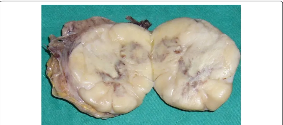
A nephrectomy specimen (15 × 9 × 7 cm, 670 g) with attached ureter and perirenal fibroadipose tissue was received. The specimen was bisected to reveal a 9 × 9 × 6 cm circumscribed but unencapsulated tumor occupy-ing the perirenal space of the upper and middle poles of kidney. The tumor was firm and showed a yellowish white to tan-gray, myxoid and lobulated cut surface with prominent hemorrhage and necrosis (Figure 2). Microscopically, the tumor showed proliferation of spin-dle cells arranging in a patternless architecture (Figure
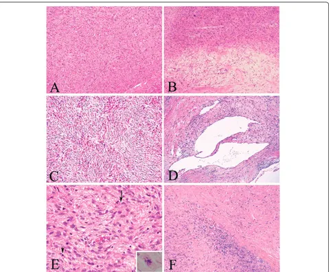
3A) with a combination of alternating hypercellular and hypocellular areas (Figure 3B). Haphazard, storiform, or short fascicular arrangements of spindle cells in a loose myxoid to fibrous stroma containing dense collagen fibers were also seen (Figure 3C). Dilated and branching hemangiopericytoma-like vessels were frequently observed (Figure 3D). Tumor cells had plump, fusiform, or elongated hyperchromatic nuclei with mild to moder-ate pleomorphism and indistinct cell borders and frequent mitoses up to 8 per 10 high power fields. Abnormal mitoses were occasionally seen (Figure 3E). Tumor necrosis was evidently present (Figure 3F). We did not find any areas of dedifferentiation after extensive tumor sampling.
Immunohistochemically, the tumor showed weak CD34 positivity (Figure 4A) and diffusely strong CD99 (Figure 4B) and vimentin staining. They stained nega-tively for bcl-2 protein, S-100 protein, NSE, muscle mar-kers, cytokeratin, CD117(C-kit), p53 and HMB-45. Ki-67 immunostaining, analyzed by ImmuoRatio quantitative image software [9], showed a 20% proliferative index. The immunohistochemical findings were highly consis-tent with SFT. Based on the microscopic features including increased cellularity, cellular atypia, frequent mitoses, a high proliferative index, necrosis and absence of dedifferentiation, a diagnosis of de novomalignant solitary fibrous tumor was established.
Discussion
SFT is a relatively uncommon but distinctive mesenchy-mal neoplasm, originally described in the pleura cavity and later reported to occur ubiquitously [1]. Although
Figure 1CT of the abdomen. Arterial phase images of dynamic computed topography scan showed a highly necrotic tumor compressing the renal parenchyma without either invasion to surrounding tissues or local lymphadenopathy.
the histogenesis of SFT remains undetermined, recent studies strongly favor a primitive mesenchymal or peri-vascular cell origin [10]. The kidney is a relatively infre-quent site for SFT, with approximately at least 36 cases of renal SFT reported in a review article [7]. Clinically, these cases were frequently considered to be malignant due to their large tumor size by physical examinations and radiographic studies. Symptoms do not differ from those reported by patients with renal cell carcinoma. Hypoglycemia, which is a rare symptom in intrathoracic and extrathoracic SFTs, was not reported in any renal SFTs including our case [2].
Malignant behaviors in the form of recurrence and/or metastases can occur in 10% to 15% of intrathoracic SFTs and up to 10% of extrathoracic SFTs [2,3]. Malig-nant SFT is postulated to develop via two pathways: (1)
de novooccurrence or (2) dedifferentiation or sarcoma-tous overgrowth from a pre-exsisting histologically benign SFT [1,4]. Most renal SFTs were classified as his-tologically benign and showed a favorable prognosis, with no evidence of recurrence during a follow-up period ran-ging from 2 to 89 months [7]. To our knowledge, two cases of malignant renal SFTs developing via dedifferen-tiation or sarcomatous overgrowth from a pre-existing
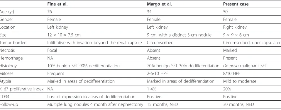
benign SFT have been reported by Margo et al. and Fine et al., respecvievely [7,8]. A detailed comparison of the clinicopatholgoic features of these two cases and ours is summarized in Table 1. The tumor reported by Fine et
al. had infiltrative borders and focal necrosis, and invaded beyond the renal capsule. The tumor described by Margo et al. was a 9-cm circumscribed mass devoid of either hemorrhage or necrosis. Notably, there was a 3-cm nodu-lar area within the main tumor, microscopically respond-ing to sarcomatous overgrowth. In contrast, our tumor showed a homogeneous cut surface with prominent necrosis and hemorrhage. Microscopically, the tumor reported by Fine et al. and Margo et al. showed typical features of benign SFT with 90% and 30% of dedifferen-tiation or sarcomatous overgrowth, respectively. In con-trast, our tumor appeared to developde novosince we did not find any areas of dedifferentiation after extensive tumor sampling.
The criteria for clinical malignancy in intrathoracic SFT, first proposed by England et al. in 1989, include increased cellularity, pleomorphism, mitotic count more than 4 per 10 high power fields, necrosis, hemorrhage, size more than 10 cm, non-pedunculated and atypical locations (parietal pleura, pulmonary parenchyma) [5]. The diagnostic criteria for malignant extrathoracic SFTs are purely microscopic and include increased cellularity, pleomorphism and mitotic count more than 4 per 10 high power fields [4,11]. Currently the impact of tumor characteristics (size, hemorrhage, necrosis and location) in predicting clinical malignancy in extrathoracic SFTs remains to be investigated. Our case fulfilled the diagnos-tic criteria for malignant extrathoracic SFTs. Addition-ally, the presence of hemorrhage and necrosis and a 20% Ki-67 proliferative index further supports a diagnosis of malignant renal SFT.
SFTs show a wide variety of microscopic growth pat-terns and should be distinguished from benign and malignant spindle cell tumors. Positive immunoreactivity for CD34 and CD99 is characteristic of SFT, and highly valuable in differentiating from other mesenchymal tumors [2,4]. However the expression of CD34 and CD99 may be decreased or absent in areas with marked atypia or dedifferentiation [8,12]. Genetic analyses of SFT to date have not found consistent and characteristic cytogenetic abnormalities that can be used an ancillary diagnostic marker. Missense mutation of platelet-derived growth factor receptor-b(PDGFR-b) has been reported in 2 of 88 pleuro-pulmonary SFTs [13], but we did not detect PDGFR-bmutation in our case (data not shown).
The prognosis of extrapleural SFTs is more unpredict-able than that of pleural tumors due to lack of large-scale studies. The clinical outcomes were rather strik-ingly different in the two malignant renal SFTs with dedifferentiation. While a patient developed multiple small lung nodules 4 months post-operatively [8], the other patient was disease-free after 15 months of follow-up [7]. Our patient has been well without evidence of recurrence or metastasis for 30 months. Some studies
indicated that extrapleural SFTs had similar prognosis as the pleural counterpart, and a tumor with malignant histological feature was associated with recurrence and metastasis [3,14], but other studies showed that extra-pleural SFTs tended to have a favorable outcome than pleural ones, even those with malignant histological fea-tures [11,15]. Studies also showed that deep-seated loca-tions, over-expression of p53 and p16, loss of CD34 immunostaining and the presence of dedifferentiated areas may indicate more aggressive behaviors [6]. Com-plete surgical excision and long-term follow-up are gen-erally recommended for patients with extrapleural SFTs [15]. Besides, due to the rich vascularity and possible origin from pericytes, antiangiogenic therapy, especially for the advanced cases is also under clinical investiga-tion [16].
Consent
Written informed consent was obtained from the patient for publication of this case report and accompanying images. Submission of this case report was approved by Institutional Review Board (IRB) of Chang Gung Mem-orial Hospital, Tao-Yuan, Taiwan. A copy of the written consent is available for review by the Editor-in-Chief of this journal.
List of abbreviations
SFT: solitary fibrous tumor
Acknowledgements
The authors appreciate Drs. Jonathan I. Epstein and Elizabeth Montgomery at the Department of Pathology, Johns Hopkins Medical Institutions, Baltimore, MD, USA for confirming our diagnosis.
Author details
1
Department of Pathology, Chang Gung Memorial Hospital, Keelung, Taiwan.
2Department of Urology, Chang Gung Memorial Hospital, Keelung, Taiwan.
3Department of Radiology, Chang Gung Memorial Hospital, Keelung, Taiwan. 4
College of Medicine, Chang Gung University, Tao-yuan, Taiwan.
Authors’contributions
TYH was responsible for data collection, literature search and manuscript preparation. YCCC carried out gross and microscopic examinations. WHC performed the surgery and clinical follow-up of the patient. CSC interpreted pre-operative and follow-up imaging studies. CLC, CCH and HPC participated in the microscopic analyses and helped making a final diagnosis. JRC participated in manuscript preparation and approved the final manuscript. All authors read and approved the final manuscript.
Competing interests
The authors declare that they have no competing interests.
Received: 10 August 2011 Accepted: 5 October 2011 Published: 5 October 2011
References
1. Chan JK:Solitary fibrous tumour–everywhere, and a diagnosis in vogue.
Histopathology1997,31:568-76.
2. Weiss SW, Goldblum JR:Soft tissue tumors of intermediate malignancy of uncertain type.InSoft Tissue Tumor..5 edition. Edited by: Weiss SW, Goldblum JR. Philadephia: Mosby Elsevier; 2008:1093-1160. 3. Vallat-Decouvelaere AV, Dry SM, Fletcher CD:Atypical and malignant
solitary fibrous tumors in extrathoracic locations: evidence of their comparability to intra-thoracic tumors.Am J Surg Pathol1998,22:1501-11. 4. Guillou LFJ, Fletcher CDM, Mandahi N:Extrapleural solitary fibrous tumour
and hemangiopericytoma.InWorld Health Organization Classification of Tumours: Pathology and Genetics of Tumours of Soft Tissue and Bone.Edited by: Fletcher CDM, Unni KK, Mertens F. Lyon: IARCPress; 2002:86-90. 5. England DM, Hochholzer L, McCarthy MJ:Localized benign and malignant
fibrous tumors of the pleura. A clinicopathologic review of 223 cases.
Am J Surg Pathol1989,13:640-58.
6. Mosquera JM, Fletcher CD:Expanding the spectrum of malignant progression in solitary fibrous tumors: a study of 8 cases with a discrete anaplastic component–is this dedifferentiated SFT?Am J Surg Pathol
2009,33:1314-21.
7. Magro G, Emmanuele C, Lopes M, Vallone G, Greco P:Solitary fibrous tumour of the kidney with sarcomatous overgrowth. Case report and review of the literature.APMIS2008,116:1020-5.
8. Fine SW, McCarthy DM, Chan TY, Epstein JI, Argani P:Malignant solitary fibrous tumor of the kidney: report of a case and comprehensive review of the literature.Arch Pathol Lab Med2006,130:857-61.
9. Tuominen VJ, Ruotoistenmaki S, Viitanen A, Jumppanen M, Isola J:
ImmunoRatio: a publicly available web application for quantitative
Table 1 Comparsion of the clinicopathologic features of malignant renal solitary fibrous tumor
Fine et al. Margo et al. Present case
Age (yr) 76 34 50
Gender Female Female Female
Location Left kidney Left kidney Right kidney
Size 12 × 10 × 7.5 cm 9 cm, with a distinct 3-cm nodule 9 × 9 × 6 cm
Tumor borders Infiltrative with invasion beyond the renal capsule Circumscribed Circumscribed, unencapsulated
Necrosis Focal Absent Marked
Hemorrhage NA Absent Present
Histology 10% benign SFT 90% dedifferentiation 70% benign SFT 30% dedifferentiation De novomalignant SFT
Mitoses Frequent 2-6/10 HPF 8/10 HPF
Atypia Marked in areas of dedifferentiation Marked in areas of dedifferentiation Mild to moderate
Ki-67 proliferative index NA 1-4% 20%
CD34 Loss of expression in areas of dedifferentiation Positive Positive
Follow-up Multiple lung nodules 4 month after nephrectomy 15 months, NED 30 months, NED
image analysis of estrogen receptor (ER), progesterone receptor (PR), and Ki-67.Breast Cancer Res2010,12:R56.
10. Ide F, Obara K, Mishima K, Saito I, Kusama K:Ultrastructural spectrum of solitary fibrous tumor: a unique perivascular tumor with alternative lines of differentiation.Virchows Arch2005,446:646-52.
11. Hasegawa T, Matsuno Y, Shimoda T, Hasegawa F, Sano T, Hirohashi S:
Extrathoracic solitary fibrous tumors: their histological variability and potentially aggressive behavior.Hum Pathol1999,30:1464-73.
12. Yokoi T, Tsuzuki T, Yatabe Y, Suzuki M, Kurumaya H, Koshikawa T, Kuhara H, Kuroda M, Nakamura N, Nakatani Y,et al:Solitary fibrous tumour: significance of p53 and CD34 immunoreactivity in its malignant transformation.Histopathology1998,32:423-32.
13. Schirosi L, Lantuejoul S, Cavazza A, Murer B, Yves Brichon P, Migaldi M, Sartori G, Sgambato A, Rossi G:Pleuro-pulmonary solitary fibrous tumors: a clinicopathologic, immunohistochemical, and molecular study of 88 cases confirming the prognostic value of de Perrot staging system and p53 expression, and evaluating the role of c-kit, BRAF, PDGFRs (alpha/ beta), c-met, and EGFR.Am J Surg Pathol2008,32:1627-42.
14. Brunnemann RB, Ro JY, Ordonez NG, Mooney J, El-Naggar AK, Ayala AG:
Extrapleural solitary fibrous tumor: a clinicopathologic study of 24 cases.
Mod Pathol1999,12:1034-42.
15. Morimitsu Y, Nakajima M, Hisaoka M, Hashimoto H:Extrapleural solitary fibrous tumor: clinicopathologic study of 17 cases and molecular analysis of the p53 pathway.APMIS2000,108:617-25.
16. Park MS, Araujo DM:New insights into the hemangiopericytoma/solitary fibrous tumor spectrum of tumors.Curr Opin Oncol2009,21:327-31. doi:10.1186/1746-1596-6-96
Cite this article as:Hsiehet al.:De novomalignant solitary fibrous tumor of the kidney.Diagnostic Pathology20116:96.
Submit your next manuscript to BioMed Central and take full advantage of:
• Convenient online submission
• Thorough peer review
• No space constraints or color figure charges
• Immediate publication on acceptance
• Inclusion in PubMed, CAS, Scopus and Google Scholar
• Research which is freely available for redistribution