C A S E R E P O R T
Open Access
Infantile myofibromatosis treated by
mandibulectomy and staged reconstruction
with submental flap and free fibula flap: a
case report
Alexandra Maby
1, Benoit Guay
1,2and François Thuot
1,2*Abstract
Background:Infantile myofibromatosis is the most common benign fibrous tumor in infants. Three different types
have been reported in the literature. The most commonly affected areas are the head, the neck and the trunk. Our patient showed a very high level of mandibular destruction resistant to all mandibular sparing treatment strategies requiring segmental mandibulectomy and complex reconstruction.
Case presentation:We describe a rare case of multicentric infantile myofibromatosis with mandibular bone destruction. The treatment required a succession of chemotherapy, a subtotal transoral resection and a hemi-mandibulectomy. The mandibular reconstruction was staged with initial bridging titanium plate with a submental flap, followed later by a fibula free flap.
Conclusion:Mandibular involvement by myofibromatosis is rare, and the extend of bone destruction and
reconstruction make this case unique. To our knowledge, this is the only reported case of fibula free flap mandibular reconstruction in a patient with infantile myofibromatosis , as well as one of the youngest reported submental island flaps for any pathology. We describe the clinical presentation and management, including relevant imaging, histopathology, medical and surgical treatment as well as a review of relevant literature.
Keywords:Infantile myofibromatosis, Myofibroma, Mandibular reconstruction, Submental island flap, Fibula free flap
Introduction
Infantile myofibromatosis (IM) is characterized by benign tumoral proliferations of fibroblasts and/or myo-fibroblasts. IM was first described in 1954 by Stout as a “generalized congenital fibromatosis” [1]. In 1981, Chung and Enzinger discovered its myofibroblastic origin and used the term infantile myofibromatosis for
the first time [2]. Although IM is rare, it is the most
common fibrous tumor in the first year of life [3]. IM
usually presents as firm, purple or flesh-colored nodules in the skin and subcutaneous tissue. The most com-monly affected body areas are the head, the neck, and
the trunk [4, 5]. The exact etiology of the condition is unknown, and most cases reported are sporadic. We present a unique case of IM with mandibular destruc-tion which appears unique for 2 reasons. First, although this pathology is benign and frequently indolent, our pa-tient showed a very high level of mandibular destruction resistant to all mandibular sparing treatment strategies. Second, his very young age at presentation (6 months) and resection (18 months) made reconstruction highly challenging.
Case report
Case presentation
A 6-month-old boy was referred for a right lower cheek mass and a left thoracic subcutaneous mass both present for 3 months. He was asymptomatic and healthy with no significant medical, surgical or familial history. The
© The Author(s). 2019Open AccessThis article is distributed under the terms of the Creative Commons Attribution 4.0 International License (http://creativecommons.org/licenses/by/4.0/), which permits unrestricted use, distribution, and reproduction in any medium, provided you give appropriate credit to the original author(s) and the source, provide a link to the Creative Commons license, and indicate if changes were made. The Creative Commons Public Domain Dedication waiver (http://creativecommons.org/publicdomain/zero/1.0/) applies to the data made available in this article, unless otherwise stated. * Correspondence:francois.thuot@fmed.ulaval.ca
1
Département d’ophtalmologie et d’oto-rhino-laryngologie–chirurgie cervico-faciale, Faculté de Médecine, Université Laval, 1050, avenue de la Médecine, Québec, QC G1V 0A6, Canada
thoracic lesion was small (1,0 × 1,6 cm) and mobile. The cheek lesion presented as a deep and firm soft tissue
submucosal mass adherent to the mandible (Fig.1).
Radiological findings
Ultrasound (US) examination of both masses showed hypoechoic lesions with small calcifications and scant vascularization. Magnetic resonance imaging (MRI) of the neck showed a soft tissue mass of 3.4 (AP) × 2.2 (T)
sied. The histopathology was similar and showed spindle
cell tumors with a storiform pattern (Fig.3) compatible
with infantile myofibroma.
Management
Throughout the treatment, the conduct was coordinated
by an adult head and neck oncology – reconstructive
otolaryngologist and a pediatric oncologist. On multiple occasions, the patient was presented at a multidisciplin-ary pediatric oncology clinic for medical aspects and at a multidisciplinary head and neck oncology clinic for surgical aspects. The first surgery (debulking) was done jointly by an adult head and neck oncology - recon-structive otolaryngologist and a pediatric otolaryngolo-gist. All other ablative and reconstructive surgeries were performed jointly by two adult head and neck oncology - reconstructive otolaryngologists. Surgical treatment of the mandibular tumor was initially judged too morbid and chemotherapy was started with Methotrexate and Vinblastine. After six cycles, the patient presented feed-ing difficulties. A computerized tomography (CT) Scan was performed at this time and showed a progression of the lesion with extension to the retromolar trigone and deep mandibular erosion (Fig.4).
Fig. 1Soft tissue submucosal mass of the right mandible
Chemotherapy was suspended, and a conservative trans-oral resection of the tumor was done. Only the intraoral exophytic portion of the tumor was excised to allow jaw closure and occlusion on the contralateral side. No other structures were resected, and the main speci-men size was 3.5 X 2.8 X 2.5 cm. The surgery was well tolerated with no complications. One month later, an MRI showed progression of the tumor reaching 4.0 (AP) × 3.2 (T) × 4.1 (CC) cm with extension to the medial pterygoid muscle and infiltration of the alveolar nerve. Chemotherapy with Methotrexate and Vinblastine was pursued. Four months later, despite the chemotherapy, the patient had weight loss because of recurrence and progression of the intraoral mass affecting the oral phase of swallowing as well as preventing contralateral occlusion contact. The control MRI demonstrated progression of the tumor now reaching 5.3 (AP) × 3.9 (T) × 4.9 (CC) cm with new tumor extension along the right maxilla and an increased recruitment of peripheral vasculature (Fig.5).
Faced with tumor progression refractory to chemo-therapy and conservative surgery, radical excision with segmental mandibulectomy was planned. We decided to stage the reconstruction because of the patient’s age (18
months) and the local aggressiveness of the disease. It was resected with conservative margins and we planned the final reconstruction later when local control was achieved. Also, there are very few precedents of mandibular free flap before 2 years, and the impact on mandibular development at this early age is not well documented. The risk of valgus ankle deformity is significant before 8 years and decreases with age. It can be prevented or corrected by a synostosis. This was considered optional by pediatric orthopedics, only if late deformity would occur. Delaying the free flap to 42 months was therefore judged a good compromise, min-imizing the risk of weight-baring plate complications on a solid diet. A combined trans-oral trans-cervical segmental mandibulectomy was done with preservation
of the condyle (Fig. 6). A temporary mandibular
recon-struction was achieved with a bridging titanium plate for the bony defect and intraoral reconstruction with a submental island flap. The plate was adapted to the outer mandibular cortex before osteotomies without preoperative 3D planning. There were no complications and the evolution and function were excellent until definitive bony reconstruction.
Fig. 3a: Mixture of spindle (fibrous) cells and round (histiocytic) cells arranged in a storiform pattern.b: Immunostaining: positive for smooth muscle actin and Hhf35
Pathology confirmed a 5.6 (CC) × 4.8 (AP) × 3.6 (T) cm myofibroma with gross mandibular invasion. Mar-gins were close but negative. Despite the invasive nature of the tumor, there was no evidence of cancer. The immunostaining was positive for smooth muscle actin (strong and diffuse) and Hhf35 (moderate to strong and local). Rare cells were positive for desmin while the markers caldesmon, MYOD-1, myogenin, CD34 and AE1-AE3 were negative. Subsequently, a control MRI showed no recurrence of the lesion. Because the mandibu-lectomy spared the condylar growth center, vertical and horizontal remodeling occurred within this region and no drift occurred between 18 and 42 months. The patient retained a functional occlusion on the left side (Fig.7).
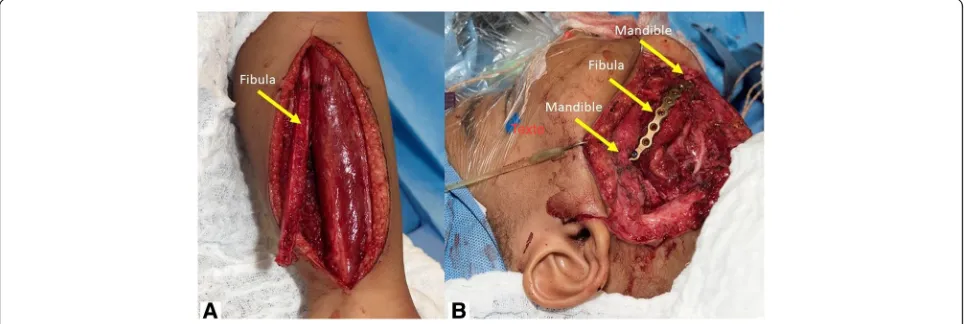
At 42-month-old, a delayed microvascular bony recon-struction with a fibula free flap was completed, surgical
exploration confirming remission of the tumor (Fig. 8).
wound care. He later had 2 revision surgeries without complications, one for titanium plate removal and one for skin paddle thinning. At the time of plate removal, a complete and solid bony union was found at both oste-otomy sites well as new bone formation along the
recon-struction plate (Fig. 9). At the last follow-up at 53/12
years-old and 4 years after the resection, he was doing well and free of disease with an excellent function.
Figure 10 is a timeline that summarizes the patient’s
evolution and treatments.
Discussion
A scoping review was performed in Medline/Pubmed database. English and French articles were searched
using the keywords “myofibromatosis” “myofibroma”
“infantile” “head and neck” and“mandible” for the
path-ology and “mandibular reconstruction” “pediatric” “free
fibula flap” and “submental island flap” for the
recon-struction. The snowballing method was also applied to selected articles. Despite being a rare pathology, IM is
the most common fibrous tumor of infancy [3]. In the
past, this disease has also been called congenital fibrosar-coma [6], congenital generalized fibromatosis, generalized harmartomatosis, multiple congenital mesenchymal tu-mors, diffuse congenital fibromatosis, and multiple
vascu-lar leiomyomas of the newborn [2]. Approximately 300
cases of IM have been reported in the English literature [5]. 30 to 50% of cases are diagnosed at birth or during the
neonatal period [2, 7, 8]. IM is characterized by the
Fig. 5CT scan shows progression of the tumor with new extension along the right maxilla despite oral excision and chemotherapy (coronal view)
formation of nodules or masses in skin, subcutaneous tissue, muscle, bone and viscera (mainly gastrointestinal, pulmonary, and cardiac) [9–11]. According to Chung, the main anatomical sites affected are the head and neck region (33%), the trunk (33%) and the limbs (31%) [2].
Three different clinical forms of IM have been defined: solitary and multicentric, with or without visceral in-volvement. Solitary IM is characterized by a single nodule and is the most frequent presentation [2]. Multicentric IM without visceral lesions involves several nodules in the skin, subcutaneous tissues, muscles and bones. The prognosis of these two forms is generally excellent with conservative surgery and can show spontaneous
regres-sion [12]. IM with visceral involvement represents 15–
20% cases and is defined by visceral lesions in addition to skin nodules. The prognosis is associated with high morbidity and mortality despite surgery and
chemother-apy [2]. Rapidly growing tumor cause visceral
compres-sion leading to gastrointestinal and cardiopulmonary
compromise, whereas perivascular nodules interfere with
organs blood supply [13, 14]. Familiarity with the
recognition of the three clinical forms is important, requiring different management strategies. Most of these tumors are sporadic and isolated. Rare familial cases of IM have been described and mutations of 2 genes (PDGFRB and NOTCH3) have been identified causing the disease [15–17].
Diagnosis can be suspected based on family history and physical examination but is made chiefly by biopsy
[2]. Histopathology examination reveals interlacing
fascicles of spindle cells (myofibroblasts) in the periph-ery, forming nodules separated by collagen tissue with
no nuclear atypia [3, 18]. Characteristics on imaging
include a mass with an anechoic center on ultrasound, low signal on T1-weighted imaging and high or low signal intensity areas on T2-weighted imaging on MRI and a mass with peripheral enhancements and calcifica-tions in contrast enhanced CT scan [19].
Fig. 7Post-operative panoramic radiograph after ablation
Due to the benign nature of IM, therapies producing the least long-term sequels and toxicity are preferred. Conservative surgery is the treatment of choice for the solitary form when morbidity and complications are minimal. In cases of incomplete resection, re-excision
can be proposed later [20]. Treatment for multicentric
IM is not well defined. For lesions affecting the skin and/or muscles only, a wait-and-see policy is often pro-posed because of a tendency towards spontaneous re-gression [21]. Radical surgical excision is required if the lesions are symptomatic or potentially life-threatening.
Chemotherapy is considered for solitary lesions when surgery is judged too morbid or for multicentric pro-gressive disease. Standard regimen is a combination of
methotrexate and vinblastine [1, 22]. As summarized by
Levine et all [12], several reports describe response and long-term success with this protocol, initially used to treat desmoid tumor allowing regression or stabilization of the lesions with no severe toxicity. This regimen is often chosen as no late effects have been described with these drugs. No large or multicentric series are available. Other treatments such as IFN-alpha or conventional chemotherapy (vincristine, actinomycin D, and cyclophos-phamide) should be considered only for disease refractory to standard protocols or with rapid progression because of the long-term risks of secondary malignancy [14,22].
Myofibroma of the oral cavity occurs mainly in the mandible (38%) and less frequently in the lips, cheeks
and tongue [7]. It is typically diagnosed in children in
the first decade of life (mean 7.2 years) with a male
predominance (male/female ratio 2.1:1) [23]. These
features vary from those found in myofibromas of the oral mucosa, which is diagnosed in an older age group (mean 21.7 years) with a female predominance (female/ male ratio 1,6:1) [24].
Fig. 9Postoperative panoramic radiograph after fibular free flap reconstruction
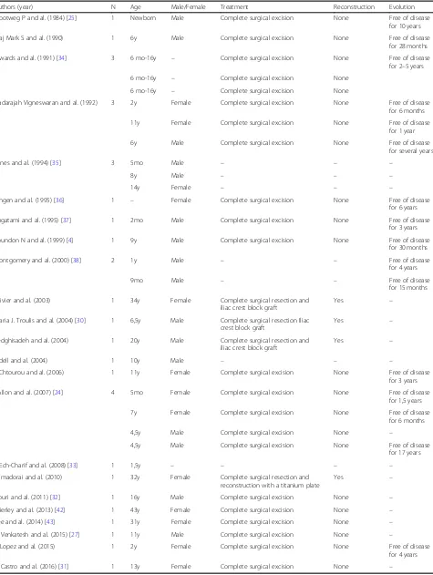
Table 1Review of Literature of infantile myofibromatosis of the mandible
Authors (year) N Age Male/Female Treatment Reconstruction Evolution
Slootweg P and al. (1984) [25] 1 Newborn Male Complete surgical excision None Free of disease for 10 years
Maj Mark S and al. (1990) 1 6y Male Complete surgical excision None Free of disease for 28 months
Inwards and al. (1991) [34] 3 6 mo-16y – Complete surgical excision None Free of disease for 2–5 years
6 mo-16y – Complete surgical excision None
6 mo-16y – Complete surgical excision None
Nadarajah Vigneswaran and al. (1992) 3 2y Female Complete surgical excision None Free of disease for 6 months
11y Female Complete surgical excision None Free of disease for 1 year
6y Male Complete surgical excision None Free of disease for several years
Jones and al. (1994) [35] 3 5mo Male – – –
8y Male – – –
14y Female – – –
Lingen and al. (1995) [36] 1 – Female Complete surgical excision None Free of disease for 6 years
Sugatami and al. (1995) [37] 1 2mo Male Complete surgical excision None Free of disease for 3 years
Loundon N and al. (1999) [4] 1 9y Male Complete surgical excision None Free of disease for 30 months
Montgomery and al. (2000) [38] 2 1y Male – – Free of disease
for 4 years
9mo Male – – Free of disease
for 15 months
Olivier and al. (2003) 1 34y Female Complete surgical resection and iliac crest block graft
Yes –
Maria J. Troulis and al. (2004) [30] 1 6,5y Male Complete surgical resection Iliac crest block graft
Yes –
Sedghisadeh and al. (2004) 1 20y Male Complete surgical resection and iliac crest block graft
Yes –
Odell and al. (2004) 1 10y Male – – –
I. Chtourou and al. (2006) 1 11y Female Complete surgical excision None Free of disease for 3 years
I Allon and al. (2007) [24] 4 5mo Female Complete surgical excision None Free of disease for 1,5 years
7y Female Complete surgical excision None Free of disease for 6 months
4,5y Male Complete surgical excision None –
4,5y Male Complete surgical excision None Free of disease for 17 years
S Ech-Charif and al. (2008) [33] 1 1,5y – – – –
Ramadorai and al. (2010) 1 32y Female Complete surgical resection and reconstruction with a titanium plate
Yes –
Nouri and al. (2011) [32] 1 16y Male Complete surgical excision None –
Brierley and al. (2013) [42] 1 43y Female Complete surgical excision None –
Lee and al. (2014) [43] 1 31y Female Complete surgical excision None –
V. Venkatesh and al. (2015) [27] 1 11y Male Complete surgical excision None –
R Lopez and al. (2015) 1 2y Female Complete surgical excision None Free of disease for 4 years
data. Twenty-two patients were treated with complete primary local resection without reconstruction. Three cases of mandibular reconstruction with iliac crest bone
grafting were reported in two adults and one child [30].
No recurrence during the follow-up period (6 months to 17 years) was observed in any patient.
Before the advent of bony free flaps and rigid recon-struction plates, children with benign or malignant jaw tumors were preferentially reconstructed by the place-ment of a bone graft and immobilization with
maxillo-mandibular fixation [44, 45]. Although this strategy
provided a favorable and functional result in a single op-eration, it was also associated with high infection rates and insufficient bone stock for dental rehabilitation [46,
47]. The use of rigid reconstruction plates allows a nega-tive margin resection while preserving the occlusion until a definitive bony reconstruction is planned in the
absence of tumor recurrence [30]. For the present case,
it also allowed growth to an age permitting the success of a free flap. Since the intraoral defect was extensive, a submental island flap was done at the time of the man-dibulectomy for soft tissue reconstruction.
The strategy of staging mandibular reconstruction using a plate and submental island flap followed by a de-layed fibula free flap is novel. To our knowledge, this is the only reported case of fibula free flap mandibular re-construction in a patient with IM, as well as one of the youngest reported submental island flaps for any path-ology. The later was first described by Martin et al. in 1993 [48]. In a review in 2014, Rahpeyma et al. described
several variants based on 90 published studies [49]. Its
use is rare in the pediatric population, the youngest be-ing at the age of 6 weeks for closure of a skull base
de-fect from resection of a teratoma [50, 51]. In 1993,
Posnick et al. reported the first free fibula flap in the
re-construction of pediatric mandibular defect [52]. Since
then, cases in patients as young as 10-month-old have been reported [53]. Stelnicki et al. even reported the bi-lateral mandibular reconstruction with two fibula free flap in a 2 ½ year-old patient with severe craniofacial malformation [54]. Several papers testify the reliability of free fibula flap in children, with more than 50 patients reported under the age of 18 years old [55–60].
growth pattern of the lower and mid face. Preservation of the condylar epiphyseal plate should be a priority before its fusion at the age of 18 to maximize proper craniofacial development. Although experimental con-cerns of lower limb growth discrepancies have been raised from pediatric fibula harvesting, there is no clin-ical demonstration of such a phenomenon in the litera-ture. There is a significant risk of valgus ankle deformity before 8 years, which can be prevented or corrected by
performing an immediate or delayed synostosis [65].
Conclusions
Despite being a rare disease and described anecdotally in the mandible, infantile myofibromatosis is the most common fibrous tumor of infancy. We presented a case of myofibroma of the mandible with very aggressive behavior resistant to all mandibular sparing treatment strategies including chemotherapy and debulking. A hemi-mandibulectomy with initial bridge plating and coverage with submental island flap and delayed recon-struction with fibula free-flap reconrecon-struction was done successfully. This reconstruction strategy is novel and
was chosen due to the patient’s very young age at the
resection (18 months) and aggressiveness of the disease. To our knowledge, this is the only reported case of fibula free flap mandibular reconstruction in a patient with IM, as well as one of the youngest reported sub-mental island flaps for any pathology. Segsub-mental man-dibular deficits are very rare in children and surgical reconstruction is a significant challenge.
Abbreviations
AP:Anteroposterior; CC: Cephalocaudal; CT: Computerized tomography; IM: Infantile myofibromatosis; MRI: Magnetic resonance imaging; T: Transverse; US: Ultrasound
Acknowledgements
We would like to acknowledge Dr. Sébastien Labonté from CHU de Québec
–Université Laval for pathology slides photography.
Funding
This research did not receive any specific grant from funding agencies in the public, commercial, or not-for-profit sectors.
Availability of data and materials
Authors’contributions
FT and BG performed patient diagnosis, investigations and treatments. AM and FT completed the literature review. All three analyzed and interpreted patient data. AM and FT were major contributors in writing the manuscript. All authors read and approved the final manuscript.
Ethics approval and consent to participate
The procedures were in accordance with the ethical standards of the CHU de Québec–Université Laval. Ethical approval by the research ethics boards was obtained (Request #2018–4099).
Consent for publication
Informed consent was obtained from the patient’s parents.
Competing interests
The authors have no potential competing interest with respect to the research, authorship, and/or publication of this article.
Publisher’s Note
Springer Nature remains neutral with regard to jurisdictional claims in published maps and institutional affiliations.
Received: 14 April 2018 Accepted: 4 March 2019
References
1. Stout AP. Juvenile fibromatoses. Cancer. 1954;7:953–78.
2. Chung EB, Enzinger FM. Infantile myofibromatosis. Cancer. 1981;48:1807–18. 3. Wiswell TE, Davis J, Cunningham BE, Solenberger R, Thomas PJ. Infantile
myofibromatosis: the most common fibrous tumor of infancy. J Pediatr Surg. 1988;23:315–8.
4. Loundon N, Dedieuleveult T, Ayache D, Roger G, Josset P, Garabedian EN. Head and neck infantile myofibromatosis—a report of three cases. Int J Pediatr Otorhinolaryngol. 1999;51:181–6.
5. Gopal M, Chahal G, Al-Rifai Z, Eradi B, Ninan G, Nour S. Infantile myofibromatosis. Pediatr Surg Int. 2008;24:287–91.
6. Williams JO, Schrum D. Congenital fibrosarcoma: report of a case in a newborn infant. AMA Arch Pathol. 1951;51:548–52.
7. Foss RD, Ellis GL. Myofibromas and myofibromatosis of the oral region: a clinicopathologic analysis of 79 cases. Oral Surg Oral Med Oral Pathol Oral Radiol Endod. 2000;89:57–65.
8. Nishioka K, Seguchi T, Yamamura Y, Tatsumura M, Sou H, Gondo T, Hoshii Y, Iwata T. Infantile myofibromatosis identified by fetal ultrasound. Br J Dermatol. 1999;140:538–68.
9. Stanford D, Rogers M. Dermatological presentations of infantile
myofibromatosis: a review of 27 cases. Australas J Dermatol. 2000;41:156–61. 10. Short M, Dramis A, Ramani P, Parikh DH. Mediastinal and pulmonary
infantile myofibromatosis: an unusual surgical presentation. J Pediatr Surg. 2008;43:29–31.
11. Hausbrandta PA, Leithnera A, Behamc A, Bodoc K, Raith J, Windhagera R. A rare case of infantile myofibromatosis and review of literature. J Pediatr Orthop. 2010;19:122–6.
12. Levine E, Freneaux P, Schleiermacher G, Brisse H, Pannier S, Teissier N, Mesples B, Orbach D. Risk-adapted therapy for infantile Myofibromatosis in children. Pediatr Blood Cancer. 2012;59:115–20.
13. Mashiah J, Hadj-Rabia S, Dompmartin A, Harroche A, Laloum-Grynberg E, Wolter M, Amoric JC, Hamel-Teillac D, Guero S, Fraitag S, Bodemer C. Infantile myofibromatosis: a series of 28 cases. J Am Acad Dermatol. 2014; 71:264–70.
14. Auriti C, Kieran MW, Deb G, Devito R, Pasquini L, Danhaive O. Remission of infantile generalized Myofibromatosis after interferon alpha therapy. J Pediatr Hematol Oncol. 2008;30:179–81.
15. Lee JW. Mutations in PDGFRB and NOTCH3 are the first genetic causes identified for autosomal dominant infantile myofibromatosis. Clin Genet. 2013;84:340–3.
16. Martignetti JA, Tian L, Li D, Ramirez MCM, Camacho-Vanegas O, Camacho SC, et al. Mutations in PDGFRB cause autosomal-dominant infantile myofibromatosis. Am J Hum Genet. 2013;92:1001–7.
17. Jennings TA, Duray PH, Collins FS, Sabetta J, Enzinger FM. Infantile myofibromatosis, evidence for an autosomal dominant disorder. Am J Surg Pathol. 1984;8:529–38.
18. Fletcher CD, Achu P, Van Noorden S, McKee PH. Infantile myofibromatosis: a light microscopic, histochemical and immunohistochemical study suggesting true smooth muscle differentiation. Histopathology. 1987;11: 245–58.
19. Koujok K, Ruiz RE, Hernandez RJ. Myofibromatosis: imaging characteristics. Pediatr Radiol. 2005;35:374–80.
20. Beck JC, Devaney KO, Weatherly RA, Koopmann CF, Lesperance MM. Pediatric myofibroblastosis of the head and neck. Arch Otolaryngol Head Neck Surg. 1999;125:39–44.
21. Hausbrandt PA, Leithner A, Beham A. A rare case of infantile myofibromatosis and review of literature. Pediatr Radiol. 2005;19:122–6. 22. Gandhi MM, Nathan PC, Weitzman S, Levitt GA. Successful treatment of
life-threatening generalized infantile Myofibromatosis using low-dose chemotherapy. J Pediatr Hematol Oncol. 2003;25:750–4.
23. Lopes RN, de Abreu Alves F, Rocha AC, Suassuna TM, Kowalski LP, de Castro JFL, Cruz Perez DE. Head and neck solitary infantile myofibroma:
Clinicopathological and immunohistochemical features of a case series. Acta Histochem. 2015;117:431–6.
24. Allon I, Vered M, Buchner A, Dayan D. Central (intraosseous) myofibroma of the mandible: clinical, radiologic, and histopathologic features of a rare lesion. Oral Surg Oral Med Oral Pathol Oral Radiol Endodontol. 2007;103:45–53. 25. Slootweg P, Muller H. Localized infantile myofibromatosis. Report of a case
originating in the mandible. J Maxillofac Surg. 1984;12:86–9. 26. Matthews MMS, Tabor MW, Thompson MSH, Gross D. Infantile
Myofibromatosis of the mandible. J Oral Maxillofac Surg. 1990;48:884–9. 27. Venkatesh V, Kumar BP, Kumar KAJ, Mohan AP. Myofibroma—a rare entity
with unique clinical presentation. J Maxillofac Oral Surg. 2015;14:64–8. 28. Chtourou I, Makni SK, Dhouib M, Khabir A, Fakhfakh I, Ayadi L, Mnif H,
Abdelmoula M, Boudawara TS. Myofibromatose infantile de la mandibule. Rev Stomatol Chir Maxillofac. 2007;108:461–4.
29. Vigneswaran N, Boyd DL, Waldron CA. Solitary infantile myofibroma of the mandible. Oral med oral pathol. 1992;73:84–8.
30. Troulis MJ, Bradford Williams W, Kaban LB. Staged protocol for resection, skeletal reconstruction, and oral rehabilitation of children with jaw tumors. J Oral Maxillofac Surg. 2004;62:335–43.
31. Castro HHO, Gomes HE, Tassara LFR, de Freitas JB, de Andrade Marigo Grandinetti H, Capistrano HM. Myofibroma of the mandible–case report. Pediatr Dent J. 2016;26:38–41.
32. Nouri H, Aderdour L, Maliki O, Bassi L, Baallal H, Brahimi M, Belaabidia B, Raji A. Myofibroma of the mandibule: a case report. Rev Laryngol Otol Rhinol. 2011;132:115–7.
33. Ech-Charif S, Benhammou A, Maher M, Séfiani S. Solitary myofibroma of the mandible: a case report. Rev Laryngol Otol Rhinol. 2008;129:337–40. 34. Inwards CY, Unni KK, Beabout JW, Shives TC. Solitary congenital fibromatosis
(infantile myofibromatosis) of bone. Am J Surg Pathol. 1991;15:935–41. 35. Jones AC, Freedman PD, Kerpel SM. Oral myofibromas: a report of 13 cases
and review of the literature. J Oral Maxillofac Surg. 1994;52:870–5. 36. Lingen MW, Mostofi RS, Solt DB. Myofibromas of the oral cavity. Oral Surg
Oral Med Oral Pathol Oral Radiol Endod. 1995;80:297–302.
37. Sugatami T, Inui M, Tagawa T, Seki Y, Mori A, Yoneda J. Myofibroma of the mandible. Clinicopathologic study and review of the literature. Oral Surg Oral Med Oral Pathol Oral Radiol Endod. 1995;80:303–9.
38. Montgomery E, Speight PM, Fisher C. Myofibromas presenting in the oral cavity: a series of 9 cases. Oral Surg Oral Med Oral Pathol Oral Radiol Endod. 2000;89:343–8.
39. Oliver RJ, Coulthard P, Carre C, Sloan P. Solitary adult myofibroma of the mandible simulating an odontogenic cyst. Oral Oncol. 2003;39:626–9. 40. Sedghizadeh PP, Allen CM, Kalmar JR, Miloro M, Suster S. Solitary central
myofibroma presenting in the gnathic region. Ann Diag Pathol. 2004;8:284–9. 41. Ramadorai A, Rajsekaran A, Narayanan V. A case report of solitary,
intraosseous, adult-onset myofibroma of the mandible. J Maxillofac Oral Surg. 2010;9:280–3.
42. Brierley DJ, Khurram SA, Speight PM. Solitary myofibroma of the adult mandible: a case report. Oral Surg Oral Med Oral Pathol Oral Radiol. 2013; 115:40–3.
43. Lee YM, Son SM, Won Kim K, Lee OJ. Solitary Myofibroma of the adult mandible: a case report and review of literature. Korean J Pathol. 2014;48: 307–10.
51. Rahpeyma A. Submental flap in intraoral reconstruction after pathologic resections: indications and limitations. J Maxillofac Oral Surgery. 2015;14:57–62. 52. Posnick JC, Wells MD, Zuker RM. Use of the free fibular flap in the
immediate reconstruction of pediatric mandibular tumors: report of cases. J Oral Maxillofac Surg. 1993;51:189–96.
53. Guo L, Ferraro NF, Padwa BL, Kaban LB, Upton J. Vascularized fibular graft for pediatric mandibular reconstruction. Plast Reconstr Surg. 2008;121:2095–105. 54. Stelnicki EJ, Boyd JB, Nott RL, Barnavon Y, Uecker C, Henson T. Early treatment
of severe mandibular hypoplasia with distraction mesenchymogenesis and bilateral free fibula flaps. J Craniofac Surg. 2001;12:337–48.
55. Zhang WB, Liang T, Peng X. Mandibular growth after paediatric mandibular reconstruction with the vascularized free fibulaflap: a systematic review. Int J Oral Maxillofac Surg. 2016;45:440–7.
56. Smith AM, Petersen DK, Samant S, Halen JP. Pediatric mandibular reconstruction following resection of oral squamous cell carcinoma: a case report. Am J Otolaryngol. 2014;35:826–8.
57. Nkenke E, Agaimy A, von Wilmowsky C, Eitner S. Mandibular reconstruction using intraoral microvascular anastomosis following removal of an ameloblastoma. J Oral Maxillofac Surg. 2013;71:1983–92.
58. Li JS, Chen WL, Huang ZQ, Zhang DM. Pediatric mandibular reconstruction after benign tumor ablation using a vascularized fibular flap. J Craniofac Surg. 2009;20:431–4.
59. Akakpo K, Iobst C, Old M, Grischkan J. Long-term follow-up with mention of complications in pediatric microvascular mandibular reconstruction. Int J Pediatr Otorhinolaryngol. 2018;105:154–7.
60. Phillips JH, Rechner B, Tompson BD. Mandibular growth following reconstruction using a free fibula graft in the pediatric facial skeleton. Plast Reconstr Surg. 2005;116:419–24.
61. Arkas LG, Posnick JC, Hreczko TM. Growth patterns of the face: a morphometric study. Cleft Palate Craniofac J. 1992;29:308–15. 62. Hans MG, Enlow DH, Noachtar R. Age-related differences in mandibular
ramus growth: a histologic study. Angle Orthod. 1995;65:335–40. 63. Goerke D, Sampson DE, Tibesar RJ, Sidman JD. Rib reconstruction of the
absent mandibular condyle in children. Otolaryngol Head Neck Surg. 2013; 149:372–6.
64. Crosby MA. Pediatric mandibular reconstruction using a vascularized fibula flap. Head Neck. 2008;30:311–9.