R E S E A R C H
Open Access
KRAS, EGFR, PDGFR-
α
, KIT and COX-2 status in
carcinoma showing thymus-like elements (CASTLE)
Lothar Veits
1*, Rupert Schupfner
2, Petra Hufnagel
3, Roland Penzel
4, Jens Freitag
5, Philipp Ströbel
6, Michael A Kern
4,
Sören Schröder
7, Nikolaus Neuhold
8, Kurt W Schmid
9, Peter Schirmacher
4, Arndt Hartmann
10and Ralf J Rieker
10Abstract
Background:CASTLE (Carcinoma showing thymus-like elements) is a rare malignant neoplasm of the thyroid
resembling lymphoepithelioma-like and squamous cell carcinoma of the thymus with different biological behaviour and a better prognosis than anaplastic carcinoma of the thyroid.
Methods:We retrospectively investigated 6 cases of this very rare neoplasm in order to investigate the mutational status ofKRAS,EGFR,PDGFR-αandKIT, as well as the immunohistochemical expression pattern of CD117, EGFR and COX-2, and possibly find new therapeutic targets.
Results:Diagnosis was confirmed by a moderate to strong expression of CD5, CD117 and CK5/6, whereas thyroglobulin, calcitonin and TTF-1 were negative in all cases. Tumors were also positive for COX-2 and in nearly all cases for EGFR. In four cases single nucleotide polymorphisms (SNPs) could be detected in exon 12 of thePDGFR-αgene (rs1873778), in three cases SNPs were found in exon 20 of theEGFRgene (rs1050171). No mutations were found in theKITandKRASgene.
Conclusions:All tumors showed a COX-2 expression as well as an EGFR expression except for one case and a
wild-typeKRASstatus. No activating mutations in theEGFR, KITandPDGFR-αgene could be detected. Our data may indicate a potential for targeted therapies, but if these therapeutic strategies are of benefit in CASTLE remains to be determined.
Virtual Slides:The virtual slide(s) for this article can be found here: http://www.diagnosticpathology.diagnomx.eu/vs/ 1658499296115016
Keywords:CASTLE, Thymic carcinoma, Mutational analysis, Immunohistochemistry, Thyroid gland
Background
Carcinoma showing thymus-like elements (CASTLE) is a rare malignant tumor of the thyroid gland. It was first described by Miyauchi et al. in 1985 as “intrathyreoidal epithelial thymoma”[1], is thought to arise from ectopic thymic tissue within the thyroid gland or rudimentary branchial pouches along the thymic line and was named CASTLE by Chan and Rosai in 1991 [2]. CASTLE-tumors belong to a group of ectopic tumors of thymic differenti-ation: Ectopic hamartomatous thymoma, ectopic cervical thymoma, spindle epithelial tumors with thymus like dif-ferentiation (SETTLE) and carcinoma showing
thymic-like elements (CASTLE). Although CASTLE shows some tendencies for squamous differentiation, it has newly been adopted as an independent entity by the recent WHO classification [3]. In contrast to ectopic thymomas, SET-TLE and CASSET-TLE are always malignant and therefore the exact diagnosis is crucial in consideration of prognosis and treatment. CASTLE tumors have characteristic clinical and histological features. They are tumor of the adults in the fifth decade of life and often invade the adjacent soft tissue. Metastases into the regional lymph nodes are common. The largest study yet performed comprised 25 cases [4]. According to this study it is likely that some thyroid can-cers, especially those with squamous differentiation may have been treated incorrectly based on an improper classi-fication. For our study we collected 6 cases of this rare ma-lignancy and evaluated possible therapeutic options. * Correspondence:[email protected]
1
Institute of Pathology, Klinikum Bayreuth, Preuschwitzerstraße 101, Bayreuth 95445, Germany
Full list of author information is available at the end of the article
Methods
In our study we included 6 patients suffering from CASTLE, 3 women and 3 men, diagnosed between 1997 and 2007. The median age was 60 years ranging from 30 to 68 years.
Tissue microarrays and immunohistochemistry
Tissue microarrays (TMA) were performed from the paraffin-wax-embedded blocks of the tumor speci-mens. A tissue arrayer device (Beecher Instruments, Sun Prairie, WI, USA) was used. All the investigated cases were reviewed and representative tumor areas were marked in the corresponding paraffin wax blocks, of which at least two were sampled. The diameter of the cylinders was 1.2 mm. For immunohistochemistry 5μm sections on poly-L-lysine-coated slides were used after drying in an oven, followed by dewaxing and per-oxidase blocking with 1% H2O2. The specificity of im-munostaining was tested by replacing primary antisera with normal rabbit serum. No additional pre-treatment procedure for antigen retrieval was done. The immunor-eactions were performed with the following primary mono-clonal antibodies: Anti-COX-2 (monomono-clonal rabbit IgG; Santa Cruz, USA; 1:50), anti-CD5 (monoclonal mouse IgG; Novocastra, Leica Microsystems, Wetzlar, Germany; 1:25), anti-TTF-1 (monoclonal mouse, IgG, SPT24; Menarini, Florence, Italy; 1:100), anti-CK 5/6 (monoclonal mouse IgG, M7237; Dako Cytomation, Glostrup, Denmark; 1:50), anti-Ki67 (monoclonal mouse IgG, K-2, Zytomed, Berlin, Germany; ready-to-use). Primary polyclonal antibodies used were: CD117 (polyclonal rabbit, A4502; Dako Cyto-mation, Glostrup, Denmark; 1:50), anti-thyroglobulin (poly-clonal rabbit, A0251; Dako Cytomation, Glostrup, Denmark; 1:80000) and anti-calcitonin (polyclonal rabbit, A0576; Dako Cytomation, Glostrup, Denmark; 1:4000). After wash-ing steps signal detection was performed (EnVision + system-HRP, AEC, Dako Hamburg, Germany; 1:200) followed by counterstaining in hemalaun. Detection of EGFR was performed with the EGFR pharmDX kit (Dako Hamburg, Germany) according to manufacturer’s instructions.
The presence of clearly visible dark brown precipita-tion was considered as immunoreacprecipita-tion, evaluated by a score for staining intensity (0: no staining; 1: weak; 2: moderate; 3: strong reaction intensity).
Mutational analysis ofKIT, PDGR-α, EGFR, KRAS
Cores of tumor tissue selected from regions on corre-spondent Hematoxylin-Eosin slides were manually punched out from the donor blocks. DNA was extracted from paraffin-embedded tissue sections using EZ1 DNA tis-sue Kit (Qiagen, Hilden Germany) after pre-treatment with proteinase K (Qiagen, Hilden Germany). 150 ng DNA were used for PCR amplification and all PCR
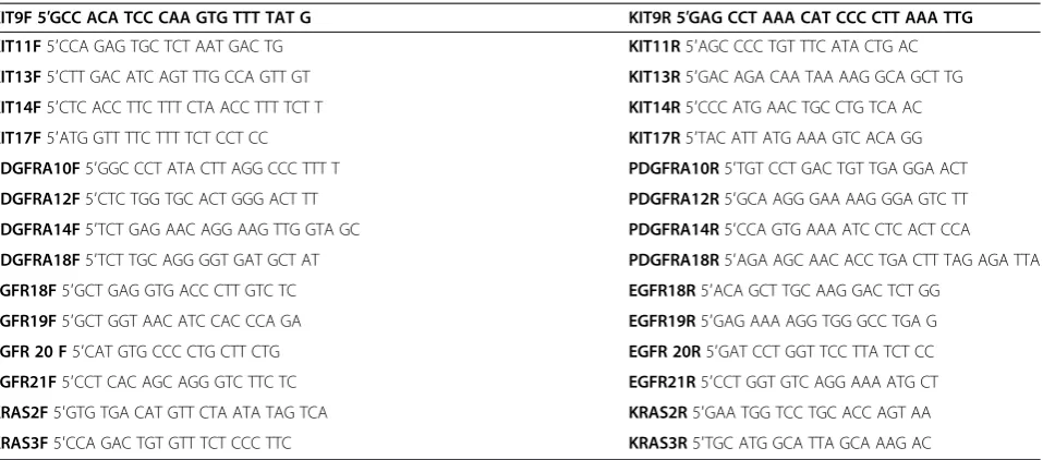
reactions were run at a final volume of 25μl (Red Jump MasterMix, Sigma-Aldrich). Exons 9, 11, 13 and 17 of KIT, exons 10, 12, 14 und 18 ofPDGFR-α, exons 18, 19, 20 and 21 of EGFR and exons 2 and 3 of KRAS were evaluated for the presence of mutations by PCR ampli-fication and direct sequencing. The primer pairs used for PCR amplification and direct sequencing are shown in Table 1. After estimation of DNA concentration by spectroscopy (GeneQuant) relevant exons were ampli-fied by PCR under standard conditions with an anneal-ing temperature of 50°C for Exon 17 (KIT), 55°C for Exon 2 and 3 (KRAS), Exon 9 (KIT) and Exon 18 (PDGFR), 60°C for Exon 18, 19, 20 and 21 (EGFR) and Exon 11 and 13 (KIT) as well as Exon 10, 12 and 14 (PDGFR) with the corresponding primers. The PCR products were then separated on agarose gel containing ethidiumbromide and visualized under ultraviolet light. Afterward PCR products were purified using high pure PCR product purification kit (Roche Diagnostics, Penzberg, Germany) and then sent to Qiagen (Hilden, Germany) for sequencing according to Sanger method.
Ethical approval
This study was approved by the ethics committee of the University Hospital Heidelberg (reference number: 206/ 2005).
Results
Clinical and histological characteristics
The primary symptom was a palpable node in 2 patients and an adenomatous goitre as well as a paresis of the re-current nerve in 1 case, respectively. In 3 cases no case history was available. 5 tumours were located in the thy-roid and 1 was partially extrathythy-roidal, the latter with the majority of the tumor mass in the thyroid gland.
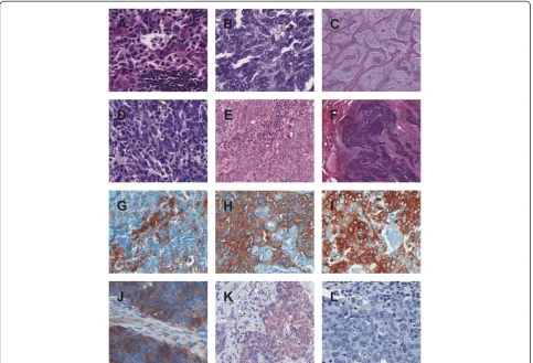
Tumor cells were varying in shape: Irregular cells as well as spindle-shaped and epithelioid cells were observed. Oc-casionally, squamoid or spindle-shaped epithelial cells with whorls resembling Hassall’s corpuscles were noticed. The cytoplasm of the tumor cells was pale and nuclei were clearly evident and slightly oval (see Figure 1A - F).
Immunohistochemistry
All CASTLE cases expressed CD5 in a moderate to strong reaction and CD117 in predominantly weak to moderate intensity in the epithelial component. Only in one case a strong staining signal for CD117 could be de-tected. (see Figure 1G + H). Staining with CK5/6 showed weak to strong reactions in all samples (see Figure 1I). EGFR showed a strong reactivity in three, weak staining intensity in two samples and no staining pattern in one case (see Figure 1J). COX-2 was detected positively in all cases with mainly moderate expression patterns (see
Figure 1 K). The expression of Ki67 showed varying re-sults between 2% and 85%. The staining rere-sults for CD117, EGFR, COX-2 and Ki67 are shown summarized in Table 3. All cases were negative for calcitonin, thyro-globulin and TTF-1 (Figure 1L).
Mutational analysis of KIT, PDGFR-α, EGFR, KRAS
Mutational analysis of KIT, PDGFR-α, EGFRand KRAS was possible in only 4 cases due to inapplicable DNA in case number 2 and 3.
Mutation analysis of all four investigated cases re-vealed the absence of an activating mutation in the PDGFR-αgene of exon 10, 12, 14 and 18. Nevertheless, in all four cases a silent single nucleotide polymorphism with a base substitution (CCA > CCG) in exon 12 at codon 567 (p.P567P) of the PDGFR-α gene was ob-served. This change has already been described in the SNP database with the number rs1873778 (Figure 2A).
Table 1 Primer sequences for KIT, PDGR, EGFR, KRAS PCR amplification
KIT9F 5′GCC ACA TCC CAA GTG TTT TAT G KIT9R 5′GAG CCT AAA CAT CCC CTT AAA TTG KIT11F5′CCA GAG TGC TCT AAT GAC TG KIT11R5′AGC CCC TGT TTC ATA CTG AC
KIT13F5′CTT GAC ATC AGT TTG CCA GTT GT KIT13R5′GAC AGA CAA TAA AAG GCA GCT TG
KIT14F5′CTC ACC TTC TTT CTA ACC TTT TCT T KIT14R5′CCC ATG AAC TGC CTG TCA AC
KIT17F5′ATG GTT TTC TTT TCT CCT CC KIT17R5′TAC ATT ATG AAA GTC ACA GG
PDGFRA10F5′GGC CCT ATA CTT AGG CCC TTT T PDGFRA10R5′TGT CCT GAC TGT TGA GGA ACT
PDGFRA12F5′CTC TGG TGC ACT GGG ACT TT PDGFRA12R5′GCA AGG GAA AAG GGA GTC TT
PDGFRA14F5′TCT GAG AAC AGG AAG TTG GTA GC PDGFRA14R5′CCA GTG AAA ATC CTC ACT CCA
PDGFRA18F5′TCT TGC AGG GGT GAT GCT AT PDGFRA18R5′AGA AGC AAC ACC TGA CTT TAG AGA TTA
EGFR18F5′GCT GAG GTG ACC CTT GTC TC EGFR18R5′ACA GCT TGC AAG GAC TCT GG
EGFR19F5′GCT GGT AAC ATC CAC CCA GA EGFR19R5′GAG AAA AGG TGG GCC TGA G
EGFR 20 F5′CAT GTG CCC CTG CTT CTG EGFR 20R5′GAT CCT GGT TCC TTA TCT CC
EGFR21F5′CCT CAC AGC AGG GTC TTC TC EGFR21R5′CCT GGT GTC AGG AAA ATG CT
KRAS2F5′GTG TGA CAT GTT CTA ATA TAG TCA KRAS2R5′GAA TGG TCC TGC ACC AGT AA
KRAS3F5′CCA GAC TGT GTT TCT CCC TTC KRAS3R5′TGC ATG GCA TTA GCA AAG AC The different exons ofKIT, PDGFR, EGFRandKRASare written in bold letters.
Table 2 Patient characteristics and tumor progression
No. Stage Therapy Progression
1 pT4, N1b (18/21), MX, R0 Total thyroidectomy, left central neck dissection, resection of the left recurrent laryngeal nerve and the left internal jugular vein, chemotherapy, radiation (50 Gy) After recurrence: Tumorectomy, bilateral neck dissection, transsternal lymphadenectomy
Local recurrence after 2 months
2 pT3, N0 (0/5), MX, R0 Thyroidectomy, cervico-central lymphadenectomy No recurrence (time of
observation: 36 months)
3 pT3, N0, MX Subtotal thyroidectomy, local telecobalttherapy of the neck (60 Gy) No recurrence (time of observation: 19 months)
4 pT2, N0 (0/7), MX Hemithyroidectomy right, then restthyroidectomy, microdissection of the cervicocentral compartment, radiation of the thyroidbed (59,4 Gy)
No recurrence (time of observation: 15 months)
5 pT3, N1 (1/19), MX En bloc resection with the right lobe, neurolysis N.laryngeus recurrens right, neck dissection right, supraclavicular cervical lateral dissection, radiation (50 Gy)
No recurrence (time of observation: 17 months)
6 pT4a, N1 (1/5), M0, R0 Total thyroidectomy, neck dissection, radiation of the neck region (50 Gy) No recurrence (time of observation: 22 months)
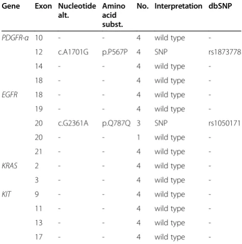
In 3 (3/4) patients a silent single nucleotide polymorph-ism with a base substitution (CAG > CAA) in theEGFR gene of exon 20 at position 787 (p.Q787Q) was ob-served, which has also already been described in the SNP database with the number rs1050171 (Figure 2B). No mutation associated with drug sensitivity in the hot-spot exons 18, 19, 20 and 21 was detected. No patient had detectable sequence variants forKITin the hotspot region of exon 9, 11, 13 and 17. Also no sequence vari-ants were found in the sequenced exons of the KRAS gene (Table 4).
Discussion
CASTLE tumors are rare malignant tumors occurring in the thyroid, but the prognosis and the concomitant long term survival rate is better than for other aggressive ad-vanced thyroid neoplasms [1,2,5]. Among other things and according to the so far largest study of CASTLE-tumors comprising 25 cases [4,6] a significant number may have been incorrectly classified, especially as squa-mous cell carcinoma of the thyroid. In this study Ito et al. showed a survival rate of 90% after 5 years and 82% after 10 years. Patients without lymph node metas-tasis had an overall survival rate of 100% after 5 years, those with lymph node metastasis 76% after 5 and 57% after 10 years. One of our patients with initial lymph node metastasis had recurrence after 2 months. No recur-rence during an observation period of up to 73 months occurred in the other patients. Interestingly, the expres-sion of Ki67 showed varying results between 2% and 85%, but could not be linked to the presence of lymph node metastases or advanced tumor stages, as the only case in which a tumor recurrence was observed showed a low proliferation rate (compared to e.g. striking 85% in case
Figure 1HE and immunohistochemical stainings. A-F. HE staining case 1–6.A. HE case 1,B. HE case 2,C. HE case 3,D. HE case 4,E. HE case 5,
F. HE case 6.G-L. Immunohistochemical staining case 1–6 for CD5, CD117, CK5/6, COX-2, EGFR and TTF-1.G. CD5 case 1.H. CD117 case 5.I. CK5/6 case 4.
J. EGFR case 6.K. COX-2 case 2.L. TTF-1 case 3.
Table 3 Immunohistochemical expression pattern
No. CD117 EGFR COX-2 Ki67 (%)
1 1 1 2 17
2 2 3 2 7
3 1 0 3 2
4 1 3 1 12
5 3 1 2 53
6 2 3 2 85
number 6 with no recurrence during an observation period of 22 months).
Because a diagnosis of CASTLE is difficult before re-section of the tumor, patients are usually treated accord-ing to the standards of thyroid gland carcinoma with surgical removal of all or part of the thyroid gland with
or without neck dissection. Depending on the tumor en-tity and stage postoperative therapy is often indicated. 5 of our patients underwent radiotherapy, 1 combined radiochemotherapy.
We have recently described chromosomal imbalances in CASTLE similar to those found in thymomas and thymic carcinomas [7]. Given the morphological, im-munohistochemical and genetic similarities with thymic carcinomas, it seems reasonable to assume that treat-ment of advanced CASTLE should follow the guidelines for thymic tumors and patients may benefit also from evolving therapeutic options for patients with thymic tumors [8-13].
CD117 is expressed in about 86% of thymic carcin-omas [14], but KITgene mutations are uncommon. So far, only few cases of metastasizing thymic carcinoma with an activating KIT mutation and a partial response to treatment with Imatinib have been described [15,16]. In our analysis, all CASTLE tumors showed positive staining results for CD117, but no mutations of theKIT gene.
Membrane associated EGFR expression has been seen in thymomas as well as in thymic carcinomas. Girard summarized eight studies [17], which investigated EGFR expression levels in thymic malignancies [18-25] using immunohistochemistry. In this meta-analysis, EGFR was overexpressed in 70% of thymomas and 53% of thymic carcinomas, but there was no correlation between EGFR expression and thymic tumor type. ActivatingEGFR mu-tations seem to be exceptionally rare in thymic carcin-omas with only few reported EGFR missense mutations
Figure 2SNPs inPDGFRandEGFRgenes. A. Forward chromatogram showing c.A1701G, predicting p.P567P in thePDGFRgene (db SNP = rs1873778).
B. Forward chromatogram showing c.G2361A, predicting p.Q787Q in theEGFRgene (db SNP = rs1050171).
Table 4 Sequence variants in the PDGFR-α, EGFR, KRAS and KIT genes
Gene Exon Nucleotide alt.
Amino acid subst.
No. Interpretation dbSNP
PDGFR-α 10 - - 4 wild type
-12 c.A1701G p.P567P 4 SNP rs1873778
14 - - 4 wild type
-18 - - 4 wild type
-EGFR 18 - - 4 wild type
-19 - - 4 wild type
-20 c.G2361A p.Q787Q 3 SNP rs1050171
20 - - 1 wild type
-21 - - 4 wild type
-KRAS 2 - - 4 wild type
-3 - - 4 wild type
-KIT 9 - - 4 wild type
-11 - - 4 wild type
-13 - - 4 wild type
-17 - - 4 wild type
in exon 21 (p.L858R in two cases and p.G863D in one case) [23,26]. In the present series, five tumors showed weak to strong immunohistochemical staining results for EGFR. Two of the four cases, in which DNA-extraction was successful, showed a weak positivity for EGFR, the other two cases showed a strong signal. In three cases mu-tational analysis revealed a single nucleotide polymorph-ism (SNP) in theEGFR gene of Exon 20 (p.Q787Q), but no activatingEGFRgene mutation could be found.
It has been shown that a therapeutic effect can be seen after the use of EGFR-inhibitors in non-small cell lung cancers, even if there is no activating mutation in the EGFRgene [27]. In a small series of advanced thymic tu-mours only a slight therapeutic effect could be demon-strated with Gefitinib [28].
Randomised clinical trials have demonstrated the po-tential of COX-inhibitors in tumor prevention [29]. Malka et al. showed that inhibition of COX-2 could play a possible therapeutic role in other tumor entities, for instance hepatocellular carcinoma [30]. In particular, the combination of COX-2 inhibitors and EGFR-inhibitors seems to increase the therapeutic effect in colon or cer-vical carcinoma [31-34]. COX-2 is frequently upregu-lated in thymomas and thymic carcinomas [11,12]. In our study all tumors expressed COX-2 raising the possi-bility to use COX-2 inhibitors in advanced cases.
Although in our investigation all four cases showed SNPs in the PDGFR-α gene of exon 12, no activating mutations in the PDGFR-α gene and no mutations in Codon 12 and 13 of theKRASgene were detected.
Conclusion
As already mentioned above, CASTLE shows some simi-larity to thymic carcinoma. However, in the latter targeted therapy is difficult and somehow limited in advanced tu-mors as there are only few reported cases in which targeted therapy was of some therapeutic benefit. Ströbel et al., for example, suggest that in cases with advanced thymic car-cinoma treatment with the multi-kinase inhibitor Sunitinib, that targets VEGFR1–3, PDGFR-α, c-KIT, FLT3, colony stimulating factor-1 (CSF1) and the RET receptor, is more effective than therapeutic regimes using single-target mo-lecular therapies like Gefitinib, even though there are no activating mutations in theEGFR, KITandPDGFR-αgene [35]. In conclusion, our data may indicate a potential for targeted therapies, but if these therapeutic strategies are of benefit in CASTLE remains to be determined.
Competing interests
The authors declare that they have no competing interests.
Authors’contributions
LV generated the study concept and design, evaluated the HE- and immunohistochemical stains, analysed and interpreted the data, confirmed the diagnosis and drafted the manuscript. RS critically revised the manuscript for important intellectual content and participated in the design of the study.
PH carried out the molecular genetic studies, participated in the sequence alignment and analysed and interpreted the data. RP participated in the molecular genetic studies and critically revised the manuscript for important intellectual content. JF critically revised the manuscript for important intellectual content and participated in the analysis of the data. PS critically revised the manuscript for important intellectual content and participated in the study design and concept. MK carried out the immunoassays. SS critically revised the manuscript for important intellectual content and participated in the study design and concept. NN critically revised the manuscript for important intellectual content and participated in the study design and concept. KS participated in the study design and concept and critically revised the manuscript for important intellectual content. PS critically revised the manuscript for important intellectual content. AH critically revised the manuscript for important intellectual content. RR critically revised the manuscript for important intellectual content, participated in the design of the study and evaluated the HE- and immunohistochemical stains, confirmed the diagnosis. All authors read and approved the final manuscript.
Authors’information
Lothar Veits and Rupert Schupfner share first authorship.
Author details
1Institute of Pathology, Klinikum Bayreuth, Preuschwitzerstraße 101, Bayreuth
95445, Germany.2Clinic of Trauma and Reconstructive Surgery, Klinikum Bayreuth, Preuschwitzerstraße 101, Bayreuth 95445, Germany.
3Pathologielabor Zams, Klostergasse 1, Zams 6511, Austria.4Department of
Pathology, University Hospital, INF 220/221, Heidelberg 69120, Germany. 5General practitioner, Moselstraße 1, Oberbettringen 73529, Germany. 6Department of Pathology, University Hospital, Robert-Koch-Straße 40,
Göttingen 37075, Germany.7Institute of Pathology/Lademannbogen Laboratories, Lademannbogen 61-63, Hamburg 22339, Germany. 8Pathologisch-Bakteriologisches Institut, Kaiserin-Elisabeth-Spital, Huglgasse
1-3, Vienna 1150, Austria.9Department of Pathology and Neuropathology, University Hospital Essen, Hufelandstraße 55, Essen 45122, Germany. 10Department of Pathology, University Hospital Erlangen, Krankenhausstr.
10-12, Erlangen 91054, Germany.
Received: 3 December 2013 Accepted: 15 May 2014 Published: 16 June 2014
References
1. Miyauchi A, Kuma K, Matsuzuka F, Matsubayashi S, Kobayashi A, Tamai H, Katayama S:Intrathyroidal epithelial thymoma: an entity distinct from squamous cell carcinoma of the thyroid.World J Surg1985,9:128–135. 2. Chan JK, Rosai J:Tumors of the neck showing thymic or related branchial
pouch differentiation: a unifying concept.Hum Pathol1991,22:349–367. 3. Cheuk W, Chan JKC, Dorfmann DM, Giordano T:Spindle Cell Tumour With
Thymus-Like Differentiation.InWorld Health Organization Classification of Tumours. Tumours of Endocrine Organs.Edited by DeLellis RA, Lloyd RV, Heitz PU, Eng C. Lyon: IARC Press; 2004:96–97.
4. Ito Y, Miyauchi A, Nakamura Y, Miya A, Kobayashi K, Kakudo K:Intrathyroid Epithelial Thymoma/Carcinoma Showing Thymus-Like Differentiation (CASTLE): Its Clinicopathological Characteristics.InRecent Advances in Thyroid Cancer Research.Kerala: Transworld Research Network; 2006:151–164. 5. Carcangiu ML, Steeper T, Zampi G, Rosai J:Anaplastic thyroid carcinoma: a
study of 70 cases.Am J Clin Pathol1985,83:135–158.
6. Alexiev BA, Drachenberg CB, Burke AP:Thymomas: a cytological and immunohistochemical study, with emphasis on lymphoid and neuroendocrine markers.Diagn Pathol2007,2:13.
7. Veits L, Mechtersheimer G, Steger C, Freitag J, Mikuz G, Schmid KW, Hofmann W, Schirmacher P, Hartmann A, Rieker RJ:Chromosomal imbalances in carcinoma showing thymus-like elements (CASTLE). Virchows Arch2011,459:221–226.
8. Henley JD, Cummings OW, Loehrer EJ Sr:Tyrosin kinase receptor expression in thymomas.J Cancer Res Clin Oncol2004,130:222–224. 9. Giaccone G:Treatment of malignant thymoma.Curr Opin Oncol2005,
17:140–146.
10. Ströbel P, Marx A, Zettl A, Muller-Hermelink HK:Thymoma and thymic carcinoma: an update of the WHO classification 2004.Surg2004,
11. Rieker RJ, Joos S, Mechtersheimer G, Blaeker H, Schnabel PA, Morresi-Hauf A, Hecker E, Thomas M, Dienemann H, Schirmacher P, Kern MA:COX-2 upregulation in thymomas and thymic carcinomas.Int J Cancer2006,
119:2063–2070.
12. Rieker RJ, Schnabel PA, Mechtersheimer G, Thomas M, Dienemann H, Schirmacher P, Kern MA:COX-2 expression in thymomas and thymic carcinomas: a novel therapeutic target?Diagn Pathol2007,2(Suppl 1):S3. 13. Ma Y, Li Q, Cui W, Miao N, Liu X, Zhang W, Zhang C, Wang J:Expression of
c-Jun, p73, Casp9, and N-ras in thymic epithelial tumors: relationship with the current WHO classification systems.Diagn Pathol2012,7:120. 14. Pan CC, Chen PC, Chiang H:KIT (CD117) is frequently overexpressed in
thymic carcinomas but is absent in thymomas.J Pathol2004,20:375–381. 15. Ströbel P, Hartmann M, Jakob A, Mikesch K, Brink I, Dirnhofer S, Marx A:
Thymic carcinoma with overexpression of mutated KIT and the response to imatinib.N Engl J Med2004,350:2625–2626.
16. Buti S, Donini M, Sergio P, Garagnani L, Schirosi L, Passalacqua R, Rossi G:
Impressive response with imatinib in a heavily pretreated patient with metastatic c-KIT mutated thymic carcinoma.J Clin Oncol2011,29:803–805. 17. Girard N:Thymic tumors: relevant molecular data in the clinic.J Thorac
Oncol2010,5:291–295.
18. Pescarmona E, Pisacane A, Pignatelli E, Baroni CD:Expression of epidermal and nerve growth factor receptors in human thymus and thymomas. Histopathology1993,23:39–44.
19. Gilhus NE, Jones M, Turley H, Gatter KC, Nagvekar N, Newsom-Davis J, Willcox N:
Oncogene proteins and proliferation antigens in thymomas: increased expression of epidermal growth factor receptor and Ki67 antigen.J Clin Pathol1995,48:447–455.
20. Henley JD, Koukoulis GK, Loehrer PJ Sr:Epidermal growth factor receptor expression in invasive thymoma.J Cancer Res Clin Oncol2002,128:167–170. 21. Suzuki E, Sasaki H, Kawano O, Endo K, Haneda H, Yukiue H, Kobayashi Y, Yano
M, Fujii Y:Expression and mutation statuses of epidermal growth factor receptor in thymic epithelial tumors.Jpn J Clin Oncol2006,36:351–356. 22. Meister M, Schirmacher P, Dienemann H, Mechtersheimer G, Schnabel PA,
Kern MA, Herpel E, Xu EC, Muley T, Thomas M, Rieker RJ:Mutational status of the epidermal growth factor receptor (EGFR) gene in thymomas and thymic carcinomas.Cancer Lett2007,248:186–191.
23. Yoh K, Nishiwaki Y, Ishii G, Goto K, Kubota K, Ohmatsu H, Niho S, Nagai K, Saijo N:Mutational status of EGFR and KIT in thymoma and thymic carcinoma.Lung Cancer2008,62:316–320.
24. Aisner SC, Dahlberg S, Hameed MR, Ettinger DS, Schiller JH, Johnson DH, Aisner J, Loehrer PJ:Epidermal growth factor receptor, C-Kit, and Her2/ neu immunostaining in advanced or recurrent thymic epithelial neoplasms staged according to the 2004 World Health Organization in patients treated with octreotide and prednisone: an Eastern Cooperative Oncology Group Study.J Thorac Oncol2010,5:885–892.
25. Girard N, Shen R, Guo T, Zakowski MF, Heguy A, Riely GJ, Huang J, Lau C, Lash AE, Ladanyi M, Viale A, Antonescu CR, Travis WD, Rusch VW, Kris MG, Pao W:Comprehensive genomic analysis reveals clinically relevant molecular distinctions between thymic carcinomas and thymomas. Clin Cancer Res2009,15:6790–6799.
26. Yamaguchi H, Soda H, Kitazaki T, Tsukamoto K, Hayashi T, Kohno S:Thymic carcinoma with epidermal growth factor receptor gene mutations. Lung Cancer2006,52:261–262.
27. Janne PA, Engelman JA, Johnson BE:Epidermal growth factor receptor mutations in nonsmall-cell lung cancer: implications for treatment and tumor biology.J Clin Oncol2005,23:3227–3234.
28. Kurup A, Burns M, Dropcho S, Pao W, Loehrer PJ:Phase II study of gefitinib treatment in advanced thymic malignancies [abstract].J Clin Oncol2005,
23:s7068.
29. Sandler RS, Halabi S, Baron JA, Budinger S, Paskett E, Keresztes R, Petrelli N, Pipas JM, Karp DD, Loprinzi CL, Steinbach G, Schilsky R:A randomized trial of aspirin to prevent colorectal adenomas in patients with previous colorectal cancer.N Engl J Med2003,348:883–890.
30. Malka D, Pacault V, De Baere T, Ducreux M, Boige V:Antitumoral effect of celecoxib in hepatocellular carcinoma.J Clin Oncol2005,23:4805–4806. 31. Kulkarni S, Rader JS, Zhang F, Liapis H, Koki AT, Masferrer JL, Subbaramaiah
K, Dannenberg AJ:Cyclooxygenase-2 is overexpressed in human cervical cancer.Clin Cancer Res2001,7:429–434.
32. Pai R, Soreghan B, Szabo IL, Pavelka MP, Baater D, Tarnawski AS:
Prostaglandin E2 transactivates EGF receptor: a novel mechanism for
promoting colon cancer growth and gastrointestinal hypertrophy. Nat Med2002,8:289–293.
33. Pai R, NakamuraT MWS, Tarnawski AS:Prostaglandins promote colon cancer invasion; signalling by cross-talk between two distinct growth factor receptors.FASEB J2003,17:1640–1647.
34. Huh YH, Kim SH, Kim SJ, Chun JS:Differentiation status-dependent regulation of cyclooxygenase-2 expression and prostaglandin E2 production by epidermal growth factor via mitogen-activated protein kinase in articular.J Biol Chem2003,278:9691–9697.
35. Ströbel P, Bargou R, Wolff A, Spitzer D, Manegold C, Dimitrakopoulou-Strauss A, Strauss L, Sauer C, Mayer F, Hohenberger P, Marx A:Sunitinib in metastatic thymic carcinomas: laboratory findings and initial clinical
experience.Br J Cancer2010,103:196–200.
doi:10.1186/1746-1596-9-116
Cite this article as:Veitset al.:KRAS, EGFR, PDGFR-α, KIT and COX-2 status
in carcinoma showing thymus-like elements (CASTLE).Diagnostic Pathology
20149:116.
Submit your next manuscript to BioMed Central and take full advantage of:
• Convenient online submission
• Thorough peer review
• No space constraints or color figure charges
• Immediate publication on acceptance
• Inclusion in PubMed, CAS, Scopus and Google Scholar
• Research which is freely available for redistribution