R E S E A R C H
Open Access
Chimeric antigen receptor containing ICOS
signaling domain mediates specific and efficient
antitumor effect of T cells against EGFRvIII
expressing glioma
Chan-Juan Shen
1†, Yu-Xiu Yang
1†, Ethan Q Han
2, Na Cao
1, Yun-Fei Wang
1, Yi Wang
1, Ying-Ying Zhao
1,
Li-Ming Zhao
1, Jian Cui
3, Puja Gupta
3, Albert J Wong
3and Shuang-Yin Han
1*Abstract
Background:Adoptive transfer of chimeric antigen receptor (CAR)-modified T cells appears to be a promising immunotherapeutic strategy. CAR combines the specificity of antibody and cytotoxicity of cytotoxic T lymphocytes, enhancing T cells’ability to specifically target antigens and to effectively kill cancer cells. Recent efforts have been made to integrate the costimulatory signals in the CAR to improve the antitumor efficacy. Epidermal growth factor receptor variant III (EGFRvIII) is an attractive therapeutic target as it frequently expresses in glioma and many other types of cancers. Our current study aimed to investigate the specific and efficient antitumor effect of T cells modified with CAR containing inducible costimulator (ICOS) signaling domain.
Methods:A second generation of EGFRvIII/CAR was generated and it contained the EGFRvIII single chain variable fragment, ICOS signaling domain and CD3ζchain. Lentiviral EGFRvIII/CAR was prepared and human CD3+T cells were infected by lentivirus encoding EGFRvIII/CAR. The expression of EGFRvIII/CAR on CD3+T cells was confirmed by flow cytometry and Western blot. The functions of EGFRvIII/CAR+T cells were evaluated using in vitro and in vivo methods including cytotoxicity assay, cytokine release assay and xenograft tumor mouse model.
Results:Chimeric EGFRvIIIscFv-ICOS-CD3ζ(EGFRvIII/CAR) was constructed and lentiviral EGFRvIII/CAR were made to titer of 106TU/ml. The transduction efficiency of lentiviral EGFRvIII/CAR on T cells reached around 70% and expression of EGFRvIII/CAR protein was verified by immunoblotting as a band of about 57 kDa. Four hour51Cr release assays demonstrated specific and efficient cytotoxicity of EGFRvIII/CAR+T cells against EGFRvIII expressing U87 cells. A robust increase in the IFN-γsecretion was detected in the co-culture supernatant of the EGFRvIII/CAR+ T cells and the EGFRvIII expressing U87 cells. Intravenous and intratumor injection of EGFRvIII/CAR+T cells inhibited the in vivo growth of the EGFRvIII expressing glioma cells.
Conclusions:Our study demonstrates that the EGFRvIII/CAR-modified T cells can destroy glioma cells efficiently in an EGFRvIII specific manner and release IFN-γin an antigen dependent manner. The specific recognition and effective killing activity of the EGFRvIII-directed T cells with ICOS signaling domain lays a foundation for us to employ such approach in future cancer treatment.
Keywords:Adoptive immunotherapy, Chimeric antigen receptor, Glioma
* Correspondence:shuangyinhan@zzu.edu.cn
†Equal contributors
1
Translational Research Center, Zhengzhou University People’s Hospital, #7 Weiwu Road, Zhengzhou, Henan 450003, China
Full list of author information is available at the end of the article
Adoptive immunotherapy (AIT) has received much at-tention as a form of cancer treatment in the past de-cades. The adoptive transfer of the ex vivo cultured tumor-infiltrating lymphocytes or lymphokine-activated killer cells has been shown to mediate tumor regression in certain type of cancers [1]. However, AIT has only been used as adjuvant therapy in clinical practice, in-stead of the first-line treatment, due to some limitations. With advances in genetic engineering, immunotherapy using genetically modified antigen specific T cells, has become more attractive in the treatment of human ma-lignancies. Chimeric antigen receptor (CAR), consisting of a single chain variable fragment (scFv) of a tumor antigen specific antibody and the signaling domains of T cell receptor (TCR), is one of the strategies to genetically modify T cells [2]. This strategy combines the antigen specificity of antibodies and the cytotoxicity of cytotoxic T lymphocytes (CTLs). CAR bypasses many of the mecha-nisms through which tumor cells escape immunorecog-nition, e.g., down-regulation of MHC and costimulatory molecules, induction of suppressive cytokine and regu-latory T cells, etc. Recent studies have generated some encouraging preclinical and clinical data regarding the CAR-mediated adoptive immunotherapy in a variety of cancers including chronic lymphocytic leukemia, neuro-blastoma and melanoma [3-5].
According to two-signal model of T cell activation [6], complete T cell activation and the prevention of activa-tion induced cell death require costimulatory signal like CD28-B7 in addition to TCR/CD3 complex. The new generation of CAR with the addition of costimulatory molecules greatly strengthens its antitumor effects in vitro and in vivo. Many reports showed that the en-hanced antitumor activity is due to the activation, pro-liferation and survival of the CAR T cells containing costimulatory molecules [7]. To date, the most com-monly used costimulatory molecule in CAR is CD28, one of the best-characterized costimulatory molecules. CD28 costimulation is essential for IL-2 production, proliferation and survival of T cells, but thought to be less important in memory and effector T cell responses [8]. Inducible costimulator (ICOS), a B7 receptor family member similar to CD28 in structure, is expressed on activated T cells. Some studies have demonstrated high cytotoxicity and more favorable Th1/Th2 cytokine pro-duction due to costimulation by ICOS [9].
Both optimal design of the CAR architecture and care-ful choice of the tumor associated antigen are important prerequisite for attaining significant response in the CAR-mediated immunotherapy. Epidermal growth factor receptor variant III (EGFRvIII) is an oncogenic variant frequently expressed in glioma and many other types of cancers [10]. EGFRvIII is made of an in-frame deletion
tracellular ligand binding domain and a constitutively activated protein in a ligand-independent manner. The expression of EGFRvIII is associated with survival, inva-sion, angiogenesis and resistance to radiation and che-motherapy in cancers, making it an attractive target for cancer immunotherapy [11]. Currently, targeting of EGFRvIII using strategies such as immunotoxin, vacci-nation, and/or small molecular inhibitor are actively car-ried out in both preclinical studies and clinical trials in many laboratories [12,13].
In this study, a second generation CAR, namely,
EGFRvIII scFv-ICOS-CD3 ζ (hereafter named as
EGFRvIII/CAR), was constructed based on our previ-ously generated EGFRvIII specific recombinant antibody
[14]. Lentivirus–mediated T cell transduction was used to
modify human T cells. The genetically engineered T cells demonstrate a specific and efficient cytotoxicity against EGFRvIII expressing glioma in vitro and in vivo.
Methods
Construction of chimeric antigen receptor and generation of EGFRvIII expressing U87cell line
Chimeric EGFRvIII/CAR is composed of EGFRvIII scFv
and ICOS-CD3ζexpression cassette. The EGFRvIII scFv
was derived from a high-affinity EGFRvIII monoclo-nal antibody described previously with the order of a
light chain -(GGGS)3- a heavy chain (726 bp) [14].
ICOS-CD3ζexpression cassette consisting a hinge and a
trans-membrane (TM) region was designed and synthesized by Anji Biotechnology Company, as shown in Figure 1A.
The two fragments of EGFRvIII scFv and ICOS-CD3ζ
were connected in-frame by overlap PCR. The generated EGFRvIII/CAR was verified by DNA sequencing and cloned into EcoRI and BamHI sites of lentiviral vector pCDH-EF1-T2A-puro (System Biosciences, CA). The new vector was named pCDH-EGFRvIII/CAR. The sequences of all PCR primers are provided on request.
Human glioma cell line U87 was bought from Chinese Academy of Science in Shanghai and maintained in
Dulbecco’s modified Eagle’s medium (DMEM, Invitrogen,
CA) supplemented with 10% FBS, 50 U/ml penicillin and
50 μg/ml streptomycin. The EGFRvIII expressing U87
cell line was generated by selecting stably transfected pcDNA3.1-EGFRvIII of U87 cells and cultured in DMEM
containing 10% FBS and 400μg⁄ml G418.
Preparation of lentiviral EGFRvIII/CAR
were determined by QuickTiter™ Lentivirus Quantitation Kit (Cell BioLabs).
Transduction of activated CD3+T cells with lentiviral EGFRvIII/CAR
The CD3+ T cells were activated and transduced with
lentiviral EGFRvIII/CAR as described previously [16]. Briefly, peripheral blood mononuclear cells (PBMCs) were isolated by Lymphoprep (Solarbio) gradient centrifugation. After washing and equilibrating with MACS buffer, T
cells were isolated using CD3+ magnetic beads (Miltenyi
Biotec). CD3+ T cells at [5×10⁵ cells/ml] were then
acti-vated with CD3/CD28 Dynabeads (Life Technologies) for
three days. The activated CD3+T cells were harvested and
hereafter expanded in the presence of IL-2 (30 units/ml)
for 7 days. Then, the activated CD3+T cells were infected
with lentiviral EGFRvIII/CAR at MOI of 5 in the presence of IL-2 (30 units/ml). After 96h, cells were collected for further analysis. All blood samples were obtained from healthy volunteers under an institutional review board-approved protocol.
Evaluation of EGFRvIII/CAR expression on CD3+T cells Flow cytometric analysis was used to detect surface
ex-pression of EGFRvIII/CAR on CD3+ T cells. Cells were
washed once with PBS containing 2% FBS/0.1% sodium azide and then incubated with the corresponding
fluor-escent antibody (2μl/3×105 cells) for 30 min at 4°C in
the dark. After that, the cells were washed again and
fixed in 0.5% paraformaldehyde/FACS buffer before analysis. Flow cytometric analyses were performed on BD FACSAria II with CellQuest Pro software. In all cases, >10,000 events was analyzed with antibody isotype control.
Western blot was done to verify the expression of
EGFRvIII/CAR protein. T cells (3×10⁶) were lysed in
100μl lysis buffer. After centrifugation, cell lysates were
denatured under reducing condition and electrophoresed by 12% SDS-PAGE. The sample was then transferred to PVDF membrane (Millipore) and immunoblotted with
goat anti-human CD3ζantibody (Santa Cruz
Biotechnol-ogy). The blot was incubated with horseradish peroxidase-conjugated rabbit anti-goat IgG (Sigma) and detected by ECL Western Blotting Analysis System (Alpha Innotech).
Functional analysis of EGFRvIII/CAR+T cells
Cytotoxicity assay was done as described [15]. Briefly,
1×10⁶target cells were labeled with 0.1 mCi (3.7 MBq)
51
Cr and mixed with decreasing numbers of effector cells at effector-to-target ratios (E:T) of 40:1, 20:1, 10:1 and 5:1. After 4 h incubation, supernatants were col-lected and radioactivity was measured in a WIZARD2 gamma counter (Perkin-Elmer). The mean percentage of specific lysis of triplicate wells was calculated according to the following formula: (test release - spontaneous re-lease)/(maximal release - spontaneous release) × 100.
GFP+T cells and non-transduced (NT) T cells were used
as control of effector cells.
FITC isotype control Anti CD3-FITC Anti F(ab)2-FITC
PE
iso
ty
pe
c
ont
ro
l
B
A
nti CD8
-P
E
Anti CD8-PE
C
Endogenous CD3ζ
14kDa
C T
40kDa 62kDa
CAR
Intracellular region 336 6
6 105
726 141 bp
EGFRvIII scFv Hinge TM ICOS CD3ζ
EGFRvIII scFv Hinge TM ICOS CD3ζ
EGFRvIII scFv Hinge TM ICOS CD3ζ
EGFRvIII scFv Hinge TM ICOS CD3ζ
A
EGFRvIII scFv Hinge TM ICOS CD3ζ
SP Extracellular region
EGFRvIII scFv Hinge TM ICOS CD3ζ
EGFRvIII scFv Hinge TM ICOS CD3ζ
EGFRvIII scFv Hinge TM ICOS CD3ζ
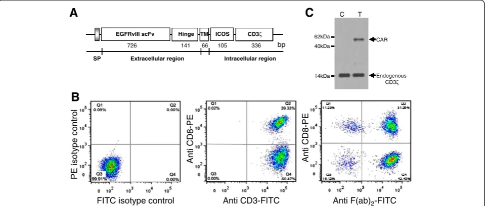
Figure 1Evaluation of EGFRvIII/CAR expression on CD3+T cells.(A) Schematic representation of EGFRvIII/CAR. It consists of EGFRvIII scFv, the hinge and transmenbrane (TM) region of human CD8α, ICOS signaling domain, and human CD3ζchain. IgGκchain was used as signal peptide (SP). (B) Surface expression of EGFRvIII/CAR on CD3+T cells. Left, isotype control; Middle, anti-human CD3-FITC and anti-human CD8-PE staining (BD Biosciences); Right, anti-mouse F(ab)2-FITC (eBiocience) and anti-human CD8-PE staining. (C) Immunoblot analysis of EGFRvIII/CAR expression.
of 2×10⁵ T cells with 1×10⁶ target cells in 200 μl me-dium per well in 96-well-plate triplicately. After 24 h,
supernatants were assayed for IFN-γ production using
ELISA (R&D Systems). The control of effector cells is the same as above cytotoxicity assay; the control of tar-get cells is EGFRvIII-negative parental U87 cells.
In vivo antitumor activity of EGFRvIII/CAR+T cells
Xenograft tumor mouse model was established by
sub-cutaneous (s.c.) flank injections of 5×10⁶ EGFRvIII
ex-pressing U87 cells in 6-week-old female BALB/cA-nude mice (Chinese Academy of Science Shanghai Experi-mental Animal Center). When the tumor burden
rea-ched about 500 mm3 in about 10–14 days after tumor
cells inoculation, the mice were assigned to different
groups (5 in each group) and injected with 1×10⁷
differ-ent T cells/100μl (EGFRvIII/CAR-transduced T cells,
GFP-transduced T cells, and control PBS) either either systemically to tail vein or locally to the tumor mass. Tumor growth was subsequently monitored by caliper measurement and tumor volume was calculated using
the formula: 1/2 × length × (width)2. The mice were
killed when tumor volume reached >2,000 mm3. This
study was carried out in strict accordance with the re-commendations in the Guide for the Care and Use of Laboratory Animals. The protocols were approved by the Animal Care Committee of Zhengzhou University
(Protocol No. 011–026).
Statistical analysis
ANOVA was used to identify the possible difference among different treatment groups. Once the difference
is confirmed, Student’sttest was applied to calculate the
significance in the difference between two treatment
groups (P values). P-Values less than 0.05 were
consi-dered statistically significant.
Results
EGFRvIII/CAR was constructed and T cells were modified successfully by lentiviral EGFRvIII/CAR
To generate EGFRvIII-specific T cells, chimeric EGFRvIII/ CAR was constructed As shown in Figure 1A, EGFRvIII/
CAR encodes a fusion protein consist of IgG κ leader
peptide, EGFRvIII scFv, the hinge and TM region of
hu-man CD8α(amino acids 135–205), intracellular signal
do-main of ICOS (amino acids 165–199) and the CD3ζchain
(amino acids 52–163). No extra linker or space was used
between gene fragments since it may increase the im-munogenicity of EGFRvIII/CAR leading to immune de-struction of the transduced T cells in vivo.
Lentiviral EGFRvIII/CAR was prepared for
transduc-tion of CD3+ T cells. The titers of Lentiviral EGFRvIII/
CAR ranged from 1×10⁶to 10×106transducing units/ml
(Cell BioLabs). After CD3+ beads selection of human
PBMCs, the purity of CD3+ T cells were almost 100%,
with 39.33% CD8+ T cells and 60.47% CD4+ T cells as
indicated in Figure 1B (middle). The surface expression of EGFRvIII/CAR on T cells was confirmed by flow
cyto-metric analysis using anti-mouse F(ab)2-FITC. As shown
in Figure 1B (right), EGFRvIII/CAR expression efficiency
reached 73.65% of CD3+ T cells, of which 31.25% were
CD8+ T cells (CTLs). The transduction efficiency was
usually about 70%. The whole population of EGFRvIII/
CAR transduced T cells was treated as EGFRvIII/CAR+
T cells for subsequent experiments.
The EGFRvIII/CAR protein expression was verified by immunoblotting. Cell lysates of EGFRvIII/CAR trans-duced and untranstrans-duced T cells were separated by SDS-PAGE under reducing condition and immunoblotted
with goat anti-human CD3ζ antibody. As shown in
Figure 1C, under reducing conditions, endogenous CD3ζ
chain was detected as a 15 kDa band in both transduced and untransduced T cell lysates. Additional band of ap-proximate 57 kDa were observed in EGFRvIII/CAR transduced T cells but absent in untransduced T cells, consistent with the calculated size of EGFRvIII/CAR protein.
EGFRvIII/CAR+T cells demonstrated specific and efficient cytotoxicity against EGFRvIII expressing glioma cells
A standard 4-hour 51Cr release assay was performed to
determine whether the EGFRvIII/CAR+ T cells could
re-cognize and kill the EGFRvIII expressing U87 cells. A robust enhancement in the cytotoxicity of the EGFRvIII/
CAR+ T cells against the EGFRvIII expressing glioma
cells was detected as an increase in E:T ratio, A sig-nificant difference was noticed in the EGFRvIII specific killing at each E:T ratio (P<0.05, Figure 2A, Left)
be-tween EGFRvIII/CAR+T cells and control GFP+ or
non-transduced T cells. The killing activity exceeded 60% at 10:1 of E:T ratio. In contrast, no evident killing activity was found among T cells of three different groups to-ward control target cells (EGFRvIII-negative U87 cells, Figure 2A, Right). This result confirmed the specificity and efficiency of cytotoxic T cell response against EGFRvIII expressing glioma cells when EGFRvIII/CAR was grafted onto T cells.
EGFRvIII/CAR+T cells secreted IFN-γin an EGFRvIII dependent mechanism
To determine whether EGFRvIII/CAR+ T cells become
activated and acquire effector cell functions when en-countering EGFRvIII target, we performed a cytokine re-lease assay. When co-cultured with EGFRvIII expressing
U87 cells, EGFRvIII/CAR+ T cells released a substantial
contrast, IFN-γrelease remained unchanged in control
ef-fector cells (GFP+and non-transduced T cells) or control
target cells (parental U87 cells) groups (P<0.05, Figure 2B).
These results indicate that EGFRvIII/CAR+T cells can be
triggered and exert effector cell functions in an EGFRvIII
dependent manner, which was consistent with previous findings that the CAR modified T cells expressing a cos-timulatory signaling domain release increased amount of cytokines [9].
EGFRvIII/CAR+T cells inhibited the in vivo growth of EGFRvIII expressing glioma cells
We developed a xenograft model by inoculating the EGFRvIII-expressing U87 cells in the flanks of BALB/ cA-nude mice. The tumor-loaded mice received injections
of 1×10⁷EGFRvIII/CAR+ T cells, GFP+ T cells and PBS,
respectively. Tumor sizes in mice receiving EGFRvIII/
CAR+ T cells started to shrink three weeks after adoptive
cell transfer, while tumors in other two groups continued to grow. Statistical analysis of the tumor growth curves re-vealed significant differences between the EGFRvIII/CAR group and control groups (P<0.05, Figure 3), confir-ming the potent antitumor property of the inoculated
the EGFRvIII/CAR+ T cells in vivo. We excluded the
possibility that the antitumor effect of the EGFRvIII/
CAR+ T cells resulted from their allogeneic effect
be-cause the inoculated GFP+ T cells did not show any
evident effects on tumor growth. In addition, we were CAR+T cells
GFP+T cells NT T cells
Effector : Target
5:1 20
40 60 80 100
0
10:1 20:1
40:1
% Target Cell Lysis
A
Effector : Target
5:1 20
40 60 80 100
0
10:1 20:1
40:1
% Target Cell Lysis
CAR+T cells GFP+T cells
NT T cells
*
*
*
*
EGFRvIII-expressing U87 Cell
EGFRvIII-negative U87 Cell
B
CAR+T cells GFP+T cells NT T cells
IFN-γ
(pg/ml)
0 500 1000 1500 2000 2500 3000
*
Figure 2Functional analysis of EGFRvIII/CAR+T cells.(A) Cytotoxic activity of EGFRvIII/CAR+T cells. (Left) Target cells were EGFRvIII-expressing U87 cells which can be lysed by EGFRvIII/CAR+T cells, not by GFP+and NT T cells. (Right) Target cells were EGFRvIII-negative U87 cells, which can
not be lysed by either EGFRvIII/CAR+or control T cells. (B) Cytokine release of EGFRvIII/CAR+T cells. Only EGFRvIII/CAR+T cells released significant amount of IFN-γwhen co-cultured with EGFRvIII-expressing U87 cells. No increased IFN-γexpression was detected when co-cultured with EGFRvIII-negative U87 cells, nor from control GFP+and NT T cells. Results are the mean and the SD from experiments in triplicate. * indicate
P<0.05.
Time (days post tumors injection)
Group A Group B
Group D Group C
Tu
m
o
r v
o
lu
me
(
m
m
3)
10 20 30 40 50 60
0 200 1000 2000
400 600 800 1200 1600 1400 1800
Figure 3In vivo antitumor activity of EGFRvIII/CAR+T cells.
EGFRvIII expressing U87 cells were used for xenograft mouse model. EGFRvIII-bearing BALB/cA-nude mice received different treatments: group A, EGFRvIII/CAR+T cells (IT); group B, EGFRvIII/CAR+T cells (IV);
group C, GFP+T cells (IV); group D, PBS (IV). Results are expressed as
a mean tumor volume (mm3±SD) withn= 5 for all groups. The
had a similar efficacy to that of local intratumor injection.
Discussions
Adoptive transfer of genetically modified T cells shows some advantages over the mobilization of the endo-genous T cell repertoire in cancer immunotherapy. The theoretical advantages and technical feasibility of CAR facilitate the development of cancer immunotherapy. The notions that CAR endows T cells antigen specific recognition, activation and proliferation in an MHC independent manner have been consolidated by some pre-clinical studies showing that retargeted T cells can recognize and kill cancer cells expressing tumor associ-ated antigen or specific antigen in vitro and in vivo. Cel-lular immunotherapy adopting such retargeted T cells has shown significant potential in the treatment of ma-lignant diseases. The mounting data have provided solid support for future clinical application of such therapy in cancers such as leukemia, colorectal colon cancer, and prostate cancer [17-19].
Recent efforts to improve the antitumor efficacy of CAR-based therapy are mainly based on the theory of the two-step T cell activation. Major progress has been made since the introduction of the costimulatory signal-ing into architecture of CAR. With the in-depth under-standing of costimulatory receptors in T cell immune response, several costimulatory molecules were embed-ded in the CAR and their roles in coordinating anti-tumor immunity were explored [20]. The observations from other groups and our own have thus far established that the inclusion of costimulatory molecule from B7 re-ceptor family (CD28 or ICOS) results in an increased
production of of IFN-γ, TNF-γ, and GM-CSF compared
with the CAR with the inclusion of either CD134 or CD137 of TNFR family. CD28 is more potent than other costimulatory molecules with respects to enhanced IL-2 production, improved clonal expansion and persistence of CAR T cells. Finney and colleagues have demon-strated that ICOS in the CAR induces the maximal ef-fect on cell lysis [9]. Thus, we hypothesized that the incorporation of ICOS into CAR favors the antitumor properties of CAR-armed T cells. Our results support our hypotheses: CAR-armed T cells demonstrate effi-cient killing of tumor cells and abundant Th1 cytokine
IFN-γis released in an EGFRvIII-specific manner.
There-fore, our results are consistent with the previous findings and consolidate the notion that the presence of ICOS as an intracellular costimulatory signaling is crucial for en-hanced T cell response to tumor cells.
The efficacy of CAR can be affected by many factors including the affinity of the selected scFv, the size of hinge region, the combination of signaling domain(s), the type of modified T cell subsets, etc. The major
tion of the antigen expressed on normal cells by CAR T cells. Such off-site on-target immune injury causes ad-verse effects, some of which may be fatal. Therefore, it is important to carefully select the target antigens that are specifically expressed in cancer cells, but not in normal cells. EGFRvIII is a commonly found mutant of EGFR and exclusively expressed in a wide range of cancers. In addition to its tumor specific expression, EGFRvIII is also involved in oncogenic phenotypes and changes the properties of tumorigenicity. Since its discovery, EGFRvIII has become an increasingly attractive molecule for cancer therapy. The EGFRvIII scFv from antibody 3C10 and MR1 was used for CAR construction and the CAR modified T cells demonstrated EGFRvIII-specific tumor cell lysis
in vitro and in vivo [21,22]. Recently, Rosenberg’s group
analyzed scFv sequences of seven EGFRvIII specific mAbs and assembled the third generation of chimeric antigen receptor (139-28BBZ CAR). The T cells transduced with retroviral EGFRvIII/CAR have been shown an EGFRvIII-specific cell lytic activity [23]. In this study,
lentivirus-mediated transduction enriched the EGFRvIII/CAR+ T
cells to about 70%. Functionality assay demonstrated that these redirected T cells exert efficient cytotoxic T cell re-sponse in an EGFRvIII specific manner and secret
cyto-kine IFN-γin an antigen dependent way. The EGFRvIII/
CAR engrafted T cells pave a way for antitumor in animal model as well as in clinical settings.
In our study, we used CD3+T cells, instead of purified
CD8+CTLs only, to investigate the performance of CAR
because CD4+ T cells have been shown to augment the
function of CD8+ T cells. The results of our
EGFRvIII-bearing mouse model demonstrates that CD3+ T cells
transduced with EGFRvIII/CAR have significantly higher antitumor activity than the T cells in control groups, which supports the theory that adoptive transfer of
mixed populations of antigen-specific CD8+and CD4+T
cells promotes overall antitumor immunity. With re-gards to the administration route of T cells, both intratu-mor injection and venous injection show similar efficacy. For clinical purpose, engraftment of adoptively trans-ferred T cells in host is a major challenge for achieving therapeutic benefit. Ongoing studies are exploring opti-mal combinations of costimulatory molecules and T cell subsets with long-term cytotoxicity [24]. Also, most of
mice in groups of the EGFRvIII/CAR+T cells and GFP+
T cells suffered from graft versus host diseases (GVHD) to some extent. The symptoms of GVHD usually started 5 weeks after T cells infusion, including less activity, ruf-fled fur, skin rash, hunched back and weight loss. The le-thality due to the transfusion-associated GVHD was rare and there was no difference between mice treated with
EGFRvIII/CAR+ T cells and GFP+T cells with regard to
Conclusion
Our study demonstrates that the EGFRvIII/CAR-modified T cells are capable of destroying glioma cells efficiently in
an EGFRvIII specific manner and release IFN-γ in an
antigen dependent manner. The specific recognition and effective killing activity of the EGFRvIII-directed T cells with ICOS signaling domain provides a basis for further studies in clinical application of cancer treatment.
Abbreviations
CAR:Chimeric antigen receptor; ICOS: Inducible costimulator;
EGFRvIII: Epidermal growth factor variant III; AIT: Adoptive immunotherapy; scFv: single chain variable fragment; TCR: T cell receptor; CTLs: Cytotoxic T lymphocytes; GFP: Green fluorescent protein; MHC: Major histocompatibility complex; TNFR: Tumor necrosis factor receptor; GVHD: Graft versus host diseases.
Competing interests
The authors declare no competing financial interests in relation to this work.
Authors’contributions
SCJ and YYX carried out most of the molecular and cellular experiments and drafted the manuscript. HEQ and CN performed the flow cytometry and Western blot analysis. WYF, WY and ZYY did the in vivo experiments in animal model. ZLM carried out the statistical analysis. CJ, GP and WAJ finished experiments related to EGFRvIII scFv and participated in discussion of the research. HSY designed the research and wrote the manuscript. All authors read and approved the manuscript.
Acknowledgments
The authors thank Dr. Yang-bing Zhao (Abramson Family Cancer Research Institute, University of Pennsylvania School of Medicine) for technical instruction and Dr. Tian-fang Li (Rush University Medical Center) for helpful discussion. This work was supported by National Natural Science Foundation of China (No. 81172415).
Author details
1Translational Research Center, Zhengzhou University People’s Hospital,
#7 Weiwu Road, Zhengzhou, Henan 450003, China.2Drexel University College of Medicine, Philadelphia, PA 19129, USA.3Department of Neurosurgery, Brain Tumor Research Laboratories, Stanford University Medical Center, Stanford, CA 94305, USA.
Received: 19 February 2013 Accepted: 4 May 2013 Published: 9 May 2013
References
1. Grupp SA, June CH:Adoptive cellular therapy.Curr Top Microbiol Immunol
2011,344:149–172.
2. Cheadle EJ, Sheard V, Hombach AA, Chmielewski M, Riet T, Berrevoets C, Schooten E, Lamers C, Abken H, Debets R, Gilham DE:Chimeric antigen receptors for T-cell based therapy.Methods Mol Biol2012,907:645–666. 3. Porter DL, Levine BL, Kalos M, Bagg A, June CH:Chimeric antigen
receptor-modified T cells in chronic lymphoid leukemia.N Engl J Med2011,365:725–733. 4. Louis CU, Savoldo B, Dotti G, Pule M, Yvon E, Myers GD, Rossig C, Russell HV,
Diouf O, Liu E, Liu H, Wu MF, Gee AP, Mei Z, Rooney CM, Heslop HE, Brenner MK:Antitumor activity and long-term fate of chimeric antigen receptor-positive T cells in patients with neuroblastoma.Blood2011,118:6050–6056. 5. Lo AS, Ma Q, Liu DL, Junghans RP:Anti-GD3 chimeric sFv-CD28/T-cell
receptor zeta designer T cells for treatment of metastatic melanoma and other neuroectodermal tumors.Clin Cancer Res2010,16:2769–2780. 6. Fontana MF, Vance RE:Two signal models in innate immunity.Immunol Rev
2011,243:26–39.
7. Carpenito C, Milone MC, Hassan R, Simonet JC, Lakhal M, Suhoski MM, Varela-Rohena A, Haines KM, Heitjan DF, Albelda SM, Carroll RG, Riley JL, Pastan I, June CH:Control of large, established tumor xenografts with genetically retargeted human T cells containing CD28 and CD137 domains.Proc Natl Acad Sci USA2009,106:3360–3365.
8. Savoldo B, Ramos CA, Liu E, Mims MP, Keating MJ, Carrum G, Kamble RT, Bollard CM, Gee AP, Mei Z, Liu H, Grilley B, Rooney CM, Heslop HE, Brenner
MK, Dotti G:CD28 Costimulation improves expansion and persistence of chimeric antigen receptor-modified T cells in lymphoma patients.
J Clin Invest2011,121:1822–1826.
9. Finney HM, Akbar AN, Lawson AD:Activation of resting human primary T cells with chimeric receptors: costimulation from CD28, inducible costimulator, CD134, and CD137 in series with signals from the TCR zeta chain.J Immunol2004,172:104–113.
10. Moscatello DK, Holgado-Madruga M, Godwin AK, Ramirez G, Gunn G, Zoltick PW, Biegel JA, Hayes RL, Wong AJ:Frequent expression of a mutant epidermal growth factor receptor in multiple human tumors.
Cancer Res1995,55:5536–5539.
11. Del Vecchio CA, Li G, Wong AJ:Targeting EGF receptor variant III: tumor-specific peptide vaccination for malignant gliomas.Expert Rev Vaccines
2012,11:133–144.
12. Gan HK, Burgess AW, Clayton AH, Scott AM:Targeting of a
conformationally exposed, tumor-specific epitope of EGFR as a strategy for cancer therapy.Cancer Res2012,72:2924–2930.
13. Sampson JH, Archer GE, Mitchell DA, Heimberger AB, Herndon JE 2nd, Lally-Goss D, McGehee-Norman S, Paolino A, Reardon DA, Friedman AH, Friedman HS, Bigner DD:An epidermal growth factor receptor variant III-targeted vaccine is safe and immunogenic in patients with glioblastoma multiforme.Mol Cancer Ther2009,8:2773–2779.
14. Gupta P, Han SY, Holgado-Madruga M, Mitra SS, Li G, Nitta RT, Wong AJ:
Development of an EGFRvIII specific recombinant antibody.BMC
Biotechnol2010,10:72.
15. Wu L, Martin TD, Vazeux R, Unutmaz D, KewalRamani VN:Functional evaluation of DC-SIGN monoclonal antibodies reveals DC-SIGN interactions with ICAM-3 do not promote human immunodeficiency virus type 1 transmission.J Virol2002,76:5905–5914.
16. Tammana S, Huang X, Wong M, Milone MC, Ma L, Levine BL, June CH, Wagner JE, Blazar BR, Zhou X:4-1BB And CD28 signaling plays a synergistic role in redirecting umbilical cord blood T cells against B-cell malignancies.Hum Gene Ther2010,21:75–86.
17. Kalos M, Levine BL, Porter DL, Katz S, Grupp SA, Bagg A, June CH:T cells with chimeric antigen receptors have potent antitumor effects and can establish memory in patients with advanced leukemia.Sci Transl Med2011,3:95ra73. 18. Sasaki T, Ikeda H, Sato M, Ohkuri T, Abe H, Kuroki M, Onodera M, Miyamoto
M, Kondo S, Nishimura T:Antitumor activity of chimeric immunoreceptor gene-modified Tc1 and Th1 cells against autologous carcinoembryonic antigen-expressing colon cancer cells.Cancer Sci2006,97:920–927. 19. Kloss CC, Condomines M, Cartellieri M, Bachmann M, Sadelain M:
Combinatorial antigen recognition with balanced signaling promotes selective tumor eradication by engineered T cells.Nat Biotechnol2012,31:71–75. 20. Zhong XS, Matsushita M, Plotkin J, Riviere I, Sadelain M:Chimeric antigen
receptors combining 4-1BB and CD28 signaling domains augment PI3kinase/AKT/Bcl-XL activation and CD8+T cell-mediated tumor eradication.Mol Ther2010,18:413–420.
21. Ohno M, Natsume A, Ichiro Iwami K, Iwamizu H, Noritake K, Ito D, Toi Y, Ito M, Motomura K, Yoshida J, Yoshikawa K, Wakabayashi T:Retrovirally engineered T-cell-based immunotherapy targeting type III variant epidermal growth factor receptor, a glioma-associated antigen.Cancer Sci2010,101:2518–2524. 22. Bullain SS, Sahin A, Szentirmai O, Sanchez C, Lin N, Baratta E, Waterman P,
Weissleder R, Mulligan RC, Carter BS:Genetically engineered T cells to target EGFRvIII expressing glioblastoma.J Neurooncol2009,94:373–382. 23. Morgan RA, Johnson LA, Davis JL, Zheng Z, Woolard KD, Reap EA, Feldman SA,
Chinnasamy N, Kuan CT, Song H, Zhang W, Fine HA, Rosenberg SA:
Recognition of glioma stem cells by genetically modified T cells targeting EGFRvIII and development of adoptive cell therapy for glioma.Hum Gene Ther2012,23:1043–1053.
24. Stroncek DF, Berger C, Cheever MA, Childs RW, Dudley ME, Flynn P, Gattinoni L, Heath JR, Kalos M, Marincola FM, Miller JS, Mostoslavsky G, Powell DJ Jr, Rao M, Restifo NP, Rosenberg SA, O'Shea J, Melief CJ:New directions in cellular therapy of cancer: a summary of the summit on cellular therapy for cancer.J Transl Med2012,10:48.
doi:10.1186/1756-8722-6-33
Cite this article as:Shenet al.:Chimeric antigen receptor containing ICOS signaling domain mediates specific and efficient antitumor effect
of T cells against EGFRvIII expressing glioma.Journal of Hematology &