Unique Features of
Aeromonas
Plasmid
pAC3 and Expression of the
Plasmid-Mediated Quinolone Resistance Genes
Dae-Wi Kim,
aCung Nawl Thawng,
aSang Hee Lee,
bChang-Jun Cha
aDepartment of Systems Biotechnology and Center for Antibiotic Resistome, Chung-Ang University, Anseong, Republic of Koreaa; Department of Biological Sciences, National Leading Research Laboratory of Drug Resistance Proteomics, Myongji University, Yongin, Republic of Koreab
ABSTRACT
A highly fluoroquinolone-resistant isolate of
Aeromonas
species was
iso-lated from a wastewater treatment plant and found to possess multiple resistance
mechanisms, including mutations in
gyrA
and
parC
, efflux pumps, and
plasmid-mediated quinolone resistance (PMQR) genes. Complete sequencing of the
IncU-type plasmid, pAC3, present in the strain revealed a circular plasmid DNA 15,872 bp
long containing two PMQR genes [
qnrS2
and
aac
(
6
=
)
-Ib-cr
]. A mobile insertion
cas-sette element containing the
qnrS2
gene and a typical miniature inverted-repeat
transposable element (MITE) structure was identified in the plasmid. The present
study revealed that this MITE sequence appears in other
Aeromonas
species
plas-mids and chromosomes. Plasmid pAC3 was introduced into
Escherichia coli
, and its
PMQR genes were expressed, resulting in the acquisition of resistance. Proteome
analysis of the recipient
E. coli
strain harboring the plasmid revealed that
aac
(
6
=
)
-Ib-cr
expression was constitutive and that
qnrS2
expression was dependent upon
flu-oroquinolone stress through regulation by regulator of sigma D (Rsd). To the best of
our knowledge, this is the first report to characterize a novel MITE sequence
up-stream of the PMQR gene within a mobile insertion cassette, as well as the
regula-tion of
qnrS2
expression. Our results suggest that this mobile element may play an
important role in
qnrS2
dissemination.
IMPORTANCE
In the present study, plasmid pAC3 isolated from a highly
fluoroquin-olone-resistant isolate of
Aeromonas
species was sequenced and found to contain
two fluoroquinolone resistance genes,
aac
(
6
=
)-
Ib-cr
and
qnrS2
. Comparative analyses
of plasmid pAC3 and other
Aeromonas
sp. IncU-type plasmids revealed a mobile
in-sertion cassette element with a unique structure containing a
qnrS2
gene and a
typi-cal miniature inverted-repeat transposable element (MITE) structure. This study also
revealed that this MITE sequence appears in other
Aeromonas
species plasmids and
chromosomes. Our results also demonstrate that the fluoroquinolone-dependent
ex-pression of
qnrS2
is associated with
rsd
in
E. coli
DH5
␣
harboring plasmid pAC3. Our
findings suggest that the mobile element may play an important role in
qnrS2
dis-semination and that
Aeromonas
species constitute an important reservoir of
fluoro-quinolone resistance determinants in the environment.
KEYWORDS
Aeromonas
, plasmid-mediated quinolone resistance,
aac
(
6’
)
-Ib-cr
,
miniature inverted-repeat transposable element,
qnrS2
, regulator of sigma D
F
luoroquinolones are broad-spectrum antimicrobial agents that have been widely
used to treat bacterial infections (1). Residual fluoroquinolones have been detected
at various environmental sites (2), including wastewater treatment plants (WWTPs) (3),
where antibiotics may act as a selective pressure that allows a potential genetic
exchange of resistance genes (4, 5).
Bacterial resistance to fluoroquinolones has been demonstrated to be caused by (i)
Received2 May 2017Accepted7 May
2017 Published24 May 2017
CitationKim D-W, Thawng CN, Lee SH, Cha
C-J. 2017. Unique features ofAeromonas
plasmid pAC3 and expression of the plasmid-mediated quinolone resistance genes. mSphere 2:e00203-17.https://doi.org/10.1128/ mSphere.00203-17.
EditorAna Cristina Gales, Escola Paulista de
Medicina, Universidade Federal de São Paulo
Copyright© 2017 Kim et al. This is an
open-access article distributed under the terms of theCreative Commons Attribution 4.0 International license.
Address correspondence to Chang-Jun Cha, cjcha@cau.ac.kr.
D.-W.K. and C.N.T. contributed equally to this work.
Applied and Environmental Science
crossm
on September 8, 2020 by guest
http://msphere.asm.org/
chromosomal mutations in the genes encoding target proteins such as DNA gyrase or
topoisomerase IV (6), (ii) efflux pumps (7), (iii) Qnr family proteins (8), and (iv)
inacti-vation by fluoroquinolone
N
-acetyltransferase AAC(6
=
)-Ib-cr (9). Plasmid-mediated
quin-olone resistance (PMQR) is known to involve
aac
(
6
=
)
-Ib-cr
,
qnr
family genes, and genes
encoding efflux pumps (
qepA
and
oqxAB
) (10). The prevalence of
qnr
and
aac
(
6
=
)
-Ib-cr
genes in plasmids of clinical and environmental isolates has been reported worldwide,
including in Korea (11, 12). Qnr family proteins interact with their target proteins, thus
blocking the action of fluoroquinolones and reducing their inhibitory effect (8).
AAC(6
=
)-Ib-cr is an aminoglycoside acetyltransferase variant with two amino acid
substitutions (W102R and D179Y) that confer extended substrate specificity for
fluoro-quinolones, thus leading to resistance (9).
Members of the genus
Aeromonas
are known to be autochthonous to aquatic
environments, although they have been isolated from a wide variety of habitats (13).
Some
Aeromonas
species have been recognized as opportunistic human pathogens, as
well as primary fish pathogens (13); these species exhibited multidrug resistance,
including to fluoroquinolones (14–16). The presence of quinolone resistance
determi-nants has been reported in various
Aeromonas
spp.; several strains contain multiple
resistance mechanisms, including mutations at quinolone resistance-determining
re-gions (QRDRs) (17–21), efflux pumps (18, 21, 22), and PMQR genes (23–26). In some
Aeromonas
sp. plasmids,
aac
(
6
=
)-
Ib-cr
and
qnrS2
are colocalized with other resistance
genes (26–28). Comparative analysis of IncU-type plasmids from
Aeromonas
spp.
re-vealed the conservation of PMQR genes, as well as high genetic plasticity in the region
of PMQR genes (26).
In this study,
Aeromonas
sp. strain C3, which displays high-level fluoroquinolone
resistance, was isolated from a WWTP and the multiple resistance mechanisms,
includ-ing PMQR, were characterized in this strain. Comparative analysis of the plasmid from
strain C3 revealed a novel mobile insertion cassette element (MICE) related to
trans-position. The role of the plasmid and the regulation of PMQR in a recipient
Escherichia
coli
strain were elucidated by proteome analysis.
RESULTS
Isolation and identification of a bacterium with high-level fluoroquinolone
resistance.
A bacterial strain isolated from a WWTP displayed unusually high-level
resistance to various fluoroquinolones (Table 1). The strain was able to transform
fluoroquinolones to their
N
-acetylated metabolites, which were structurally
identi-fied by liquid chromatography (LC)-tandem mass spectrometry (MS/MS) and proton
nuclear magnetic resonance (NMR) analyses (see Tables S1 and S2) (29). On the
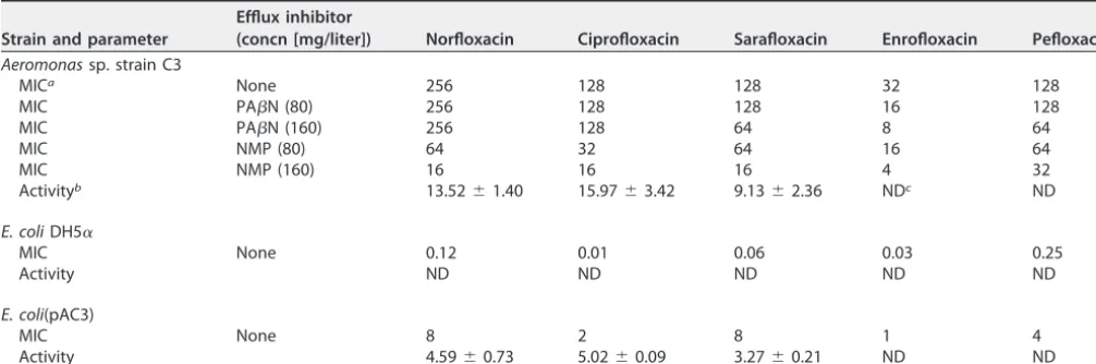
TABLE 1 Fluoroquinolone susceptibilities andN-acetylation activities ofAeromonassp. strain C3,E. coliDH5␣, andE. coli(pAC3)
Strain and parameter
Efflux inhibitor
(concn [mg/liter]) Norfloxacin Ciprofloxacin Sarafloxacin Enrofloxacin Pefloxacin
Aeromonassp. strain C3
MICa None 256 128 128 32 128
MIC PAN (80) 256 128 128 16 128
MIC PAN (160) 256 128 64 8 64
MIC NMP (80) 64 32 64 16 64
MIC NMP (160) 16 16 16 4 32
Activityb 13.52⫾1.40 15.97⫾3.42 9.13⫾2.36 NDc ND
E. coliDH5␣
MIC None 0.12 0.01 0.06 0.03 0.25
Activity ND ND ND ND ND
E. coli(pAC3)
MIC None 8 2 8 1 4
Activity 4.59⫾0.73 5.02⫾0.09 3.27⫾0.21 ND ND
aMICs are expressed in mg/liter.
bAverageN-acetyltransferase activity⫾the standard deviation is expressed in mU/mg of protein. cND, not detected.
on September 8, 2020 by guest
http://msphere.asm.org/
basis of its 16S rRNA gene sequence, the isolate showed 99.9% similarity with
Aeromonas hydrophila
subsp.
hydrophila
ATCC 7966
T. We therefore designated the
isolate
Aeromonas
sp. strain C3.
The presence of multiple fluoroquinolone resistance mechanisms in strain C3.
To elucidate this high-level quinolone resistance, the resistance determinants present
in the strain were characterized. QRDR sequence analysis showed that point mutations
related to resistance were found in the QRDRs of GyrA (D87N) and ParC (S80I).
Considering the MICs of
Aeromonas
spp. harboring these mutations (20), the extremely
high level of fluoroquinolone resistance exhibited by strain C3 suggested the presence
of additional resistance mechanisms. Of the PMQR genes tested, only
aac
(
6
=
)-
Ib-cr
and
qnrS
were detected by PCR; these two genes were found to be located in plasmid pAC3
of the strain. The fluoroquinolone resistance of strain C3 was also examined by
monitoring changes in MICs in the presence of the efflux pump inhibitors PA

N and
NMP, which are known to inhibit a broad range of efflux pumps (30, 31). NMP caused
a significant reduction (4- to 16-fold) in the MICs of all of the fluoroquinolones tested
(Table 1), while PA

N resulted in minor effects on the MICs of enrofloxacin, sarafloxacin,
and pefloxacin.
Comparative analysis of plasmid pAC3.
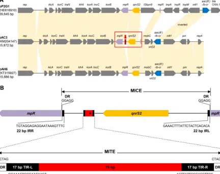
The plasmid harboring the PMQR genes,
designated pAC3, was isolated, sequenced, and annotated. The total length of the
complete plasmid sequence is 15,872 bp and includes 21 protein-coding genes
(Fig. 1A). The plasmid was identified as a member of the IncU-type plasmid family and
contained two PMQR genes,
aac
(
6
=
)-
Ib-cr
and
qnrS2
(Fig. 1A). For comparative plasmid
FIG 1 Comparative analysis ofAeromonassp. IncU-type plasmids and the MICE of plasmid pAC3 containing a MITE. (A) Plasmids pAH6 and pP2G1 were used for comparative analysis with plasmid pAC3. Shaded connections between the plasmids show the conserved and shared regions (⬎99% identity). The red box and arrow indicate the MICE and the MITE, respectively. (B) Genetic map of the MICE region carrying the MITE sequence in plasmid pAC3. IRR, right inverted repeat; IRL, left inverted repeat.
on September 8, 2020 by guest
http://msphere.asm.org/
analyses, the six IncU-type plasmids phylogenetically closest to plasmid pAC3 were
selected (see Fig. S1 in the supplemental material) and local colinear block (LCB)
analysis was performed with MAUVE software (see Fig. S1). Plasmid pAC3 exhibited the
closest relationship with plasmids pAH6 and pP2G1, which possess the identical PMQR
genes (Fig. 1A). These plasmids have the same backbone structure, from the replication
gene
rep
to the metallopeptidase gene
mpR
(Fig. 1A) (26).
A MICE bracketed by 22-bp left and right inverted repeats was found to contain the
qnrS2
gene. Insertion of the MICE disrupted the
mpR
gene encoding a putative zinc
metalloprotease. IncU plasmids carrying the
qnrS2
gene from other
Aeromonas
spp.
have been found in Europe and Asia (26–28). The presence of the
qnrS2
-carrying MICE
structure in different
Aeromonas
species from geographically distant aquatic
environ-ments suggests that this PMQR determinant is widespread in this genus, and these
MICE-type structures are potential vehicles of PMQR determinants (32). Interestingly, a
novel genetic structure not present in other MICE structures found in
Aeromonas
species (26–28) was discovered inside the MICE of plasmid pAC3 and exhibited a typical
miniature inverted-repeat transposable element (MITE) structure (33, 34). The MITE
consists of a 75-bp core sequence bracketed by 4-bp direct repeats (DRs) and 17-bp
terminal inverted repeats (TIRs) (Fig. 1B). Both the MICE and the MITE are
nonautono-mous derivatives of insertion sequences generated by internal deletion, and the
difference between these two elements is the presence of coding sequences (CDSs)
(35). In the present study, the MICE harbored the
qnrS2
gene while the MITE carried no
passenger gene.
MITE sequences have not been previously identified in
Aeromonas
species. The
present study revealed that these sequences are also present in the partial fragment
sequence of plasmid p42 from
A. media
A39 (24); pGNB2 from an uncultured bacterium
(36); and the chromosomes of
A. media
WS,
A. veronii
AVNIH1, and
A. hydrophila
GYK1.
The MITE of plasmid p42 is identical to that of plasmid pAC3; in contrast to plasmid
pAC3, in IncQ-type plasmid pGNB2, the MITE is found upstream of the
qnrS2
gene
(Fig. 2A). The MITE sequences were also frequently found in the genome of
A. media
WS
(Fig. 2B). All of these MITE sequences were highly conserved, except for the DRs (see
Fig. S2).
The role of PMQR proteins in fluoroquinolone resistance.
Introduction of PMQR
into other bacteria poses a potential risk of dissemination of antibiotic resistance.
Plasmid pAC3 was successfully introduced into antibiotic-susceptible
E. coli
DH5
␣
.
Transformants cultured in the presence of fluoroquinolones showed the production of
N
-acetylated fluoroquinolones. To further elucidate the resistance mechanisms of
PMQR, an antibiotic susceptibility test and an
N
-acetylation activity assay against five
different fluoroquinolones were performed with
Aeromonas
sp. strain C3, parental
E. coli
DH5
␣
, and
E. coli
DH5
␣
harboring plasmid pAC3 [
E. coli
(pAC3)]. The results demonstrate
that
E. coli
(pAC3) acquired resistance to all of the fluoroquinolones tested (Table 1).
Similar to that from strain C3, the cell-free protein extract from
E. coli
(pAC3) was also
able to transform norfloxacin, ciprofloxacin, and sarafloxacin into their
N
-acetylated
metabolites (Table 1). No activity was detected with enrofloxacin and pefloxacin, which
are structurally inaccessible to AAC(6
=
)-Ib-cr. The acetyltransferase activity present in
E. coli
(pAC3) suggests that the introduced resistance was conferred by AAC(6
=
)-Ib-cr.
Interestingly, the MICs of enrofloxacin and pefloxacin for
E. coli
(pAC3) increased
sig-nificantly, although acetyltransferase activity was not detected with these substrates.
These results imply that
qnrS2
, which is also located on the plasmid, contributes to
resistance.
Expression of PMQR proteins.
To further confirm the role of the PMQR of plasmid
pAC3, the expression of AAC(6
=
)-Ib-cr and QnrS2 was analyzed by a proteomics
approach. Proteomes were analyzed by using cells spiked at mid-exponential phase
with fluoroquinolones at concentrations based on the MICs (see Table S3). The numbers
of proteins detected by proteome analysis are detailed in Table S3. Proteomes obtained
from strain C3,
E. coli
DH5
␣
, and
E. coli
(pAC3) cells treated with ciprofloxacin (at the
on September 8, 2020 by guest
http://msphere.asm.org/
MIC) and enrofloxacin (two or three times the MIC) were compared with those of
untreated cells. AAC(6
=
)-Ib-cr was found to be constitutively expressed in both strain C3
and
E. coli
(pAC3) regardless of fluoroquinolone treatment and the host cell (Fig. 3A).
The level of AAC(6
=
)-Ib-cr expression in strain C3 was 3- to 4-fold higher than that in
E. coli
(pAC3) (Fig. 3A). These results coincide with the higher
N
-acetylation activities of
the cell-free protein extract from strain C3 than that from
E. coli
(pAC3) (Table 1). QnrS2
was expressed in
E. coli
(pAC3) when the cells were treated with fluoroquinolones
FIG 2 Genetic characterization of the MITE-harboring regions in Aeromonassp. plasmids and chromosomes. (A) Comparison of plasmids p42 and pGNB2 with plasmid pAC3. Shaded connections between the plasmids show the conserved and shared regions. (B) Genetic map of regions containing MITE sequences inAeromonassp. chromosomes. White arrowheads indicate gene truncations; the transposase-, integrase-, and hypothetical-protein-encoding genes aretnp,int, andhyp, respectively.
FIG 3 Expression of AAC(6=)-Ib-cr (A) and QnrS2 (B) inAeromonassp. strain C3 andE. coli(pAC3). C, 1⫻, 2⫻, and 3⫻indicate 0-, 1-, 2-, and 3-fold MIC fluoroquinolone treatments, respectively. Protein expres-sion levels are expressed as normalized peptide spectrum matches (PSMs).
on September 8, 2020 by guest
http://msphere.asm.org/
(Fig. 3B). These results explain why
E. coli
(pAC3) acquired resistance to enrofloxacin and
pefloxacin (Table 1). In contrast, QnrS2 expression was not detected in strain C3 under
the conditions used (Fig. 3B); QnrS2 was not expressed, even under higher
concentra-tions of fluoroquinolone treatment (up to 16 and 32 times the MIC), suggesting that it
may not be significantly involved in fluoroquinolone resistance in the
Aeromonas
strain.
The role of plasmid pAC3 in recipient cells in response to fluoroquinolone.
A
global proteome analysis revealed that a fluoroquinolone target protein (GyrA) was
upregulated in response to antibiotic treatment in
E. coli
DH5
␣
and to a lesser degree
in
E. coli
(pAC3) (see Fig. S3), suggesting that the presence of the plasmid might affect
GyrA expression. In addition, expression of AcnB, which is known to be involved in
cellular death induced by bactericidal antibiotics (37), increased in response to
fluoro-quinolone treatment in
E. coli
DH5
␣
but remained relatively constant in
E. coli
(pAC3)
(see Fig. S3). The expression of several major regulators, including AcrA, IhfA, Crp, HNS,
and LRP, decreased in response to fluoroquinolone treatment in
E. coli
DH5
␣
and to a
lesser extent in
E. coli
(pAC3) (see Fig. S3). These results suggest that the presence of the
plasmid in recipient cells may contribute to the reduction of antibiotic stress.
Regulation of QnrS2 expression in recipient cells.
While fluoroquinolone
treat-ment increased the expression of QnrS2 in
E. coli
(pAC3) (Fig. 3B), the expression of
ribonucleoside-diphosphate reductase (NrdA and NrdB) also showed a
fluoroquin-olone-induced increase in both
E. coli
DH5
␣
and
E. coli
(pAC3) (see Fig. S3). The
expression of SOS response regulator protein RecA did not increase under these
conditions (see Fig. S3). It has been previously reported that NrdA and NrdB were
upregulated by fluoroquinolone treatment (38) and that their expression was
indepen-dent of the SOS response (39). Our results also suggest that expression of QnrS2 was
induced not by the SOS response but rather by fluoroquinolone-dependent signaling,
as previously reported (40–42). In addition to the major regulators examined, the
expression of regulator of sigma D (Rsd) also decreased in response to fluoroquinolone
in
E. coli
DH5
␣
(Fig. 4A). However,
rsd
expression was upregulated in
E. coli
(pAC3)
(Fig. 4A), suggesting that
rsd
may be associated with QnrS2 expression. The proteome
of an
E. coli rsd
deletion mutant harboring pAC3 [
E. coli
Δrsd(pAC3)] in response to
fluoroquinolone was compared with that of
E. coli
(pAC3). While the expression of
AAC(6
=
)-Ib-cr did not vary greatly, the expression of QnrS2 was significantly lower in
E. coli
Δrsd(pAC3) than in
E. coli
(pAC3) when the strains were treated with the same
amount of antibiotic stress (twice the MIC) (Fig. 4B). Furthermore, the
rsd
deletion
mutant was much more susceptible than
E. coli
(pAC3), with approximately 4-fold lower
FIG 4 Differential expression of Rsd (A) and PMQR (B) proteins in E. coli strains and susceptibility of the strains to various fluoroquinolones (C). White, gray, and black bars indicateE. coliDH5␣,E. coli(pAC3), andE. coliΔrsd(pAC3), respectively. C, 1⫻, and 2⫻indicate fluoroquinolone treatments at 0-, 1-, and 2 times the MIC, respectively. Protein expression levels are expressed as normalized relative peptide spectrum matches (PSMs). Nor, Cip, Sar, Enr, and Pef indicate norfloxacin, ciprofloxacin, sarafloxacin, and pefloxacin, respectively.
on September 8, 2020 by guest
http://msphere.asm.org/
MICs than those of
E. coli
(pAC3) (Fig. 4C); although its MICs were higher than those of
E. coli
DH5
␣
(Table 1; Fig. 4C). These results suggest that the
rsd
gene plays a role in
resistance via the regulation of QnrS2 expression.
DISCUSSION
Fluoroquinolone-resistant
Aeromonas
spp. have been identified in both
environ-mental and clinical isolates; in several strains, high-level fluoroquinolone resistance was
found to be conferred by multiple resistance mechanisms (16, 18, 23). In the present
study,
Aeromonas
sp. strain C3, which was highly resistant to fluoroquinolones, was
isolated from a WWTP and found to possess mutations in
gyrA
and
parC
. Efflux pumps
were also shown to contribute to resistance, as previously reported in this genus (18,
21, 22). The presence of 10 resistance-nodulation-division efflux systems in the genome
of
A. hydrophila
subsp.
hydrophila
ATCC 7966 (22, 43) explains the complex changes in
resistance to various fluoroquinolones in the presence of efflux inhibitors (Table 1).
Sequencing analysis of plasmid pAC3 present in strain C3 identified two PMQR
deter-minants,
qnrS2
and
aac
(
6
=
)
-Ib-cr
. Recent comparative analysis studies of
Aeromonas
sp.
plasmids belonging to the same incompatibility group have revealed the conservation and
variation in their sequences and genetic structures (26, 44). In the present study, we
identified a mobile insertion cassette with a unique structure containing the PMQR gene
qnrS2
and a novel MITE structure. MITEs are small, nonautonomous mobile elements
broadly dispersed in prokaryotes, although they have been formalized as nonautonomous
transposable sequences in plants (34). Bacterial MITE sequences are primarily found in
intergenic regions of the chromosome; however, they are also present intragenically (34).
In this study, a MITE structure was identified within the MICE inserted in the
mpR
gene in
plasmid pAC3. The MITE sequence also showed typical signatures of TIRs, a target site
duplication consisting of DRs and a core sequence lacking a transposase gene. However, in
this study we demonstrate a plasmid location for the MITE associated with an antibiotic
resistance gene (ARG). There have been only a few reports on ARG-associated MITEs. A
special type of MITE, termed an integron mobilization unit, has been identified in the
bla
GES-5-carrying plasmid of carbapenem-resistant
Enterobacter cloacae
(45). MITE-flanked
integrons carrying ARGs were also revealed in several
Acinetobacter
strains (46, 47). These
studies suggested that MITEs might be associated with the mobilization of ARGs. The MITE
structures in the present study are the first characterized in an
Aeromonas
species (Fig. 2).
The frequent occurrence of MITE structures in
Aeromonas
spp. suggests that they play a role
in the evolution of this genus (Fig. 2B). In particular, the MITE sequences in
A. media
and
A. veronii
are associated with nearby transposases (Fig. 2B), raising the possibility of
transposition.
Comparative analysis of the proteomes of
E. coli
DH5
␣
and its derivative harboring
plasmid pAC3 revealed constitutive expression of AAC(6
=
)-Ib-cr and
fluoroquinolone-dependent expression of QnrS2 in the recipient strain. The proteome results also
suggest that the presence of the plasmid in the recipient strain may reduce antibiotic
stress through the expression of PMQR proteins, thus influencing the cellular regulatory
network. The promoter sequence of
aac
(
6
=
)
-Ib-cr
in pAC3 was found to be
con-served with those from many other bacteria, indicating that constitutive and
host-independent expression of
aac
(
6
=
)
-Ib-cr
could be a general feature. Differential
expres-sion of QnrS1 has been reported in a fluoroquinolone-sensitive
E. coli
strain and a
fluoroquinolone-resistant
E. coli
strain harboring a mutated
gyrA
gene (41). In the
present study,
rsd
in the recipient strain was shown to be involved in antibiotic
stress-dependent expression of
qnrS2
, independent of the SOS response. Although
qnrS2
has been identified as a fluoroquinolone resistance determinant in many
Aero-monas
species (48, 49), there is no direct evidence that the gene is actually expressed
in
Aeromonas
. A previous study has reported that
A. allosaccharophila
, which also
contains the two PMQR determinants [
qnrS2
and
aac
(
6
=
)
-Ib-cr
] on a plasmid, remained
susceptible to quinolones and that these genes might spread silently (27). The putative
promoter region of
qnrS2
was highly conserved in the plasmids of
Aeromonas
spp.,
on September 8, 2020 by guest
http://msphere.asm.org/
indicating that these features may be conserved in this genus. It is still unclear whether
the regulation system of
qnrS2
expression in
Aeromonas
is functional.
To the best of our knowledge, this is the first report of a novel mobile insertion
cassette structure in the PMQR region of an environmental
Aeromonas
species,
sug-gesting that the mobile element may play an important role in
qnrS2
dissemination.
The present study also demonstrates that the fluoroquinolone-dependent expression
of
qnrS2
is associated with
rsd
in
E. coli
DH5
␣
harboring plasmid pAC3. Our results
suggest that the genus
Aeromonas
is important as a reservoir of fluoroquinolone
resistance determinants in the environment and should be under intensive surveillance
for antibiotic resistance.
MATERIALS AND METHODS
Chemicals and media.Ciprofloxacin, norfloxacin, enrofloxacin, phenylalanine arginine-naphthylamide (PAN), and 1-(1-naphthylmethyl)-piperazine (NMP) were purchased from Sigma-Aldrich (St. Louis, MO). Pefloxacin and sarafloxacin were obtained from Santa Cruz Biotechnology (Dallas, TX). R2A, Luria-Bertani (LB), and Mueller-Hinton (MH) media were purchased from BD Science (San Jose, CA).
Isolation and identification of the bacterial strain.A sludge sample obtained from a WWTP in Anseong, South Korea, was inoculated onto R2A agar supplemented with 100 mg/liter ciprofloxacin and incubated at 30°C for 48 h to obtain bacteria with high-level fluoroquinolone resistance. The 16S rRNA gene of the isolate was amplified and sequenced with universal primers (27F and 1492R) and identified by using the EzTaxon-e database (http://www.ezbiocloud.net/) (50).
Analysis of antimicrobial susceptibility.MICs of fluoroquinolones were determined by the broth microdilution method detailed in the Clinical and Laboratory Standards Institute (CLSI) guidelines (51). Susceptibility of the strain to fluoroquinolones was assessed in the absence or presence of PAN or NMP (efflux pump inhibitors) as previously described (30, 31).
Characterization of fluoroquinolone resistance genes.The presence of fluoroquinolone resistance genes, includingqnrA,qnrB,qnrC,qnrD,qnrS,qnrVC1,qnrVC4,qepA,aac(6=)-Ib-cr,oqxA, andoqxB(49, 52–54), and mutations in the QRDRs of thegyrA,gyrB, andparCgenes (17, 18) were verified by PCR amplification and sequencing as previously described (see Table S4).
Enzyme assay.Bacterial strains were cultivated in LB medium at 30°C until cultures reached the mid-exponential growth phase. Cells were harvested 3 h following the addition of ciprofloxacin at a concentration of 20 mg/liter. Cells were harvested and disrupted by sonication in 20 mM Tris-HCl buffer (pH 7.5), and cell debris was removed by filtration (0.2m) to obtain cell-free protein extracts. The reaction mixture, consisting of 90M fluoroquinolone substrate, 200M acetyl coenzyme A, and 50 to 100g of protein extract in 500l of 50 mM Tris-HCl buffer (pH 7.5), was incubated at 30°C. Samples taken from the reaction mixtures were subjected to high-performance liquid chromatography (HPLC) analysis with an Atlantis dc18 column (4.6 by 250 mm; Waters Corp., Milford, MA) and a Varian ProStar HPLC system (Varian, Inc., Walnut Creek, CA) set at 280 nm in a diode array detector. The mobile phase consisted of a linear gradient of acetonitrile (10 to 95%) containing 0.1% formic acid at a flow rate of 1 ml/min. One enzyme unit was defined as the amount of enzyme required to convert 1 mol of substrate to itsN-acetylated product per minute at 30°C.
Genetic manipulation of E. colistrains. Plasmid pAC3 was introduced into fluoroquinolone-sensitiveE. colistrain DH5␣by the transformation method (55), and transformants were selected in the presence of norfloxacin (1 mg/liter). Precise deletion-replacement of the rsdgene fromE. coliwas conducted by the method of Datsenko and Wanner (56) to obtainE. coliΔrsd.
Sequencing and comparative analysis of the plasmid.The plasmid was isolated by the maxiprepa-ration method as previously described (57). The purified plasmid was treated with Plasmid-Safe ATP-Dependent DNase (Epicentre, Madison, WI) to remove chromosomal DNA contaminants. The plasmid DNA was fragmented with dsDNA Fragmentase (NEB, Hitchin, United Kingdom) to make a proper size for library construction. The DNA fragments were processed with the TruSeq DNA sample preparation kit v 2 (Illumina, San Diego, CA) in accordance with the manufacturer’s instructions. The library was quantified with a Bioanalyzer 2100 (Agilent Technologies, Santa Clara, CA), and the average size of the library was 300 bp. Plasmid pAC3 was sequenced with the Illumina MiSeq platform (Illumina) at Chunlab (Seoul, South Korea). The paired-end sequencing reads generated were assembled with CLC genomics work-bench 6.0 (CLC Bio, Boston, MA), and the contigs and PCR-based gap reads were combined with CodonCode aligner 3.7.1 (CodonCode Corp., Centerville, MA). The CDSs were predicted by Glimmer 3.02 (58). For functional annotation, the predicted CDSs were compared to those previously deposited with catalytic families (CatFam), the COG database, NCBI reference sequences (RefSeq), and the SEED subsystem (59–62). Comparison of plasmid pAC3 with Aeromonas sp. plasmids was conducted by phylogenetic and LCB analyses as previously described (see Fig. S1) (44). Plasmids pAH6 and pP2G1 were used for the comparative analysis of IncU-type plasmids. A partial fragment sequence of plasmid p42; the complete sequence of plasmid pGNB2; and the genome sequences ofA. mediaWS,A. veroniiAVNIH1, andA. hydrophilaGYK1 were used for comparison of transposable elements with plasmid pAC3.
Proteome analysis.Cells ofAeromonassp. strain C3,E. coliDH5␣, and derivativeE. colistrains were prepared as described above. Ciprofloxacin and enrofloxacin were added to each strain at the MIC to three times the MIC. Cells were suspended in lysis buffer (50 mM Tris-HCl buffer [pH 7.8], 6 M guanidine hydrochloride, 10 mM dithiothreitol), and cell-free protein extracts were obtained by sonication. The protein samples were reduced, alkylated, and precipitated with dithiothreitol (10 mM), iodoacetamide
on September 8, 2020 by guest
http://msphere.asm.org/
(100 mM), and trichloroacetic acid (30%, wt/vol), respectively. The dried protein pellets were dissolved in 50 mM ammonium bicarbonate and digested with trypsin (Thermo Scientific, Waltham, MA). The trypsin digests were cleaned up with Sep-Pak C18columns (Waters Corp., Milford, MA). One microgram of sample in 0.4% acetic acid was loaded onto a linear ion trap mass spectrometer (LTQ Velos; Thermo Scientific) coupled with a nanosprayer (Thermo Scientific) and a nano column (8.5 cm by 75m) packed with C18 medium (200 Å Magic; Michrom Bioresources, Auburn, CA) (63). The organic mobile phase consisted of a linear gradient of acetonitrile (5 to 30%) containing 0.1% formic acid for 380 min at a flow rate of 70l/min. The mass spectrometer was operated with the following parameters and options: a 2.0-kV nanospray distal voltage, a capillary temperature of 200°C, and full-scan mode (300 to 5,000 Da). MS/MS data were acquired and deconvoluted with Xcalibur 2.1 (Thermo Scientific), and the whole data set was searched with the SEQUEST search algorithm (64) implemented in the Proteome Discoverer 1.3 software (Thermo Scientific). For protein identification, the genome sequences ofE. coliK-12 strain MG1655 (accession no.NC_000913) and plasmid pAC3 (accession no.KM204147) were used as databases. The genome ofA. hydrophilasubsp.hydrophilaATCC 7966 (accession no.CP000462) was used as a reference genome for quantification of the relative abundance of plasmid proteins in strain C3. Filter parameters for peptide identification (medium peptide confidence [ΔCn] of⬎0.1 and false discovery rate of⬍5%) and protein identification (more than two peptides per protein) were applied to the spectra searched by SEQUEST. The shared proteome of biological duplicate samples was used for further analysis, and the protein expression level was determined by using normalized spectral counts.
Accession number(s). The nucleotide sequence of plasmid pAC3 was deposited in GenBank (http://www.ncbi.nlm.nih.gov/GenBank) under accession numberKM204147. The accession numbers of the other plasmid and genome sequences used in the study are as follows: pAH6,KT315927; pP2G1,
HE616910; p42,EU439941; pGNB2,DQ460733;A. mediaWS,CP007567;A. veroniiAVNIH1,CP014774; A. hydrophilaGYK1,CP016392.
SUPPLEMENTAL MATERIAL
Supplemental material for this article may be found at
https://doi.org/10.1128/
mSphere.00203-17
.
FIG S1,
PDF file, 0.4 MB.
FIG S2,
PDF file, 0.1 MB.
FIG S3,
PDF file, 0.1 MB.
TABLE S1,
PDF file, 0.1 MB.
TABLE S2,
PDF file, 0.1 MB.
TABLE S3,
PDF file, 0.1 MB.
TABLE S4,
PDF file, 0.1 MB.
ACKNOWLEDGMENTS
C.N.T. is grateful for a CAYSS scholarship to Chung-Ang University. This work was
supported by the National Research Foundation of Korea (NRF-2013R1A1A2012270),
Ministry of Education, Science & Technology, and funded by the Korean Ministry of
Environment (MOE) as the Environmental Health Action Program (2016001350004). The
authors declare no conflict of interest.
REFERENCES
1. Picó Y, Andreu V. 2007. Fluoroquinolones in soil—risks and challenges. Anal Bioanal Chem 387:1287–1299.https://doi.org/10.1007/s00216-006 -0843-1.
2. Rutgersson C, Fick J, Marathe N, Kristiansson E, Janzon A, Angelin M, Johansson A, Shouche Y, Flach CF, Larsson DG. 2014. Fluoroquinolones andqnrgenes in sediment, water, soil, and human fecal flora in an environment polluted by manufacturing discharges. Environ Sci Technol 48:7825–7832.https://doi.org/10.1021/es501452a.
3. Rodriguez-Mozaz S, Chamorro S, Marti E, Huerta B, Gros M, Sànchez-Melsió A, Borrego CM, Barceló D, Balcázar JL. 2015. Occurrence of antibiotics and antibiotic resistance genes in hospital and urban waste-waters and their impact on the receiving river. Water Res 69:234 –242.
https://doi.org/10.1016/j.watres.2014.11.021.
4. Marathe NP, Regina VR, Walujkar SA, Charan SS, Moore ER, Larsson DG, Shouche YS. 2013. A treatment plant receiving waste water from mul-tiple bulk drug manufacturers is a reservoir for highly multi-drug resis-tant integron-bearing bacteria. PLoS One 8:e77310.https://doi.org/10 .1371/journal.pone.0077310.
5. Ruiz J, Pons MJ, Gomes C. 2012. Transferable mechanisms of quinolone resistance. Int J Antimicrob Agents 40:196 –203.https://doi.org/10.1016/ j.ijantimicag.2012.02.011.
6. Aldred KJ, Kerns RJ, Osheroff N. 2014. Mechanism of quinolone action
and resistance. Biochemistry 53:1565–1574. https://doi.org/10.1021/ bi5000564.
7. Vila J, Fàbrega A, Roca I, Hernández A, Martínez JL. 2011. Efflux pumps as an important mechanism for quinolone resistance. Adv Enzymol Relat Areas Mol Biol 77:167–235.https://doi.org/10.1002/9780470920541.ch5. 8. Tran JH, Jacoby GA. 2002. Mechanism of plasmid-mediated quinolone resistance. Proc Natl Acad Sci U S A 99:5638 –5642.https://doi.org/10 .1073/pnas.082092899.
9. Robicsek A, Strahilevitz J, Jacoby GA, Macielag M, Abbanat D, Park CH, Bush K, Hooper DC. 2006. Fluoroquinolone-modifying enzyme: a new adaptation of a common aminoglycoside acetyltransferase. Nat Med 12:83– 88.https://doi.org/10.1038/nm1347.
10. Jacoby GA, Strahilevitz J, Hooper DC. 2014. Plasmid-mediated quinolone resistance. Microbiol Spectr 2:1– 42. https://doi.org/10.1128/ microbiolspec.PLAS-0006-2013.
11. Strahilevitz J, Jacoby GA, Hooper DC, Robicsek A. 2009. Plasmid-mediated quinolone resistance: a multifaceted threat. Clin Microbiol Rev 22:664 – 689.https://doi.org/10.1128/CMR.00016-09.
12. Kim HB, Park CH, Kim CJ, Kim EC, Jacoby GA, Hooper DC. 2009. Preva-lence of plasmid-mediated quinolone resistance determinants over a 9-year period. Antimicrob Agents Chemother 53:639 – 645.https://doi .org/10.1128/AAC.01051-08.
on September 8, 2020 by guest
http://msphere.asm.org/
13. Janda JM, Abbott SL. 2010. The genusAeromonas: taxonomy, pathoge-nicity, and infection. Clin Microbiol Rev 23:35–73.https://doi.org/10 .1128/CMR.00039-09.
14. Ko WC, Yu KW, Liu CY, Huang CT, Leu HS, Chuang YC. 1996. Increasing antibiotic resistance in clinical isolates ofAeromonasstrains in Taiwan. Antimicrob Agents Chemother 40:1260 –1262.
15. Goñi-Urriza M, Pineau L, Capdepuy M, Roques C, Caumette P, Quentin C. 2000. Antimicrobial resistance of mesophilicAeromonasspp. isolated from two European rivers. J Antimicrob Chemother 46:297–301.https:// doi.org/10.1093/jac/46.2.297.
16. Dobiasova H, Kutilova I, Piackova V, Vesely T, Cizek A, Dolejska M. 2014. Ornamental fish as a source of plasmid-mediated quinolone resistance genes and antibiotic resistance plasmids. Vet Microbiol 171:413– 421.
https://doi.org/10.1016/j.vetmic.2014.02.011.
17. Shakir Z, Khan S, Sung K, Khare S, Khan A, Steele R, Nawaz M. 2012. Molecular characterization of fluoroquinolone-resistantAeromonasspp. isolated from imported shrimp. Appl Environ Microbiol 78:8137– 8141.
https://doi.org/10.1128/AEM.02081-12.
18. Arias A, Seral C, Gude MJ, Castillo FJ. 2010. Molecular mechanisms of quinolone resistance in clinical isolates ofAeromonas caviaeand Aero-monas veroniibv. sobria. Int Microbiol 13:135–141.https://doi.org/10 .2436/20.1501.01.118.
19. Alcaide E, Blasco MD, Esteve C. 2010. Mechanisms of quinolone resis-tance inAeromonasspecies isolated from humans, water and eels. Res Microbiol 161:40 – 45.https://doi.org/10.1016/j.resmic.2009.10.006. 20. Sinha S, Chattopadhyay S, Bhattacharya SK, Nair GB, Ramamurthy T.
2004. An unusually high level of quinolone resistance associated with type II topoisomerase mutations in quinolone resistance-determining regions of Aeromonas caviae isolated from diarrhoeal patients. Res Microbiol 155:827– 829.https://doi.org/10.1016/j.resmic.2004.06.008. 21. Giraud E, Blanc G, Bouju-Albert A, Weill FX, Donnay-Moreno C. 2004.
Mechanisms of quinolone resistance and clonal relationship among Aeromonas salmonicidastrains isolated from reared fish with furunculo-sis. J Med Microbiol 53:895–901.https://doi.org/10.1099/jmm.0.45579-0. 22. Hernould M, Gagné S, Fournier M, Quentin C, Arpin C. 2008. Role of the AheABC efflux pump inAeromonas hydrophilaintrinsic multidrug resis-tance. Antimicrob Agents Chemother 52:1559 –1563.https://doi.org/10 .1128/AAC.01052-07.
23. Figueira V, Vaz-Moreira I, Silva M, Manaia CM. 2011. Diversity and antibiotic resistance ofAeromonasspp. in drinking and waste water treatment plants. Water Res 45:5599 –5611. https://doi.org/10.1016/j .watres.2011.08.021.
24. Cattoir V, Poirel L, Aubert C, Soussy CJ, Nordmann P. 2008. Unexpected occurrence of plasmid-mediated quinolone resistance determinants in environmentalAeromonasspp. Emerg Infect Dis 14:231–237.https://doi .org/10.3201/eid1402.070677.
25. Han JE, Kim JH, Choresca CH, Jr., Shin SP, Jun JW, Chai JY, Park SC. 2012. First description of ColE-type plasmid inAeromonasspp. carrying quin-olone resistance (qnrS2) gene. Lett Appl Microbiol 55:290 –294.https:// doi.org/10.1111/j.1472-765X.2012.03293.x.
26. Dobiasova H, Videnska P, Dolejska M. 2015. Complete sequences of IncU plasmids harboring quinolone resistance genesqnrS2andaac(6=)-Ib-cr inAeromonasspp. from ornamental fish. Antimicrob Agents Chemother 60:653– 657.https://doi.org/10.1128/AAC.01773-15.
27. Picão RC, Poirel L, Demarta A, Silva CS, Corvaglia AR, Petrini O, Nord-mann P. 2008. Plasmid-mediated quinolone resistance inAeromonas allosaccharophilarecovered from a Swiss lake. J Antimicrob Chemother 62:948 –950.https://doi.org/10.1093/jac/dkn341.
28. Marti E, Balcázar JL. 2012. Multidrug resistance-encoding plasmid from Aeromonassp. strain P2G1. Clin Microbiol Infect 18:E366 –E368.https:// doi.org/10.1111/j.1469-0691.2012.03935.x.
29. Parshikov IA, Freeman JP, Lay JO, Jr., Beger RD, Williams AJ, Sutherland JB. 1999. Regioselective transformation of ciprofloxacin to N-acetylciprofloxacin by the fungusMucor ramannianus. FEMS Microbiol Lett 177:131–135.https://doi.org/10.1111/j.1574-6968.1999.tb13723.x. 30. Schumacher A, Steinke P, Bohnert JA, Akova M, Jonas D, Kern WV. 2006.
Effect of 1-(1-naphthylmethyl)-piperazine, a novel putative efflux pump inhibitor, on antimicrobial drug susceptibility in clinical isolates of En-terobacteriaceae other thanEscherichia coli. J Antimicrob Chemother 57:344 –348.https://doi.org/10.1093/jac/dki446.
31. Kern WV, Steinke P, Schumacher A, Schuster S, von Baum H, Bohnert JA. 2006. Effect of 1-(1-naphthylmethyl)-piperazine, a novel putative efflux pump inhibitor, on antimicrobial drug susceptibility in clinical isolates of
Escherichia coli. J Antimicrob Chemother 57:339 –343.https://doi.org/10 .1093/jac/dki445.
32. Piotrowska M, Popowska M. 2015. Insight into the mobilome of Aero-monasstrains. Front Microbiol 6:494.https://doi.org/10.3389/fmicb.2015 .00494.
33. Delihas N. 2011. Impact of small repeat sequences on bacterial genome evolution. Genome Biol Evol 3:959 –973.https://doi.org/10.1093/gbe/ evr077.
34. Delihas N. 2008. Small mobile sequences in bacteria display diverse structure/function motifs. Mol Microbiol 67:475– 481.https://doi.org/10 .1111/j.1365-2958.2007.06068.x.
35. Siguier P, Gourbeyre E, Chandler M. 2014. Bacterial insertion sequences: their genomic impact and diversity. FEMS Microbiol Rev 38:865– 891.
https://doi.org/10.1111/1574-6976.12067.
36. Bönemann G, Stiens M, Pühler A, Schlüter A. 2006. Mobilizable IncQ-related plasmid carrying a new quinolone resistance gene,qnrS2, iso-lated from the bacterial community of a wastewater treatment plant. Antimicrob Agents Chemother 50:3075–3080.https://doi.org/10.1128/ AAC.00378-06.
37. Kohanski MA, Dwyer DJ, Hayete B, Lawrence CA, Collins JJ. 2007. A common mechanism of cellular death induced by bactericidal antibiot-ics. Cell 130:797– 810.https://doi.org/10.1016/j.cell.2007.06.049. 38. Sitjes J, Ysern P, Barbe J, Llagostera M. 1992. Induction of ribonucleoside
diphosphate reductase gene transcription by chemicals inEscherichia coli. Mutagenesis 7:47– 49.https://doi.org/10.1093/mutage/7.1.47. 39. Courcelle J, Khodursky A, Peter B, Brown PO, Hanawalt PC. 2001.
Com-parative gene expression profiles following UV exposure in wild-type and SOS-deficientEscherichia coli. Genetics 158:41– 64.
40. Wang M, Jacoby GA, Mills DM, Hooper DC. 2009. SOS regulation ofqnrB expression. Antimicrob Agents Chemother 53:821– 823.https://doi.org/ 10.1128/AAC.00132-08.
41. Okumura R, Liao CH, Gavin M, Jacoby GA, Hooper DC. 2011. Quinolone induction ofqnrVS1inVibrio splendidusand plasmid-carriedqnrS1in Escherichia coli, a mechanism independent of the SOS system. Antimi-crob Agents Chemother 55:5942–5945. https://doi.org/10.1128/AAC .05142-11.
42. Kwak YG, Jacoby GA, Hooper DC. 2013. Induction of plasmid-carried qnrS1inEscherichia coliby naturally occurring quinolones and quorum-sensing signal molecules. Antimicrob Agents Chemother 57:4031– 4034.
https://doi.org/10.1128/AAC.00337-13.
43. Seshadri R, Joseph SW, Chopra AK, Sha J, Shaw J, Graf J, Haft D, Wu M, Ren Q, Rosovitz MJ, Madupu R, Tallon L, Kim M, Jin S, Vuong H, Stine OC, Ali A, Horneman AJ, Heidelberg JF. 2006. Genome sequence of Aeromo-nas hydrophilaATCC 7966T: jack of all trades. J Bacteriol 188:8272– 8282.
https://doi.org/10.1128/JB.00621-06.
44. Del Castillo CS, Hikima J, Jang HB, Nho SW, Jung TS, Wongtavatchai J, Kondo H, Hirono I, Takeyama H, Aoki T. 2013. Comparative sequence analysis of a multidrug-resistant plasmid fromAeromonas hydrophila. Antimicrob Agents Chemother 57:120 –129.https://doi.org/10.1128/AAC .01239-12.
45. Poirel L, Carrër A, Pitout JD, Nordmann P. 2009. Integron mobilization unit as a source of mobility of antibiotic resistance genes. Antimicrob Agents Chemother 53:2492–2498.https://doi.org/10.1128/AAC.00033-09. 46. Gillings MR, Labbate M, Sajjad A, Giguère NJ, Holley MP, Stokes HW.
2009. Mobilization of a Tn402-like class 1 integron with a novel cassette array via flanking miniature inverted-repeat transposable element-like structures. Appl Environ Microbiol 75:6002– 6004. https://doi.org/10 .1128/AEM.01033-09.
47. Zong Z. 2014. The complex genetic context of blaPER-1 flanked by miniature inverted-repeat transposable elements inAcinetobacter john-sonii. PLoS One 9:e90046.https://doi.org/10.1371/journal.pone.0090046. 48. Marti E, Huerta B, Rodríguez-Mozaz S, Barceló D, Jofre J, Balcázar JL. 2014. Characterization of ciprofloxacin-resistant isolates from a waste-water treatment plant and its receiving river. Water Res 61:67–76.
https://doi.org/10.1016/j.watres.2014.05.006.
49. Flach CF, Johnning A, Nilsson I, Smalla K, Kristiansson E, Larsson DG. 2015. Isolation of novel IncA/C and IncN fluoroquinolone resistance plasmids from an antibiotic-polluted lake. J Antimicrob Chemother 70: 2709 –2717.https://doi.org/10.1093/jac/dkv167.
50. Kim OS, Cho YJ, Lee K, Yoon SH, Kim M, Na H, Park SC, Jeon YS, Lee JH, Yi H, Won S, Chun J. 2012. Introducing EzTaxon-e: a prokaryotic 16S rRNA gene sequence database with phylotypes that represent uncul-tured species. Int J Syst Evol Microbiol 62:716 –721.https://doi.org/10 .1099/ijs.0.038075-0.
on September 8, 2020 by guest
http://msphere.asm.org/
51. Wiegand I, Hilpert K, Hancock RE. 2008. Agar and broth dilution methods to determine the minimal inhibitory concentration (MIC) of antimicrobial substances. Nat Protoc 3:163–175. https://doi.org/10.1038/nprot.2007 .521.
52. Liao X, Fang L, Li L, Sun J, Li X, Chen M, Deng H, Yang Q, Li X, Liu Y. 2015. Characterization of chromosomalqnrBandampCalleles inCitrobacter freundiiisolates from different origins. Infect Genet Evol 35:214 –220.
https://doi.org/10.1016/j.meegid.2015.07.011.
53. Liu BT, Wang XM, Liao XP, Sun J, Zhu HQ, Chen XY, Liu YH. 2011. Plasmid-mediated quinolone resistance determinants oqxAB and aac(6=)-Ib-cr and extended-spectrum beta-lactamase gene blaCTX-M-24 co-located on the same plasmid in oneEscherichia colistrain from China. J Antimicrob Chemother 66:1638 –1639. https://doi.org/10.1093/jac/ dkr172.
54. Park CH, Robicsek A, Jacoby GA, Sahm D, Hooper DC. 2006. Prevalence in the United States ofaac(6=)-Ib-crencoding a ciprofloxacin-modifying enzyme. Antimicrob Agents Chemother 50:3953–3955.https://doi.org/ 10.1128/AAC.00915-06.
55. Cohen SN, Chang AC, Hsu L. 1972. Nonchromosomal antibiotic resis-tance in bacteria: genetic transformation ofEscherichia coliby R-factor DNA. Proc Natl Acad Sci U S A 69:2110 –2114.https://doi.org/10.1073/ pnas.69.8.2110.
56. Datsenko KA, Wanner BL. 2000. One-step inactivation of chromosomal genes inEscherichia coliK-12 using PCR products. Proc Natl Acad Sci U S A 97:6640 – 6645.https://doi.org/10.1073/pnas.120163297. 57. Sambrook J, Russell DW. 2006. Preparation of plasmid DNA by alkaline
lysis with SDS: maxipreparation. CSH Protoc 2006:pdb.prot4090.https:// doi.org/10.1101/pdb.prot4090.
58. Delcher AL, Bratke KA, Powers EC, Salzberg SL. 2007. Identifying bacterial
genes and endosymbiont DNA with Glimmer. Bioinformatics 23: 673– 679.https://doi.org/10.1093/bioinformatics/btm009.
59. Overbeek R, Begley T, Butler RM, Choudhuri JV, Chuang HY, Cohoon M, de Crécy-Lagard V, Diaz N, Disz T, Edwards R, Fonstein M, Frank ED, Gerdes S, Glass EM, Goesmann A, Hanson A, Iwata-Reuyl D, Jensen R, Jamshidi N, Krause L, Kubal M, Larsen N, Linke B, McHardy AC, Meyer F, Neuweger H, Olsen G, Olson R, Osterman A, Portnoy V, Pusch GD, Rodionov DA, Rückert C, Steiner J, Stevens R, Thiele I, Vassieva O, Ye Y, Zagnitko O, Vonstein V. 2005. The subsystems approach to genome annotation and its use in the project to annotate 1000 genomes. Nucleic Acids Res 33:5691–5702.https://doi.org/10.1093/nar/gki866.
60. Yu C, Zavaljevski N, Desai V, Reifman J. 2009. Genome-wide enzyme annotation with precision control: catalytic families (CatFam) databases. Proteins 74:449 – 460.https://doi.org/10.1002/prot.22167.
61. Pruitt KD, Tatusova T, Klimke W, Maglott DR. 2009. NCBI reference sequences: current status, policy and new initiatives. Nucleic Acids Res 37:D32–D36.https://doi.org/10.1093/nar/gkn721.
62. Tatusov RL, Galperin MY, Natale DA, Koonin EV. 2000. The COG database: a tool for genome-scale analysis of protein functions and evolution. Nucleic Acids Res 28:33–36.https://doi.org/10.1093/nar/28.1.33. 63. Gatlin CL, Kleemann GR, Hays LG, Link AJ, Yates JR, III. 1998. Protein
identification at the low femtomole level from silver-stained gels using a new fritless electrospray interface for liquid chromatography-microspray and nanospray mass spectrometry. Anal Biochem 263: 93–101.https://doi.org/10.1006/abio.1998.2809.
64. Eng JK, McCormack AL, Yates JR. 1994. An approach to correlate tandem mass spectral data of peptides with amino acid sequences in a protein database. J Am Soc Mass Spectrom 5:976 –989.https://doi.org/10.1016/ 1044-0305(94)80016-2.