Iran J Public Health, Vol. 49, No.1, Jan 2020, pp.77-85
Original Article
Sulforaphane Modulates Cell Migration and Expression of
β-Catenin and Epithelial Mesenchymal Transition Markers in
Breast Cancer Cells
Mehdi BAGHERI
1, Mozhgan FAZLI
2, Sara SAEEDNIA
2, Majid GHOLAMI
KHA-RANAGH
3, *Naghmeh AHMADIANKIA
2,41. Clinical Research Development Unit, Imam Hossein Hospital, Shahroud University of Medical Sciences, Shahroud, Iran 2. School of Medicine, Shahroud University of Medical Sciences, Shahroud, Iran
3. Student Research Committee, School of Medicine, Shahroud University of Medical Sciences, Shahroud, Iran 4. Cancer Prevention Research Center, Shahroud University of Medical Sciences, Shahroud, Iran
*Corresponding Author: Email: Ahmadian@Shmu.ac.ir
(Received 07 Jun 2018; accepted 12 Aug 2018)
Introduction
Breast cancer is the most common cancer in women and the leading cause of cancer-related death among females worldwide. In fact, the cause of death in many patients with breast can-cer is tumor spreading to other parts of body. Currently, there is not a cure for metastatic breast
cancer and patients live approximately five years after initial diagnosis (1).
Metastasis is an enormously complex biological process involving different genes and biomole-cules. More recently, epithelial-mesenchymal transition (EMT) has been shown to be one of
Abstract
Background: We aimed to assess the effect of sulforaphane (SFN) on breast cancer cell migration and also its effect on the expression of epithelial mesenchymal transition (EMT) markers and β-catenin.
Methods: This study was performed in Shahroud University of Medical Sciences, Shahroud, Iran from 2017-2018. In this experimental study, MDA-MB-231 cells were treated with different concentrations of SFN (5, 10, 20, 30 and 40 μM) at different time points of 24, 48, and 72 h. The control group was untreated cells. The in-hibitory effects of different concentrations of SFN on cell migration at different time points were evaluated using scratch assay. Moreover, apoptosis was assessed by using flow cytometric analysis. The expression of β-catenin and EMT markers of ZEB1, fibronectin, and claudin-1 were determined by real-time PCR. Western blotting analysis of β-catenin was applied to determine its changes after SFN treatment.
Results: SFN markedly inhibited the migration of cells at concentrations of 10, 20, 30, and 40µM after 24, 48, and 72 h. At relatively, high concentrations (30, 40µM), SFN induced apoptosis. Moreover, SFN reduced the gene expression of ZEB1, fibronectin, and claudin-1 after 72 h. The expression of β-catenin revealed a time-dependent decrease at the concentration of 40 µM SFN.
Conclusion: Downregulation of EMT markers and β-catenin showed accordance with the inhibition of migra-tion. SFN could be a promising drug candidate to reduce metastasis in breast cancer.
the key regulators of cancer metastasis. EMT is a physiological process by which epithelial cells lose their adherent junctions and apical-basal cell polarity to form spindle-shaped cells that con-tribute to their ability to migrate as single cells. Loss of epithelial markers such as E-cadherin and acquisition of mesenchymal markers like fibron-ectin is a fundamental event in EMT. This switch in cell structure and behavior is mediated by key transcription repressors such as zinc finger pro-teins of ZEB family (2). Additionally, dysregula-tion of claudin-1 both increase and decrease in expression has been reported in several cancers (3). Moreover, upregulation of Wnt/β-catenin pathway has been demonstrated to play an im-portant role in the transcription of EMT-promoting genes followed by cancer metastasis (4).
In recent years, much attention has been directed towards therapeutic strategies based on targeting β-catenin and EMT markers as the key players in cancer metastasis. There is a constant demand to develop less toxic, more efficacious, and affordable anticancer drugs with reduced side effects. In recent years, cancer prevention by natural products has received considerable attention(5). Among various natural products, sulforaphane (SFN), a chemopreventive is thio-cyanate derived from broccoli, showed cancer inhibitory properties. SFN has been shown to inhibit cell cycle progression, induce apoptotic cell death, and inhibit angiogenesis in a variety of cancer cell types (6, 7).
Considering the promising anticancer properties of SFN, the aim of this study was to evaluate the effects of various concentrations of SFN on cell migration in MDA-MB-231 human metastatic breast cancer cells at different time points of 24, 48, and 72 h. Moreover, the expression of certain key elements of EMT, including ZEB1, fibron-ectin, and claudin-1 in breast cancer cells were examined in vitro after treatment with SFN. Fur-thermore, as upregulation of the Wnt/β-catenin signaling pathway has also been shown to lead to tumor metastasis, our present study was de-signed to determine the expression level
ofβ-catenin in MDA-MB-231 breast cancer cells in response to SFN.
Materials and Methods
Cell culture
In this in vitro experimental study, human breast cancer cell line (MDA-MB-231), was obtained from the Pasteur Institute, National Cell Bank of Iran. The study was performed in Shahroud Uni-versity of Medical Sciences, Shahroud, Iran from 2017-2018. The SFN was purchased from Sigma Company. Cells were cultured in Dulbecco modi-fied Eagle’s medium (DMEM), supplemented with 10% fetal calf serum (FCS), and antibiotics (Penicillin 100 IU/ml, Streptomycin 100 µg/ml). Cells were incubated at 37 °C in a humidified at-mosphere composed of 95% air and 5% CO2.
Apoptosis assay
MDA-MB-231 cells were plated at a density of 2×10⁵ cells/well in six-well plates. Cells were treated with different concentrations of SFN (5, 10, 20, 30 and 40 μM). Untreated cells were con-sidered as control group. After time points of 24, 48, and 72 h, the cells were trypsinized and washed with PBS. Annexin-V-FITC/PI labeling was performed according to the manufacturers’ instructions. Quantification of Annexin-V/propidium iodide incorporation was per-formed using a FACScalibur flow cytometer (BD Biosciences, San Jose, CA, USA). Acquired data were analyzed using the Win-MDI software.
The cell scratch assay
The other micrographs were taken from the same region, after 24, 48 and, 72 h. The gap width of scratch was compared with the gap size at 0 h and analyzed by Image J software.
RNA extraction and Real-time PCR
By using TriPure isolation reagent (Roche, Ger-many) based on the manufacturer’s instructions, total RNA was extracted from cells. The extract-ed RNAs were treatextract-ed with DNase I enzyme to
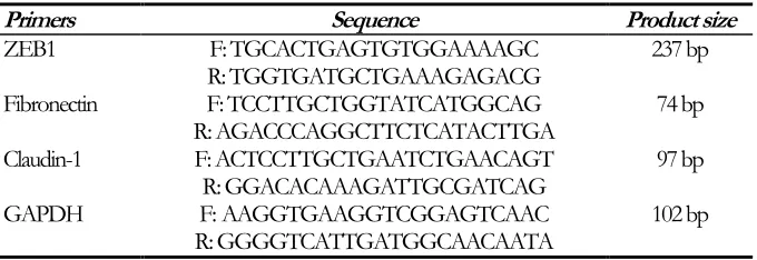
eliminate probable contamination with genomic DNA. Reverse transcription and real‐time PCR were performed following the manufacturer’s instructions using SYBR Green Master Mix (Parstoos, Iran). Primers used in this study are shown in Table 1. The conditions for all reac-tions were as follows: denaturation at 95 °C for 10 min and 40 cycles of 95 °C for 15 sec, 60 °C for 30 sec, and 72 °C for 30 sec (10, 11).
Table 1: Real-time PCR primer sequences
Primers Sequence Product size
ZEB1 F: TGCACTGAGTGTGGAAAAGC 237 bp
R: TGGTGATGCTGAAAGAGACG
Fibronectin F: TCCTTGCTGGTATCATGGCAG 74 bp
R: AGACCCAGGCTTCTCATACTTGA
Claudin-1 F: ACTCCTTGCTGAATCTGAACAGT 97 bp
R: GGACACAAAGATTGCGATCAG
GAPDH F: AAGGTGAAGGTCGGAGTCAAC 102 bp
R: GGGGTCATTGATGGCAACAATA
Western blotting analysis
The pellets of control and treated cells after the proposed time points of 24, 48, and 72 h were suspended in lysis buffer (BioBasic, PH= 8), supplemented with protease inhibitor cocktail (Sigma-Aldrich). Forty micrograms of proteins were denatured using Laemmli’s sample Buffer and boiled at 95 °C for 5 min. Proteins were sep-arated by SDS-polyacrylamide gel electrophoresis and transferred onto PVDF membranes (Amer-sham). The membranes were placed in a blocking buffer for 60 min at RT and subsequently incu-bated with anti-β-catenin primary antibody (1:1000; Cell Signaling) overnight, at 4 °C. The following day, the membranes were washed and incubated with a peroxidase conjugate secondary antibody (1:5000; Cell Signaling) for 60 min, at RT. Then membranes were washed and incubat-ed with the Bio-Rad Clarity™ western ECL sub-strate and finally exposed to X-ray film from FU-JIFILM Corporation. Band intensity was calcu-lated using Image J software.
Statistical analysis
The statistical analyses were performed using GraphPad Prism version 6.0 software. One way ANOVA with Dunnett's multiple comparison test was used to assess differences between vari-ous groups. Data are presented as mean ± stand-ard deviation (SD).
Results
SFN inhibited the migration of breast cancer cells
Fig. 1: The migration capability of SFN treated MDA-MB-231 cells was evaluated by scratch assay. (A) Scratch assay images show the extent of healing and black lines indicate the scratch borders. Scale bar: 200 𝜇m. (B) Graph repre-sents statistical analysis of scratch assay. Data show a role of the SFN as a suppressor of breast cancer cell migration.
Significance was set at *P< 0.05, **P< 0.01
SFN induced cell apoptosis in MDA-MB-231 Cells
The ability of SFN to induce apoptosis was as-sessed by flow cytometry analysis. MDA-MB-231 cells were treated with different concentrations of
SFN (0, 5, 10, 20, 30 and 40 μM) for 24, 48 and 72 h. As shown in Fig.2, compared with untreat-ed cells, SFN increasuntreat-ed the percentage of the ear-ly/late apoptotic and necrotic cells significantly, at the maximum doses of 30 and 40 µM.
Fig. 2: SFN enhanced apoptosis in breast cancer cells. Human breast cancer MDA-MB 231 cells were treated with
increasing concentrations of SFN (0-40 μM) for 24, 48 and 72 h. The flow cytometry assay was used to assess cell viability and the results were normalized to the untreated control cells. Quantitative data show a significant increase of apoptosis at 30 and 40 μM of SFN in compare with control cells. * and ** donate P<0.05 and P<0.01
SFN decreased expression of genes involved in EMT
We performed quantitative RT-PCR to examine the effect of SFN on expression levels of specific genes involved in EMT such as ZEB1, fibron-ectin and claudin-1. In the present study, MDA-MB-231 cells were treated with different concen-trations of SFN (0, 5, 10, 20, 30 and 40μM).
To-tal RNAs were collected at 24, 48 and 72 h. We found that SFN significantly downregulated the expression of ZEB1 at 40μM after 72 h com-pared with the control group. SFN also decreased the expression of fibronection at 20, 30, and 40 μM after 72 h. Decreased expression of claudin-1 was observed at 30 and 40μM after 72 h (Fig.3).
Fig. 3: Effects of SFN on transcriptional levels of specific genes involved in EMT. Total RNA was extracted from MDA-MB-231 cells treated
for 24, 48, and 72 h with 10, 20, 30 and 40μM of SFN. Various concentrations of SFN have different effects on the expression of EMT mark-ers of ZEB1, fibronectin, and claudin-1 after 72 h. Fold changes in gene expression were analyzed relative to gene expression in untreated
controls. Significance was set at *P<0.05 and **P<0.01
SFN inhibited β-catenin expression in MDA-MB-231 cells
To further investigate the possible mechanisms by which SFN exerts its anti-migratory effect, we evaluated whether the expression of β-catenin is associated with SFN treatment. Immunoblot
analysis was employed to examine the expression of β-catenin. As shown in Fig.4, there was a time-dependent decrease at the concentration of 40 µ MSFN. There were no changes in the expression of β-catenin protein in MDA-MB-231 cells with lower doses of SFN (data not shown).
Fig 4: Immunoblot analysis of β-catenin with protein extracts from MDA-MB-231 cells treated with 40μM SFN for 24, 48 and 72 h compared
Discussion
The present study illustrates that SFN exposure induces apoptosis at 30-40µMand also inhibits the migratory capabilities of MDA-MB-231 cells at 10-40µM. These results raise the possibility that SFN may be a promising candidate to pre-vent breast cancer progression and metastasis. It was revealed that the anti-cancer activity of SFN is modulated by a variety of cell-signaling path-ways including NF-κB signaling (12), Keap1– Nrf2 signaling (13), and ROS-dependent pathway (14). Further studies are needed to investigate the underlying molecular mechanisms of SFN effects to provide a rational basis for its clinical applica-tion in the future.
EMT is a crucial step for tumor cell migration and invasion in various types of human cancers.
Overexpression of ZEB1, as a transcription fac-tor, is critical for induction of EMT(15, 16). The results of our study revealed that SFN decreased cell migration at concentrations of 10-40µMSFN; however, reduced expression of ZEB1was ob-served just at 40µM of SFN. It can be assumed that ZEB1 is not involved in mediating the anti-metastatic effect of SFN at lower concentrations in MDA-MB-231 cells. Recently, expression of ZEB1 activates signaling pathways leading to en-hanced cell proliferation, and tumor growth (17). Based on the present study, SFN at high concentration may at least partly exerts its
proapoptotic and antimigratory effects by target-ing the expression of ZEB1 in MDA-MB-231 cells.
Fibronectin is another established EMT marker, which appears to be involved in extracellular ma-trix (ECM) remodeling during EMT. It is upregu-lated during development and has been linked to the growth, survival and metastasis in variety of cancers including breast (18), prostate (19) and colon (20) cancers. In accordance with previous studies, we find that decreased expression of fi-bronectin coincides with decreased cell migration in response to SFN treatment (10-40µM). Con-sidering the important role of fibronectin in
can-cer metastasis, it is possible that SFN exerts its anti migratory effects by decreasing the expres-sion of fibronectin. A more detailed study is re-quired to clarify the exact role of fibronectin in this effect.
Moreover, claudins, the integral membrane pro-teins which form the backbone of tight junctions, are aberrantly expressed in diverse types of hu-man cancers (21). Claudin-1, the first claudin family member identified, is downregulated in hepatocellular carcinoma (22), breast cancer (23) and lung adenocarcinomas (24). Conversely,
overexpression of claudin-1 increases cell inva-sion in oral squamous cell carcinoma (25), mela-noma (26), colon (27), and colorectal cancers (28). Further support for such speculations comes from studies that showed increased ex-pression of claudin-1 induced exex-pression of the EMT-regulating transcription factors such as ZEB1 in hepatocellular carcinomas, and claudin-1 can be exploited as a biomarker for liver cancer metastasis (29). Collectively, claudin-1 can both promote or suppress tumorigenesis, depending on the cancer type.MDA-MB-231, the cell line used in this study, is a basal B/triple-negative breast cancer (TNBC) cell line (30, 31). The knockdown of claudin-1 in a basal-like subtype human breast cancer cell line resulted in reduced cancer cell migration (32). Furthermore, down-regulation of claudin-1 induced apoptosis in dif-ferent breast cancer cells including MDA-MB-231(33). In accordance with these studies, results presented here indicated that decreased migration and increased apoptosis in MDA-MB-231 cells in response to SFN (30-40µM) coincided with re-duced expression of claudin-1. Apoptotic and anti-metastatic effects of SFN at 30-40µMcan be at least partly mediated by decreased expression of claudin-1. Although cell migration decreased at concentrations less than 30 µM of SFN; however, it did not change the expression of claudin-1. Claudin-1 is not involved in antimigratory effect of SFN at low concentrations.
of Wnt stimulation, cytosolic β-catenin is sequen-tially phosphorylated and degraded by glycogen synthase kinase 3β (GSK3-β) within a protein complex (35). Wnt stimulation is suggested to inhibit β-catenin phosphorylation, thus inducing the accumulation of cytosolic β-catenin. Next, the stabilized β-catenin is translocated into the nucleus, in which it activates the expression of a range of genes related to cancer progression and metastasis (36). Aberrant activation of Wnt/β-catenin pathway occurs in several tumor types, including colorectal and breast cancers (37, 38). ”Recently, there is considerable interest in devel-oping Wnt/β-catenin pathway inhibitors as anti-cancer therapeutics (39)”. In the current study, the effect of SFN on β-catenin, as the key media-tor of the Wnt pathway in human breast cancer cells was examined. The results of our survey showed that SFN at 40µMsuppressed the expres-sion of β-catenin in a time-dependent manner in breast cancer MDA-MB-231 cells. No change in the expression of β-catenin was observed at low-er concentrations of SFN. Apoptotic and anti-metastatic effects of SFN at high concentration might be at least partly mediated through its ef-fect on the expression of β-catenin. A casual mechanism of this association should be unrav-eled by more detailed study.
Conclusion
SFN decreases migration of breast cancer cells and it can be considered as a promising candidate for treatment of metastatic breast cancer. Fur-thermore, anti-migratory effects of SFN at low concentrations may be mediated in part by de-creased expression of fibronectin, not ZEB1, claudin-1 and β-catenin. Further attempts have to be made to understand more about the mecha-nisms involved in anti-metastatic effects of SFN on breast cancer cells.
Ethical considerations
Ethical issues (Including plagiarism, Informed Consent, misconduct, data fabrication and/or falsification, double publication and/or
submis-sion, redundancy, etc.) have been completely ob-served by the authors.
Acknowledgements
The authors acknowledge the financial support provided by Cancer Prevention Research Center, Shahroud University, project No 94109.
Conflict of interest
The authors declare that there is no conflict of interest.
References
1. Tevaarwerk AJ, Gray RJ, Schneider BP et al (2013). Survival in patients with metastatic recurrent breast cancer after adjuvant
chemotherapy: little evidence of
improvement over the past 30 years.
Cancer,119:1140-8.
2. Lamouille S, Xu J, Derynck R (2014). Molecular mechanisms of epithelial-mesenchymal transition. Nat Rev Mol Cell Biol,15:178-96. 3. Zhou B, Moodie A, Blanchard AA et al (2015).
Claudin 1 in Breast Cancer: New Insights. J
Clin Med,4:1960-76.
4. Ghahhari NM, Babashah S (2015). Interplay between microRNAs and WNT/β-catenin signalling pathway regulates epithelial– mesenchymal transition in cancer. Eur J
Cancer,51:1638-49.
5. Ahmadiankia N, Moghaddam HK, Mishan MA
et al (2016). Berberine suppresses migration of MCF-7 breast cancer cells through down-regulation of chemokine receptors. Iran J Basic
Med Sci,19:125-31.
6. Hecht SS (2000). Inhibition of carcinogenesis by isothiocyanates. Drug Metab Rev,32:395-411. 7. Qazi A, Pal J, Maitah M et al (2010). Anticancer
activity of a broccoli derivative, sulforaphane, in barrett adenocarcinoma: potential use in chemoprevention and as adjuvant in chemotherapy. Transl Oncol,3:389-99.
cancer cells. Cell Mol Biol (Noisy-le-grand),61:37-43.
9. Pawlik A, Wiczk A, Kaczynska A et al (2013).
Sulforaphane inhibits growth of
phenotypically different breast cancer cells.
Eur J Nutr,52:1949-58.
10. Mishan MA, Heirani-Tabasi A, Mokhberian N et al (2015). Analysis of Chemokine Receptor Gene Expression in Esophageal Cancer Cells Compared with Breast Cancer with Insights into Metastasis. Iran J Public Health,44:1353-8. 11. Bagheri M, Fazli M, Saeednia S et al (2018).
Pomegranate peel extract inhibits expression of β-catenin, epithelial mesenchymal transition, and metastasis in triple negative breast cancer cells. Cell Mol Biol
(Noisy-le-grand),64:86-91.
12. Li W, Khor TO, Xu C et al (2008). Activation of Nrf2-antioxidant signaling attenuates NFκB-inflammatory response and elicits apoptosis.
Biochem Pharmacol,76:1485-9.
13. Kensler TW, Egner PA, Agyeman AS et al (2013). Keap1-nrf2 signaling: a target for cancer prevention by sulforaphane. Top Curr
Chem,329:163-77.
14. Wang L, Tian Z, Yang Q et al (2015). Sulforaphane inhibits thyroid cancer cell growth and invasiveness through the reactive
oxygen species-dependent pathway.
Oncotarget,6:25917-31.
15. Spaderna S, Schmalhofer O, Wahlbuhl M et al (2008). The transcriptional repressor ZEB1 promotes metastasis and loss of cell polarity in cancer. Cancer Res,68:537-44.
16. Thomson S, Buck E, Petti F et al (2005). Epithelial to mesenchymal transition is a determinant of sensitivity of non–small-cell lung carcinoma cell lines and xenografts to epidermal growth factor receptor inhibition.
Cancer Res,65:9455-62.
17. Sanchez-Tillo E, Fanlo L, Siles L et al (2014). The EMT activator ZEB1 promotes tumor growth and determines differential response to chemotherapy in mantle cell lymphoma.
Cell Death Differ,21:247-57.
18. Fernandez-Garcia B, Eiro N, Marin L et al
(2014). Expression and prognostic
significance of fibronectin and matrix metalloproteases in breast cancer metastasis.
Histopathology,64:512-22.
19. Konac E, Kiliccioglu I, Sogutdelen E et al (2017). Do the expressions of epithelial-mesenchymal transition proteins, periostin, integrin-alpha4 and fibronectin correlate with clinico-pathological features and prognosis of metastatic castration-resistant prostate cancer?
Exp Biol Med (Maywood),242:1795-801.
20. Yi W, Xiao E, Ding R et al (2016). High expression of fibronectin is associated with poor prognosis, cell proliferation and malignancy via the NF-kappaB/p53-apoptosis signaling pathway in colorectal cancer. Oncol Rep,36:3145-53.
21. Escudero-Esparza A, Jiang WG, Martin TA (2011). The Claudin family and its role in cancer and metastasis. Front Biosci (Landmark
Ed),16:1069-83.
22. Higashi Y, Suzuki S, Sakaguchi T et al (2007). Loss of claudin-1 expression correlates with malignancy of hepatocellular carcinoma. J
Surg Res,139:68-76.
23. Morohashi S, Kusumi T, Sato F et al (2007). Decreased expression of claudin-1 correlates with recurrence status in breast cancer. Int J
Mol Med,20:139-43.
24. Chao YC, Pan SH, Yang SC et al (2009). Claudin-1 is a metastasis suppressor and correlates with clinical outcome in lung adenocarcinoma. Am J Respir Crit Care
Med,179:123-33.
25. Oku N, Sasabe E, Ueta E et al (2006). Tight junction protein claudin-1 enhances the invasive activity of oral squamous cell carcinoma cells by promoting cleavage of laminin-5 gamma2 chain via matrix metalloproteinase (MMP)-2 and membrane-type MMP-1. Cancer Res,66:5251-7.
26. Leotlela PD, Wade MS, Duray PH et al (2007). Claudin-1 overexpression in melanoma is regulated by PKC and contributes to melanoma cell motility. Oncogene,26:3846-56. 27. Dhawan P, Singh AB, Deane NG et al (2005).
Claudin-1 regulates cellular transformation and metastatic behavior in colon cancer. J Clin
Invest,115:1765-76.
28. Kinugasa T, Huo Q, Higashi D et al (2007). Selective up-regulation of claudin-1 and claudin-2 in colorectal cancer. Anticancer
Res,27:3729-34.
through activation of the c-Abl-ERK signaling pathway in human liver cells.
Oncogene, 32:4873-82.
30. Chavez KJ, Garimella SV, Lipkowitz S (2010). Triple negative breast cancer cell lines: one tool in the search for better treatment of triple negative breast cancer. Breast Dis,32:35-48. 31. Kao J, Salari K, Bocanegra M et al (2009).
Molecular profiling of breast cancer cell lines defines relevant tumor models and provides a resource for cancer gene discovery. PLoS
One,4:e6146.
32. Blanchard AA, Ma X, Dueck KJ et al (2013). Claudin 1 expression in basal-like breast cancer is related to patient age. BMC
Cancer,13:268.
33. Achari C, Winslow S, Larsson C (2015). Down Regulation of CLDND1 Induces Apoptosis
in Breast Cancer Cells. PLoS
One,10:e0130300.
34. Cai J, Guan H, Fang L et al (2013). MicroRNA-374a activates Wnt/beta-catenin signaling to promote breast cancer metastasis. J Clin
Invest,123:566-79.
35. Orford K, Crockett C, Jensen JP et al (1997). Serine phosphorylation-regulated ubiquitina-tion and degradaubiquitina-tion of β-catenin. J Biol Chem
,272:24735-8.
36. Valenta T, Hausmann G, Basler K (2012). The many faces and functions of beta-catenin.
Embo j,31:2714-36.
37. Nusse R, Clevers H (2017). Wnt/beta-Catenin Signaling, Disease, and Emerging Therapeutic Modalities. Cell,169:985-99.
38. Prosperi JR, Goss KH (2010). A Wnt-ow of opportunity: targeting the Wnt/beta-catenin pathway in breast cancer. Curr Drug
Targets,11:1074-88.


![catena Poly[[chlorocopper(I)] μ 1,3 di 4 pyridylpropane κ2N:N′]](data:image/gif;base64,R0lGODlhAQABAIAAAP///wAAACH5BAEAAAAALAAAAAABAAEAAAICRAEAOw==)