1535-9778/05/$08.00⫹0 doi:10.1128/EC.4.1.121–133.2005
Copyright © 2005, American Society for Microbiology. All Rights Reserved.
Role for RNA-Binding Proteins Implicated in Pathogenic
Development of
Ustilago maydis
†
Philip Becht, Evelyn Vollmeister, and Michael Feldbru
¨gge*
Department for Organismic Interactions, Max Planck Institute for Terrestrial Microbiology, Marburg, Germany
Received 26 August 2004/Accepted 26 October 2004
Ustilago maydis causes smut disease on corn. Successful infection depends on a number of morphological transitions, such as pheromone-dependent formation of conjugation tubes and the switch to filamentous dikaryotic growth, as well as different types of mycelial structures during growth within the host plant. In order to address the involvement of RNA-binding proteins during this developmental program, we identified 27 open reading frames from the genome sequence encoding potential RNA-binding proteins. They exhibit similarities to RNA-binding proteins with Pumilio homology domains (PUM), the K homology domain (KHD), the double-stranded RNA binding motif (DSRM), and the RNA recognition motif (RRM). For 18 of these genes, we generated replacement mutants in compatible haploid strains. Through analysis of growth behavior, morphology, cyclic AMP response, mating, and pathogenicity, we identified three candidates with aberrant phenotypes. Loss of Khd1, a K homology protein containing three KHDs, resulted in a cold-sensitive growth
phenotype. Deletion ofkhd4 encoding a protein with five KHDs led to abnormal cell morphology, reduced
mating, and virulence.rrm4⌬strains were affected in filamentous growth and pathogenicity. Rrm4 is an RRM
protein with a so far unique domain organization consisting of three N-terminal RRMs as well as a domain found in the C terminus of poly(A)-binding proteins. These results indicate a role for RNA-binding proteins
in regulation of morphology as well as in pathogenic development inU. maydis.
Presently, the causative agent of corn smut disease,Ustilago maydis, and the human pathogenCryptococcus neoformansare model systems of choice for pathogenic basidiomycetes (7, 25). For completion of its sexual life cycle,U. maydisdepends on a number of morphological transitions. These are exquisitely regulated and intimately connected to the presence of its host. A prerequisite for infection is mating of two compatible hap-loid sporidia that proliferate by budding. Mating involves the formation of conjugation hyphae that grow towards each other and fuse at their tips (59). Upon cell fusion, a dikaryon is generated that arrests its cell cycle and switches to fast polar growth. The dikaryon represents the infective form and is able to penetrate the plant surface after formation of appressorium-like structures. Inside the plant, the cell cycle stop is released, resulting in a multicompartmental dikaryotic mycelium. At later stages karyogamy occurs and diploid teliospores are formed (4, 27).
Mating compatibility is determined by a tetrapolar mating system consisting of two mating-type loci designatedaand b. The biallelic alocus (a1and a2) encodes the precursor and receptor of lipopeptide pheromones regulating intercellular recognition (9). The multiallelic b locus encodes a pair of homeodomain proteins, bE and bW, that are only active as heterodimeric transcription factors with subunits derived from different allelic origins (29). The formation of an active bE/bW heterodimer is necessary and sufficient for the developmental
switch to fast polar growth. The concerted action of mating-type gene products is orchestrated by cross talk between pro-tein kinase A (PKA) and mitogen-activated propro-tein kinase (MAPK) signaling (17, 36, 54).
To investigate regulation of cell polarity and morphology during pathogenic development, we tested the involvement of RNA-binding proteins, since this class of proteins determines cell polarity in higher eukaryotes during developmental pro-grams such as oogenesis and early embryogenesis inDrosophila melanogasterand sex determination inCaenorhabditis elegans, as well as mammalian stem cell proliferation (34). RNA-bind-ing proteins regulate mRNA activity at the level of maturation, nuclear export, cytoplasmic localization, stability, and transla-tion (15, 40, 42). A well-studied example is Pumilio fromD. melanogaster, which recognizes a specific response element in the 3⬘-untranslated region of hunchback mRNA. Thereby translation is repressed, resulting in a protein gradient throughout the embryo that determines the posterior polarity of the embryo (44, 69). Also regarding lower eukaryotes, the number of reports that describe connections between RNA-binding proteins and development is growing. PufA from Dic-tyostelium discoideumis involved in determining sporulation in response to starvation by regulating translation of mRNA en-coding the catalytic subunit of PKA (60). Nrd1 and Msa1 are negative regulators of sexual differentiation in Schizosaccharo-myces pombe(26), and Scp160p regulates mating in Saccharo-myces cerevisiaeby interacting with the heterotrimeric G pro-tein Gpa1p during pheromone-responsive MAPK signaling (22).
To study the function of RNA-binding proteins inU.maydis, we chose a combinatorial approach based on bioinformatics and reverse genetics, using the recently released genome
se-* Corresponding author. Mailing address: Max Planck Institute for Terrestrial Microbiology, Department of Organismic Interactions, Karl-von-Frisch-Strasse, 35043 Marburg, Germany. Phone: 49-6421-178602. Fax: 49-6421-178609. E-mail: [email protected]. † Supplemental material for this article may be found at http: //ec.asm.org/.
121
on September 8, 2020 by guest
http://ec.asm.org/
quence (http://www.broad.mit.edu/annotation/fungi/ustilago _maydis/) and an efficient gene replacement strategy (10, 28). Genes encoding potential RNA-binding proteins can be easily identified due to the presence of at least one RNA-binding domain. In eukaryotes, the majority of RNA-binding domains belong to the following four classes that are well defined on primary, secondary, and tertiary structure: the Pumilio homol-ogy domain (PUM), the K homolhomol-ogy domain (KHD), the dou-ble-stranded RNA-binding domain (DSRM), and the RNA recognition motif (RRM) (12, 41, 49).
The PUM was named after its founding member, Pumilio (69, 73). The domain recognizes RNA sequence specifically and is composed of eight tandem repeats, each ⬃40 amino acids (aa) in length. Characteristic of these repeats is a core consensus containing aromatic and basic amino acids (70). The KHD was first identified in hnRNP K (heterogeneous nuclear ribonucleoprotein K), a major pre-mRNA-binding protein (58). The domain comprises⬃60 aa and contains a character-istic Gly-X-X-Gly loop that grips RNA (38, 45). The double-stranded RNA-binding motif is an⬃65-aa domain found in various proteins that bind double-stranded RNAs such as Staufen fromD. melanogasteror RNase III fromEscherichia coli(31, 61). The RRM is by far the best-characterized and most widespread domain. At present there are 4,160 proteins with 7,079 domains known (http://smart.embl.de/). The RRM is 90 to 100 aa in length, containing two well-conserved se-quences, the RNP1 octamer and the RNP2 hexamer (6, 12). Charged and aromatic side chains of RNP1 and RNP2 are solvent exposed and contact RNA (12).
Here, we describe the identification of 27 open reading frames (ORFs) inU. maydisencoding putative RNA-binding proteins of the PUM, KHD, DSRM, and RRM types. A subset of 18 gene replacement mutants were generated and subjected to a detailed phenotypic analysis revealing that KHD and RRM candidates are important for pathogenic development.
MATERIALS AND METHODS
Strains.TheE. coliK-12 derivatives DH5␣(Bethesda Research Laboratories) and Top10 (Invitrogen) were used for cloning purposes.U. maydisstrains FB1, FB2, UM521, SG200, FBD12-17, and FBD11-7 have been published previously (5, 8).
Bioinformatic analysis.In order to identify PUM-type RNA-binding proteins, the following protein sequences (http://smart.embl-heidelberg.de/smart/do _annotation.pl?DOMAIN⫽Pumilio&BLAST⫽DUMMY&EVOLUTION
⫽Show#Evolution) have been compared to the genomic sequence from U. maydis(http://www.broad.mit.edu/annotation/fungi/ustilago_maydis/) by using BLAST (2):S. cerevisiae(7), Q12221, P47077, P39016, Q07807, P25339, Q04373, and P47135;S. pombe(9), Q10238, Q09829, Q92347, O94462, O60059, Q92359, Q9P789, Q9UU76, and O74438;Neurospora crassa(2), Q8X035, Q9P6D4;D. discoideum (1), Q9Y1J5; Arabidopsis thaliana (6), Q9LRZ3, Q9SVZ7, BAB08273, Q95547, Q9SFL0, and Q9LVC3;H. sapiens(3), AAG31807, O00234, and AAG31806;C. elegans(6), O01322, QZ3191, Q9U2G4, Q09487, Q9N3Q2, and Q9N5M6; andD. melanogaster(1), Q9VHH6.
To identify KHD-containing proteins, we used the following protein sequences from the SMART (Simple Modular Architecture Research Tool) database (http: //smart.embl-heidelberg.de/smart/do_annotation.pl?DOMAIN⫽KH&BLAST
⫽DUMMY&EVOLUTION⫽Show#Evolution):S. cerevisiae(6), Q07834, Q12186, YBD2, PBP2, RS3, and Q99216;S. pombe(7), O59810, O74359, O74919, O74555, O74777, YAJE_Schpo, and O14044;N. crassa(1), Q9P6B5;H. sapiens(5), Q13601, AAG02184, AAG45476, Q9Y307, and Vigilin;C. elegans(1), Q9U3B2; and
D. melanogaster(1), Q9V8H6.
In order to identify DSRM-type proteins, we used the following protein se-quences (http://smart.embl-heidelberg.de/smart/do_annotation.pl?DOMAIN
⫽DSRM&BLAST⫽DUMMY&EVOLUTION⫽Show#Evolution):S. cerevisiae
(2), RM03 and RNT1;S. pombe(2), O43042 and PAC1;N. crassa(2), Q7S6N0 and Q9P6D6; and D. melanogaster(23), AAQ23615, Q8MRC7, Q8MRY4, Q95YG3, Q960Y4, Q961S7, Q9GNJ2, Q9NHW9, Q9NII2, Q9U6N4, Q9V847, Q9V9V7, Q9VCU9, Q9VHB0, Q9VJY9, Q9VLW8, Q9VRL8, Q9VY45, Q9VZ88, Q9W5S7, Q9XYN5, MLE, and STAU.
To identify RRM-type proteins, we used the following sequences (http: //smart.embl-heidelberg.de/smart/do_annotation.pl?DOMAIN⫽RRM&BLAST
⫽DUMMY&EVOLUTION⫽Show#Evolution): S. cerevisiae (6), P04147, P32588, P38922, P32831, P27476, and P34217;S. pombe(2), Q09702 and P08965;
H. sapiens(3), Q9UNP9, Q15717, and Q9Y3B4; andD. melanogaster(5), P19018, P19339, P16914, O02374, and AAF54070.
For the phylogenetic analysis, sequence alignments were performed with CLUSTAL_X (63). Dendrograms were constructed with MEGA 2.1 (http: //www.megasoftware.net) (35) with the minimum evolution algorithm and an initial tree obtained by the neighbor-joining method. Two KHD proteins each have been predicted fromC. neoformans(Cn; http://www.broad.mit.edu/annotation/fungi /cryptococcus_neoformans/) andCoprinus cinereus(Cc; http://www.broad.mit-.edu/annotation/fungi/coprinus_cinereus/). KHD protein Cn1, 1,113 aa in length, is located on contig 1.77 between nucleotide positions 175691 and 172233. Two introns were predicted (intron 1, 173461 to 1733960; and intron 2, 172828 to 172779). KHD protein Cn2, 891 aa in length, is located on contig 1.12 between nucleotide positions 65877 and 61787. Eight introns were predicted (intron 1, 65225 to 65028; intron 2, 64929 to 64881; intron 3, 64745 to 64107; intron 4, 63950 to 63919; intron 5, 63720 to 63383; intron 6, 62923 to 62877; intron 7, 62675 to 62618; intron 8, 62371 to 62318). KHD protein Cc1, 1,228 aa in length, is located on contig 1.44 between nucleotide positions 260066 and 264281. Seven introns were predicted (intron 1, 260264 to 260745; intron 2, 261695 to 264754; intron 3, 262019 to 262074; intron 4, 262244 to 262303; intron 5, 262418 to 262528; intron 6, 262595 to 262654; intron 7, 263275 to 263334). KHD protein Cc2, 797 aa in length, is located on contig 1.9 between nucleotide positions 41512 and 44651. Eight introns were predicted (intron 1, 41834 to 41904; intron 2, 42041 to 42094; intron 3, 42237 to 42284; intron 4, 42550 to 42610; intron 5, 42821 to 43159; intron 6, 43619 to 43671; intron 7, 43873 to 43920; intron 8, 44306 to 44376).
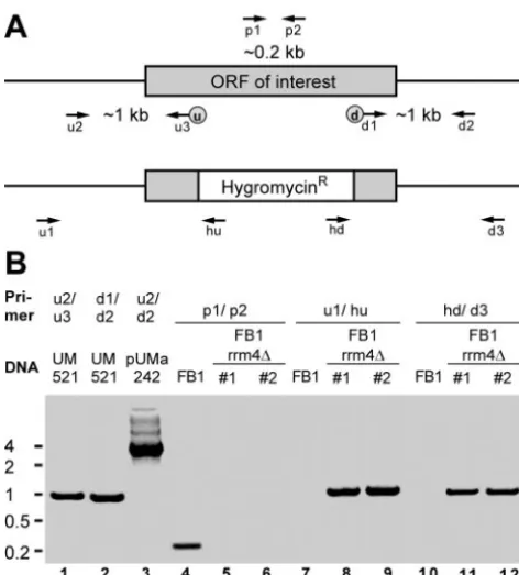
Generation of gene replacement strains in U. maydis.Gene replacement mutants were generated by a PCR-based approach (10, 28). For each gene replacement construct, eight PCR primers were designed: u1, u2, and u3 (up-stream flanking sequence); d1, d2, and d3 (down(up-stream flanking sequence); and p1 and p2 (10) (see Fig. 2A and Table 2; see Table S1 in the supplemental material). Primer u3 and d1 introduced SfiI(u) and SfiI(d) recognition sites at the 3⬘end of the upstream flank and the 5⬘end of the downstream flank, respec-tively. Initially⬃1-kb upstream and downstream flanking sequences of the ORF region of interest were amplified with genomic DNA of wild-type strain UM521 (a1b1) as a template and primer combinations u2/u3 and d1/d2, respectively (Fig. 2B, lanes 1 and 2; Table 2). PCR products were cleaved with SfiI and ligated in the presence of a compatible 1.8-kb SfiI(u)/SfiI(d) fragment containing the hy-gromycin resistance cassette (28). Gel-purified ligation products were inserted in pCR2.1-TOPO (Invitrogen; Table 2, pUMa). The resulting plasmids were used as a template for PCR with primers u2 and d2 (Fig. 2B, lane 3), and the respective products were transformed inU. maydiswild-type strains FB1 and FB2 (a1b1anda2b2, respectively). Hygromycin-resistant colonies were subjected to whole-cell PCR analysis using primers p1 and p2 (Fig. 2B, lanes 4 to 6; Table 2). Amplification of an⬃200-bp-long fragment was diagnostic for the presence of the wild-type allele indicative for a nonhomologous integration event (Fig. 2B, lane 4). Only those transformants in which the diagnostic PCR product was absent were analyzed further (Fig. 2B, lanes 5 and 6). Successful gene replace-ments were verified by detection of an⬃1-kb PCR product using primers u1 and hu (Fig. 2B, lanes 7 to 9) and by amplification of⬃1-kb PCR products with primer combinations d3 and hd (Fig. 2B, lanes 10 to 12). To validate this approach, we verified successful gene replacements in the cases ofpum1-3⌬and
khd4⌬, as well asrrm4⌬, by Southern analysis (10).
Growth on CM and minimal medium plates.Starter cultures were grown overnight on a rotary shaker at 220 rpm in liquid complete medium (CM) at 28°C (24). Using a Neubauer counting chamber, cell density was determined. An aliquot of cells was centrifuged at 3,500 rpm (5 min at room temperature), and the density of cells was adjusted to 106
cells/ml with sterile water. Two-microliter cell suspensions of fivefold serial dilutions were applied as spots in six steps on CM or nitrate minimal (NM) plates (3-g/liter KNO3, 10-g/liter glucose, and 2-g/liter agarose) (24). Plates were incubated for 2 to 5 days at 15, 28, and 34°C. Pigmentation, morphology, and growth of colonies were scored by visual inspec-tion.
Measuring doubling time during exponential growth.Starter cultures were grown overnight on a rotary shaker at 220 rpm in liquid CM at 28°C (24). A 1-l
on September 8, 2020 by guest
http://ec.asm.org/
cell suspension of stationary-phase starter cultures was transferred in 1 ml of CM and incubated on a thermomixer compact (Eppendorf) at 400 rpm in a 24-well plate at 28°C. At appropriate time intervals, optical density (OD) was measured as ODA600 with a TECAN Saphire fluorescence reader. Doubling time was calculated from the slope of a linear regression line derived from values during the exponential phase of growth (see Fig. S1 in the supplemental material).
Quantifying response to external cAMP.Starter cultures (1 ml) were grown overnight on a thermomixer compact (Eppendorf) at 1,100 rpm in liquid CM (24) at 28°C using 2-ml tubes sealed with LidBac(Eppendorf). Two-microliter cell suspensions of stationary-phase starter cultures were transferred in 1 ml of CM in the presence or absence of 15 mM cyclic AMP (cAMP) and incubated on a thermomixer compact (Eppendorf) at 1,100 rpm for 18 h at 28°C. An aliquot was transferred to a Neubauer counting chamber, and the percentage of cells in aggregates of more than two cells was counted (for each quantification,⬃100 cells; see Fig. S2 in the supplemental material).
Mating, pheromone production, and filamentation assays.Starter cultures were grown overnight on a rotary shaker at 220 rpm in PD medium (2.4% potato dextrose broth; Difco). Stationary-phase cultures were diluted in PD medium to an OD at 600 nm (OD600) of 0.2. After growth at 28°C on a rotary shaker at 220 rpm to an OD600of 0.5, a 0.5-ml aliquot of cell suspension was centrifuged at 3,500 rpm (5 min at room temperature). Cells were resuspended in 0.5 ml of water. For filamentation assays, cells were applied as spots and, for mating as well as pheromone production assays, respective strains were applied as cospots on charcoal-containing PD plates that were sealed with Parafilm and incubated at 22°C for 24 to 48 h.
Cell morphology and conjugation hypha formation. Starter cultures were grown overnight on a rotary shaker at 220 rpm in CM (24) at 28°C. Stationary-phase cultures were diluted in CM to an OD600of 0.2. After incubation as mentioned above to an OD600of 0.5, a 1-ml aliquot of cells was centrifuged at 3,500 rpm (5 min at room temperature) and cells were resuspended in CM with or without synthetic a1 or a2 pheromone dissolved in dimethyl sulfoxide (2.5
g/ml) (62). Cells were incubated for 5 h at 28°C in a 15-ml plastic tube rotating with 20 rpm. For microscopic observation, we used a Zeiss Axiophot with dif-ferential interference contrast optics. Pictures were taken with a charge-coupled device camera (AxioCam HRm; Zeiss).
Pathogenicity assay.Plant infections of corn variety Early Golden Bantam (Olds Seeds, Madison, Wis.) were performed as described previously (11). Tu-mor formation was scored after 14 days. In the case ofrrm4deletion mutants, a more detailed disease rating was adopted from earlier studies (20) using the following categories: no tumor, minor disease symptoms at leaves (disease area with tumors is not larger than 1.5 cm and the number of tumors does not exceed a cutoff of 20), major disease symptoms at leaves (disease area with tumors is wider than 1.5 cm and the number of tumors exceeds 20), minor disease symp-toms at stems (disease area with tumors is not larger than 4 cm and heights of tumors do not exceed 0.6 cm), major disease symptoms at stems (disease with tumors is larger than 4 cm and heights of tumors exceed 0.6 cm), as well as wilted or dead plants.
RESULTS
Identification of ORFs encoding potential RNA-binding
do-mains of the PUM, KHD, DSRM, and RRM types.In order to
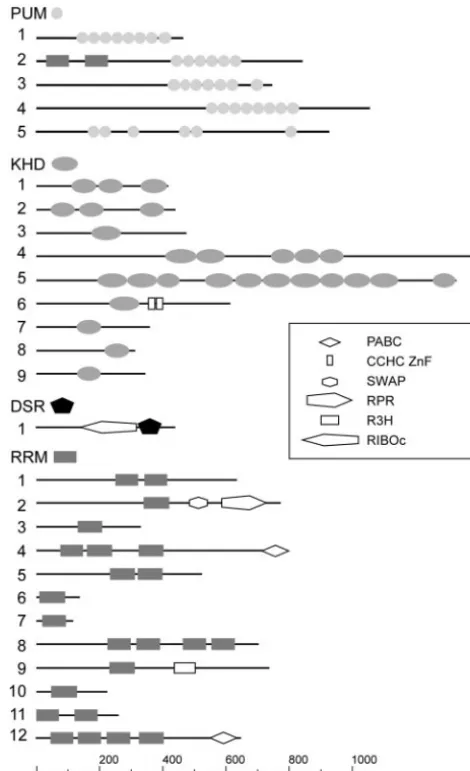
identify genes encoding RNA-binding proteins of the PUM, KHD, DSRM, and RRM types in the genome ofU.maydis, we selected known RNA-binding proteins of the respective types according to the SMART database from fungi and model or-ganisms such asA.thaliana,D.melanogaster,C.elegans, andH. sapiens(http://smart.embl.de; accession numbers are given in Materials and Methods) (37, 56). The respective protein sequences were compared to predicted ORFs fromU. maydis by using BLAST (http://www.broad.mit.edu/annotation/fungi /ustilago_maydis/) (2). Candidate ORFs were analyzed with the SMART service at http://smart.embl.de (37, 56). Only those proteins were considered positive that were predicted to contain the respective domain with scores more significant than established cutoff values (http://smart.embl.de/) (Fig. 1). All ORFs were defined by conceptual translation from the genomic sequence. In four cases, we were able to predict the
presence of introns according to sequence similarities to ho-mologous proteins (Table 1). However, the predicted se-quences might still contain surplus amino acids due to the presence of in-frame introns.
For the identification of PUM-type proteins inU.maydis, we chose 35 representative proteins containing Pumilio-like re-peats from SMART: 19 from various fungi, 6 fromA.thaliana, 6 from C. elegans, 3 from H. sapiens, and Pumilio from D. melanogaster. Five candidate ORFs were identified in the U. maydis genome, designatedpum1 to -5. The respective pro-teins contain several adjacent Pumilio-like repeats with con-served core sequences in the C-terminal half of the protein. These features are hallmarks for this class of proteins (Fig. 1; Table 1) (70). Pum2 has been predicted to contain two
addi-FIG. 1. Domain structures of PUM-, KHD-, DSRM-, and RRM-type RNA-binding proteins. Lines symbolize ORFs (length scale at the bottom), and domains are indicated by symbols. These are either labeled adjacently in cases of PUM, KHD, DSRM (DSR), and RRM (at the top of each class) or are given in the inset. PABC, poly(A)-binding protein C terminus; CCHC ZnF, CCHC-type zinc finger; SWAP, suppressor-of-white-apricot splicing regulator; RPR, regula-tion of pre-mRNA; R3H, conserved arginine histidine domain; and RIBOc, RNase III family. The numbers on the left correspond to the designations of the respective RNA-binding proteins.
on September 8, 2020 by guest
http://ec.asm.org/
tional RNA-binding domains of the RRM type in its N termi-nus. This domain composition is reminiscent of the structure of Pumilio-like proteins Puf1p/Jsn1p and Puf2p fromS. cerevisiae. Both proteins carry a single RRM domain N terminal to these PUM repeats (48). Pum5 belongs to a group of atypical PUM-type proteins, since it contains six PUM repeats that are not clustered in the C terminus (Fig. 1).
To identify KHD proteins, we chose 21 candidates from SMART: 14 from fungi, 5 fromH.sapiens, and 1 each fromD. melanogasterandC. elegans. Nine candidate ORFs were found and designatedkhd1to-9(Fig. 1; Table 1). Khd1 and Khd2 share the same domain organization consisting of three KHD regions and are similar in sequence to the Pab1p-binding pro-tein Pbp2p that is required for proper chromosome segrega-tion during meiosis (39, 51). Khd4 and Khd5 are KHD proteins that contain 5 and 11 KHD regions, respectively. Both are more than 1,000 aa in length. Khd5 is most likely the homo-logue of Scp160p fromS. cerevisiae (18, 67), since it shares substantial sequence similarity over the entire length of the protein (BLAST expect value of 9⫻e⫺42; Table 1). Khd6 is related to the branch point bridging protein Msl5p involved in splicing (1). According to their similarity to proteins fromS.
cerevisiae, Khd7, Khd8, and Khd9 are likely to be involved in ribosome maturation and function. Khd7 exhibits significant sequence similarity to Krr1p, a protein involved in ribosomal small subunit assembly and maintenance (55). The Khd8-re-lated protein Pno1p is a component of the 90S preribosome involved in ribosome maturation (57). Khd9 displays high se-quence similarity to the ribosomal protein S3 (Rps3p), an integral part of the small subunit of ribosomes (Fig. 1; Table 1) (66).
In order to identify DSRM proteins inU.maydis, we chose 29 proteins containing the DSRM: 6 from fungi and 23 fromD. melanogaster. Only one candidate ORF, termeddsr1, encodes a DSRM domain (Fig. 1; Table 1). Dsr1 exhibits high sequence similarity to the mitochondrial precursor of the 60S ribosomal protein L3 from S. cerevisiae (Mrpl3p, Table 1) (21). Both proteins share a characteristic RNase III RIBOc domain in N-terminal proximity to the DSRM (http://smart.embl.de/). The presence of only few DSRM proteins was not surprising sinceS. cerevisiae, S. pombe, and N. crassaalso contain only two each (http://smart.embl.de/). Apparently,U. maydislacks a clear homologue of Rnt1p, the second DSRM protein fromS. cerevisiaeinvolved in small nucleolar RNA metabolism (13).
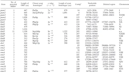
TABLE 1. RNA-binding proteins inU. maydis
Gene
No. of potential
RBDsa
Length of ORF (aa)
Closest yeast homologueb
e value (⬍e⫺15)c
Length of yeast
homologue (aa) Contig
d Nucleotide
position Deleted region Chromosome
k
pum1 1 467 Puf3p 7e⫺58 879 14 1433–2836 1779–2669 I
pum2 3 845 Jsn1p/Puf1p 2e⫺52 1,091 4 78730–76193 77494–76581 I
pum3 1 748 197 19370–21616 20583–20831 XVIII
pum4 1 1,058 Puf4p 5e⫺72 888 115 115768–118711 n.a.l
pum5 1 921 101 44697–47462 n.a.
khd1 3 422 Pbp2p 4e⫺26 413 215 157776–156339f 157387–156576 XX
khd2 3 442 Pbp2p 1e⫺23 413 124 7412–6173 7330–6459 X
khd3 1 479 139 71807–70368 71480–70994 XI
khd4 5 1,416 129 47506–43256 46492–45548 X
khd5 11 1,330 Scp160p 9e⫺42 1,222 245 18952–14960 n.a.
khd6 2 614 Ms15p 2e⫺58 476 247 93724–95568 XXIII
khd7 1 364 Krr1p 1e⫺107 316 109 110546–109452 VIII
khd8 1 313 Pno1p 4e⫺67 274 117 28298–27357 IX
khd9 1 347 Rps3p 9e⫺83 240 101 137517–136474 VIII
dsr1 1 444 Mrp13p 1e⫺24 390 103 133224–134558 VII
rrm1 2 638 83 396083–397999 396806–397324 V
rrm2 1 779 124 43778–41439 43133–41881 X
rrm3 1 332 34 72086–73354g 72518–73235 II
rrm4 3 792 112 120505–118126h 120357–119302 VIII
rrm5 2 525 101 247488–249232i 248295–248884 VII
rrm6 1 139 109 85216–84797 85182–84979 VIII
rrm7 1 121 86 175280–175645 175292–175649 VI
rrm8 4 704 Ylp184p 3e⫺96 612 157 28299–26185 27551–26442 XIV
rrm9 1 740 Pin4p 9e⫺37 668 126 64830–62611 64209–63824 X
rrm10 1 228 148 37894–37208 37726–37460 XII
rrm11 2 263 208e 732–30560j 1468–1463 XVI
rrm12 4 651 Pab1p 1e⫺132 577 117 24758–26713 IX
a
RNA-binding domain.
b
Only proteins were listed with BLAST expect value below e⫺15 .
c
BLAST expect value.
d
Genomic sequence release 1 (http://www.broad.mit.edu/annotation/fungi/ustilago_maydis/).
e
Fusion of contig 1.208 with 1.207.
f
Predicted introns at positions 157314 to 157244 and 157174 to 157077.
g
Predicted introns at positions 72310 to 72489 and 72745 to 72832.
h
Frameshift at position 119257, as predicted due to comparison to the sequence from Bayer CropScience.
i
Predicted introns at positions 248204 to 248343 and 248477 to 248557.
j
Predicted intron at positions 651 to 585.
k
According to the sequence project from Bayer CropScience.
l
Information not available, since these genes were missing in this sequence project.
on September 8, 2020 by guest
http://ec.asm.org/
To detect RRM proteins inU. maydis, we chose 16 RRM-containing proteins from SMART: 8 from fungi, 3 from H. sapiens, and 5 fromD. melanogaster. Since the number of RRM proteins is substantially higher than the number of proteins containing the other domains (e.g.,S. cerevisiaecontains 53), we chose to list only 12 candidates (Fig. 1; Table 1). Rrm1 and Rrm5 contain two central RRM domains, while Rrm11 har-bors two such motifs near the N terminus. Rrm2 exhibits a distinct domain organization consisting of an RRM domain adjacent to a SWAP (suppressor-of-white-apricot splicing reg-ulator) domain and an RPR (regulation of pre-mRNA) do-main. The latter two domains are found in proteins involved in regulation of processing nuclear pre-mRNA (http://smart.embl .de/). Proteins sharing this particular domain organization are also found inC. elegans, H. sapiens,D.melanogaster, and A. thaliana but are absent in S. pombe or S. cerevisiae (http: //smart.embl.de/). Rrm12 is most likely the functional homo-logue of the poly(A)-binding Pab1p. It contains the character-istic domain organization of poly(A)-binding proteins from other species consisting of four N-terminal RRM domains in combination with a C-terminal PABC domain [poly(A)-bind-ing protein C-terminal domain; Fig. 1; Table 1; BLAST expect value of e⫺132]. Rrm4 shares a similar domain composition. However, this potential RNA-binding protein exhibits a novel
domain architecture, since it contains only three N-terminal RRM domains that are combined with a C-terminal PABC domain (http://smart.embl.de/). Rrm8 contains four central RRM domains and is similar to Ylp184p fromS. cerevisiaeand to the negative regulator of development Nrd1 fromS. pombe (64). Rrm9 carries in addition to its RRM region an R3H domain, which exhibits a characteristic spacing of conserved arginine (R) and histidine (H) residues and is implicated in binding single-stranded nucleic acids. This domain organiza-tion has not been found in proteins of the aforemenorganiza-tioned model organisms (http://smart.embl.de). In summary, using known RNA-binding proteins as a template, we were able to identify 27 ORFs inU. maydisencoding 5 proteins of the PUM type, 9 of the KHD type, a single protein of the DSRM type, and 12 of the RRM type.
Phenotyping of mutants carrying deletions in genes
encod-ing proteins of the PUM, KHD, and RRM types.In order to
investigate the function of the identified ORFs, we chose a subset of 18 ORFs for a reverse genetic approach:pum1to-3, khd1to-4, andrrm1to-11(Tables 1 and 2). We focused on those genes that were not highly similar in sequence to essen-tial genes fromS. cerevisiae. Therefore, proteins such as Khd6 to Khd9, potentially involved in ribosomal processes, or the putative poly(A)-binding protein Rrm12 were omitted (Table 1). Respective gene replacement mutants were obtained by a PCR-based approach (Materials and Methods; Table 2 and Fig. 2) (10, 28). For each gene, two independent deletion strains were generated in strain FB1 as well as FB2 (a1b1and a2b2, respectively; Table 2).
All deletion strains were viable and were subjected to a detailed phenotypic analysis assaying morphology and pigmen-tation of colonies, growth on solid minimal medium at various temperatures, doubling time in liquid medium, cell morphol-ogy, cAMP-dependent cell aggregation, mating on plates con-taining activated charcoal, as well as disease symptoms on corn seedlings (Materials and Methods) (Tables 3 and 4). None of the deletions caused auxotrophy, heat sensitivity, abnormal morphology, or pigmentation of colonies (Table 3). To score the cAMP response, deletion strains were grown in the pres-ence of 15 mM cAMP (Materials and Methods). Wild-type strains respond to the presence of external cAMP with a char-acteristic cell separation defect resulting in cell aggregates (Materials and Methods) (20, 33). Onlykhd4⌬strains differed from the wild type by containing a large proportion of cell aggregates in the absence of external cAMP. This phenotype was not enhanced by cAMP addition (see Fig. S2 in the sup-plemental material).
Assaying deletion strains for cold sensitivity, growth rate, morphology, mating, and pathogenicity revealed mutant phe-notypes in the cases of thekhd1⌬,khd4⌬, andrrm4⌬strains (Tables 3 and 4). In comparison to the wild type, allkhd1⌬ strains were reduced in growth at 15°C on plates as well as in liquid culture, indicating a cold-sensitive growth phenotype (Table 3) (data not shown). khd4⌬ and rrm4⌬ strains were disturbed in mating and pathogenicity. In addition, khd4⌬ strains differed from the wild type in growth rate and cell morphology (Table 3).
khd4⌬strains are affected in growth, pheromone response,
filamentation, and cell morphology, as well as pathogenicity.
Analysis ofkhd4⌬strains revealed a number of aberrant
phe-FIG. 2. Strategy to generate mutants carrying deletions in genes encoding RNA-binding proteins. (A) Schematic representation of the locus encoding the gene of interest before (above) and after (below) homologous recombination with a hygromycin resistance cassette. La-beled arrows indicate the positions of oligonucleotide primers. The locations of upstream and downstream SfiI sites are given as circled u and d, respectively. (B) Verification of successful gene replacement by PCR. As an example, strains FB1rrm4⌬#1 and -#2 are shown carrying a deletion inrrm4. Primer combinations and template DNA are indi-cated at the top. PCR products were separated on a 1.5% agarose gel and stained with ethidium bromide. Size markers are given in kilobases on the left.
on September 8, 2020 by guest
http://ec.asm.org/
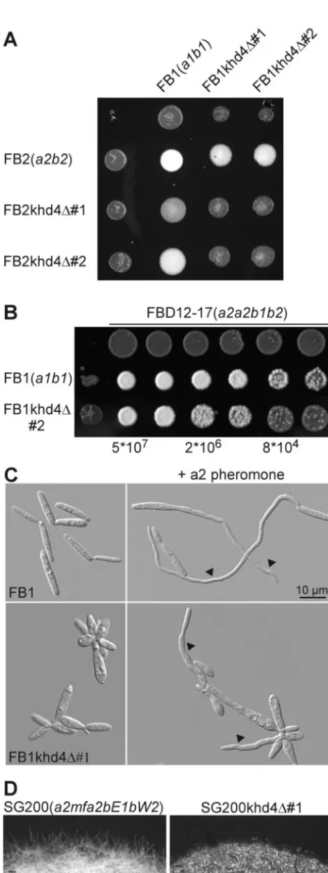
notypes. First, mutants showed a twofold longer doubling time of 330 min with respect to wild-type strains (Materials and Methods) (see Fig. S1 in the supplemental material). Second, mating assays on activated charcoal-containing plates revealed an almost complete absence of dikaryotic hyphae in crosses of compatible khd4deletion strains and a significant reduction whenkhd4⌬strains were cospotted with wild-type strains, in-dicative of defects in pheromone response and filamentation (Fig. 3A). Third, to test whether pheromone secretion was disturbed inkhd4⌬strains, serial dilutions were cospotted with the diploid pheromone tester strain FBD12-17 (a2a2b1b2) on plates containing activated charcoal. In response to a1 phero-mone, FBD12-17 switches to filament formation without prior cell fusion (5). khd4 deletion mutants were significantly re-duced in elicitation of filament formation in FBD12-17, indi-cating reduced pheromone production (Fig. 3B). Fourth, to assay conjugation tube formation, wild-type as well askhd4⌬ strains were incubated for 6 h in liquid CM with synthetic a1 or a2 pheromone (Materials and Methods) (62). Untreated khd4⌬ cells were defective in cytokinesis, often shaped like lemons or droplets, and showed an increased diameter with respect to the wild type (Fig. 3C). Upon pheromone stimula-tion, only a fewkhd4⌬cells responded by formation of conju-gation tubes, and these were substantially shorter and thicker than the wild type (Fig. 3C).
Fifth, to assay the effects of Khd4 onb-dependent filamen-tation, khd4 was deleted in strain SG200 (a1mfa2bE1bW2). This strain grows filamentously on activated charcoal-contain-ing plates, because autocrine pheromone stimulation triggers expression of an active bE1/bW2 heterodimer (8). SG200 strains carrying a deletion in khd4 exhibited a substantially decreased capability to form filaments on activated charcoal-containing plates compared to SG200 (Fig. 3D).
Sixth, to test for virulence ofkhd4⌬strains, 7-day-old corn
seedlings were infected with a cross of compatible deletion strains (Materials and Methods). Formation of tumors was scored after 2 weeks and compared to that of the wild type. Mixtures of FB1khd4⌬#1 with FB2khd4⌬#1 and FB1khd4⌬#2 with FB2khd4⌬#2 showed 16 and 27% tumor formation, respectively. This was approximately fourfold lower than the tumor formation of a mixture of wild-type strains (80% tumor formation in FB1⫻ FB2 crosses; Table 4). In summary, loss of Khd4 results in re-duced pheromone secretion, conjugation tube formation, andb -dependent filamentation, as well as pathogenicity.
rrm4⌬strains are reduced in filamentous growth and
viru-lence. According to our initial phenotypic analysis, rrm4⌬
strains were slightly affected in mating (Table 3). Since the mating defects were only subtle, we performed more elaborate assays. Cospotting of strain FB2rrm4⌬#1 with serial dilutions of either FB1 (a1b1) or FB1rrm4⌬#1 on activated charcoal-containing plates confirmed that rrm4 deletion strains were impaired in mating (Fig. 4A, top), which could be due to defects ina-dependent pheromone response or inb-dependent filamentation. To test pheromone response, respective strains were cospotted in serial dilutions with diploid pheromone tester strain FBD12-17 (a2a2b1b2) (5). We observed no dif-ference between elicitation of pheromone-induced filamenta-tion of the diploid tester strain by either the wild type or FB1rrm4⌬#1 (Fig. 4B). We also tested conjugation tube for-mation by treatment of FB1rrm4⌬#1/#2 or FB2rrm4⌬#1/#2 with synthetic a1 or a2 pheromone (62) and could not observe any difference from the wild type (data not shown). To assay for postfusion defects in filament formation, we addressed whether wild-type strains were able to complement the mating defect ofrrm4⌬strains. Therefore, we cospotted FB2 (a2b2) with serial dilutions of either FB1 (a1b1) or FB1rrm4⌬#1. The observation that there was no difference between the wild type and FB1rrm4⌬#1 (Fig. 4C) indicated that wild-type strains are
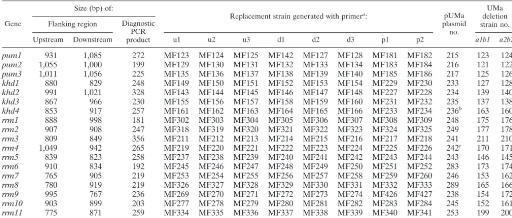
TABLE 2. Deletion of genes encoding RNA-binding proteins
Gene
Size (bp) of:
Replacement strain generated with primera: pUMa
plasmid no.
UMa deletion strain no. Flanking region Diagnostic
PCR product
Upstream Downstream u1 u2 u3 d1 d2 d3 p1 p2 a1b1 a2b2
pum1 931 1,085 272 MF123 MF124 MF125 MF142 MF127 MF128 MF181 MF182 215 123 124
pum2 1,055 1,000 199 MF129 MF130 MF131 MF132 MF133 MF134 MF183 MF184 216 121 122
pum3 1,011 1,056 225 MF135 MF136 MF137 MF138 MF139 MF140 MF185 MF186 217 125 126
khd1 880 829 248 MF149 MF150 MF151 MF152 MF153 MF154 MF229 MF230 233 127 128
khd2 991 1,021 328 MF143 MF144 MF145 MF146 MF147 MF148 MF227 MF228 234 139 140
khd3 867 966 230 MF155 MF156 MF157 MF158 MF159 MF160 MF231 MF232 235 137 138
khd4 853 917 257 MF161 MF162 MF163 MF164 MF165 MF166 MF233 MF234 236b 163 160
rrm1 888 998 181 MF302 MF303 MF304 MF305 MF306 MF307 MF308 MF309 248 175 176
rrm2 907 908 247 MF318 MF319 MF320 MF321 MF322 MF323 MF324 MF325 249 177 178
rrm3 809 849 356 MF211 MF212 MF213 MF214 MF215 MF216 MF217 MF218 241 211 210
rrm4 1,049 942 265 MF219 MF220 MF221 MF222 MF223 MF224 MF225 MF226 242c 170 171
rrm5 839 823 258 MF237 MF238 MF239 MF240 MF241 MF242 MF243 MF244 243 146 145
rrm6 910 834 192 MF245 MF246 MF247 MF248 MF249 MF250 MF251 MF252 283 173 174
rrm7 765 905 219 MF253 MF254 MF255 MF256 MF257 MF258 MF259 MF260 246 153 162
rrm8 780 919 219 MF326 MF327 MF328 MF329 MF330 MF331 MF332 MF333 289 165 166
rrm9 995 767 236 MF269 MF270 MF271 MF272 MF273 MF274 MF426 MF427 238 154 172
rrm10 903 899 203 MF277 MF278 MF279 MF280 MF281 MF282 MF283 MF284 245 152 161
rrm11 775 871 259 MF334 MF335 MF336 MF337 MF338 MF339 MF340 MF341 253 199 200
a
The sequence of the oligonucleotide primers is given in Table S1 in the supplemental material.
b
This plasmid was also used to generate UMa205#1 and -#2 (a1mfa2bE1bW2, khd4⌬), using SG200 as a progenitor.
c
This plasmid was used to generate UMa212#1 and -#2 (a1mfa2bE1bW2, rrm4⌬), using SG200 as a progenitor.
on September 8, 2020 by guest
http://ec.asm.org/
able to rescue the mating defect ofrrm4⌬strains, suggesting a role for Rrm4 in postfusion events. For verification, we deleted rrm4 in strain SG200 (a1mfa2bW2bE1) (8). SG200rrm4⌬#1 and -#2 strains produced only short filaments compared to the SG200 progenitor (Fig. 4D). These data illustrate that Rrm4 is not drastically affected in pheromone response but is crucial forb-dependent filamentation.
To assay pathogenicity, compatible rrm4⌬ strains were mixed (FB1rrm4⌬#1 with FB2rrm4⌬#1 and FB1rrm4⌬#2 with FB2rrm4⌬#2) and more than 360 corn seedlings each were infected in five independent infection experiments (Ma-terials and Methods; Fig. 5B). We observed that, in mixtures of rrm4⌬strains, the number of wilted or dead plants decreased approximately 1 order of magnitude in comparison to the wild type, whereas the number of plants without symptoms or with minor leaf tumors increased approximately threefold (Fig. 5B). Thus,rrm4⌬strains are still able to form tumors, but virulence is strongly affected.
DISCUSSION
Research on RNA-binding proteins in filamentous fungi is still in its infancy, and only a few examples of their biological roles exist (53, 65), such as regulation ofareAmRNA stability by nitrogen metabolite signaling inAspergillus nidulans(43, 50) or translational regulation by small upstream ORFs in arg-2 andcpcAmRNA fromN. crassaandA.nidulans, respectively (19, 23). Here, we addressed the roles of the PUM-, KHD-, and RRM-type RNA-binding proteins inU.maydis, through an approach that combined bioinformatics with reverse genet-ics. A substantial part of our study is a detailed phenotyping
TABLE 3. Phenotyping of strains carrying deletions in genes encoding proteins of the PUM, KHD, or RRM type
Gene
Morphology and pigmentation of
coloniesa
Growth on plates
Doubling timeb
Cell morphologyc
cAMP
responsed Matinge Pathogenicityf
CM at 28°C
NM
28°C 15°C 34°C
pum1 wt wt wt wt wt wt wt wt wt wt
pum2 wt wt wt wt wt wt wt wt wt wt
pum3 wt wt wt wt wt wt wt wt wt wt
khd1 wt wt wt Cold sensitive wt wt wt wt wt wt
khd2 wt wt wt wt wt wt wt wt wt wt
khd3 wt wt wt wt wt wt wt wt wt wt
khd4 wt wt wt wt wt Increased Aberrant Aberrant Reduced Reduced
rrm1 wt wt wt wt wt wt wt wt wt wt
rrm2 wt wt wt wt wt wt wt wt wt wt
rrm3 wt wt wt wt wt wt wt wt wt wt
rrm4 wt wt wt wt wt wt wt wt Reduced Reducedg
rrm5 wt wt wt wt wt wt wt wt wt wt
rrm6 wt wt wt wt wt wt wt wt wt wt
rrm7 wt wt wt wt wt wt wt wt wt wt
rrm8 wt wt wt wt wt wt wt wt wt wt
rrm9 wt wt wt wt wt wt wt wt wt wt
rrm10 wt wt wt wt wt wt wt wt wt wt
rrm11 wt wt wt wt wt wt wt wt wt wt
aMorphology and pigmentation were scored on CM and nitrate minimal medium plates (Materials and Methods). wt, comparable to wild type. bDoubling time was measured during exponential growth of strains in liquid CM (see Fig. S1 in the supplemental material).
cCell morphology was determined by differential interference contrast light microscopy of cells grown in liquid CM.
dcAMP response was analyzed by quantifying the cytokinesis defect of strains grown in the presence of 15 mM cAMP (see Fig. S2 in the supplemental material). eMating was investigated by cospotting of compatible haploid deletion strains on plates containing activated charcoal.
fPathogenicity was determined by infection of corn seedlings with mixtures of compatible deletion strains (Table 4). gPathogenicity was scored with a more detailed disease rating (Fig. 5B).
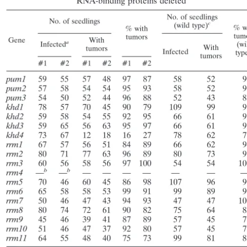
TABLE 4. Pathogenicity of strains with genes encoding RNA-binding proteins deleted
Gene
No. of seedlings
% with tumors
No. of seedlings (wild type)c
% with tumors (wild type)c
Infecteda With
tumors Infected With tumors #1 #2 #1 #2 #1 #2
pum1 59 55 57 48 97 87 58 52 90
pum2 57 58 54 54 95 93 58 52 90
pum3 54 50 52 44 96 88 52 43 83
khd1 78 57 70 45 90 79 109 99 91
khd2 59 58 54 55 92 95 66 61 92
khd3 59 65 56 63 95 97 66 61 92
khd4 73 67 12 18 16 27 78 62 79
rrm1 67 57 56 51 84 89 66 62 94
rrm2 80 71 77 63 96 89 80 73 91
rrm3 60 56 58 56 97 100 54 54 100
rrm4 —b —b — — — — — — —
rrm5 70 46 60 45 86 98 107 96 90
rrm6 65 58 58 53 99 91 99 89 90
rrm7 50 46 47 43 94 93 47 47 100
rrm8 80 74 72 61 90 82 75 64 85
rrm9 45 46 39 41 87 89 57 45 79
rrm10 51 46 47 37 92 80 57 45 79
rrm11 64 55 48 40 75 73 99 81 82
aCrossings of two compatible deletion strains: e.g., FB1khd4⌬#1 ⫻
FB2khd4⌬#1 or FB1khd4⌬#2⫻FB2khd4⌬#2.
b—, pathogenicity ofrrm4⌬strains was scored with a more elaborate disease
rating (Fig. 5B).
cCrossings of two compatible wild-type strains FB1⫻FB2 were carried out in
parallel to validate the infection rate; identical numbers indicate experiments in parallel.
on September 8, 2020 by guest
http://ec.asm.org/
regimen custom tailored for the biology of this fungus. To our knowledge, this is the first example of an extensive reverse genetic approach in filamentous fungi since the postgenomic era has been launched. We succeeded in the identification of two RNA-binding proteins implicated in pathogenic develop-ment: a KHD protein that belongs to a novel family of KHD proteins and a so far unique ELAV (embryonic lethal abnor-mal vision)-like RRM protein.
RNA-binding proteins of the PUM type in U. maydis. U.
maydiscontains five PUM-type proteins, and it is very likely that these are all members of this particular family, since the primary sequence and iterative nature of Pumilio-like repeats are very conserved (70). This notion is supported by a phylo-genetic analysis of fungal PUM-type proteins. Representatives from U. maydis are present in all four clusters and in an additional region of nonclustering sequences. All five proteins fall into separate branches of the dendrogram, indicating that they did not arise by recent gene duplication. This is likely the case for other proteins such as Puf1p and Puf2p fromS. cer-evisiae(Fig. 6A).
Our phenotypic analysis of pum1to-3deletion strains re-vealed no difference from wild-type strains. The fact that PUM protein-encoding genes are not essential for viability is consis-tent with previous work inS. cerevisiae. There, it was demon-strated that deletion mutants of each of the five PUM-type proteins as well as a corresponding quintuple-deletion mutant were viable at temperatures ranging from 18 to 37°C (48). However, the observation that none of the tested deletion strains was affected in mating, cAMP response, or pathogenic-ity was surprising since connections between pheromone re-sponse and cAMP signaling have been reported for PUM-type proteins. For example, loss of Puf5p/Mpt5p in S. cerevisiae results in a temperature-sensitive growth phenotype, an in-crease in pheromone sensitivity, and a defect in recovery from pheromone-induced cell cycle arrest (14). PufA fromD. dis-coideumhas been implicated in translational control ofpkaC mRNA encoding the catalytic subunit of PKA (60). The lack of phenotypes in our assays might be due to insufficient sensitiv-ity, redundancy, or partly overlapping functions. This has been reported for Puf4p and Puf5p during regulation of life span in S. cerevisiae(30). In addition, we cannot rule out the possibility that other PUM-type proteins fromU.maydis, such as Pum4 or Pum5, are involved in mating or cAMP signaling.
RNA-binding proteins of the KHD type in U. maydis.
Ac-cording to our analysis, nine KHD-type proteins are present in
FIG. 3.khd4⌬ strains are impaired in mating and morphology. (A) Strains indicated on the left were coinoculated on activated char-coal-containing plates. The strains are indicated at the top. Successful mating events can be scored as the amount of aerial hyphae resulting
in white, fuzzy colonies. (B) Serial fivefold dilutions (given at the bottom) of strains indicated on the left were cospotted with diploid pheromone tester strain FBD12-17 (a2a2b1b2; 5⫻107cells/ml; top).
Pheromone secretion is direct proportional to filamentation of the pheromone tester strain. (C) Formation of conjugation tubes (arrow-heads) was observed by differential interference contrast light micros-copy. FB1 (a1b1; top) or FB1kdh4⌬#1 (bottom) was incubated in the absence (left) or presence (right) of compatible synthetic a2 phero-mone (top) (62) for 5 h in liquid CM. (D) The strains indicated at the top were grown on plates containing activated charcoal. Formation of filaments was visible at the edge of the colony. These strains grew filamentously on plates containing activated charcoal due to autocrine stimulation of pheromone-induced expression of compatible bE/bW transcription factors.
on September 8, 2020 by guest
http://ec.asm.org/
U. maydis. This number is comparable to the numbers of KHD proteins fromS. cerevisiaeand S. pombe(10 and 8 proteins, respectively; http://smart.embl.de/), and most of the KHD pro-teins fromU. maydishave related counterparts inS. cerevisiae (Table 1). The phenotypic analysis of khd1 to -4 deletion strains revealed that khd1⌬ strains exhibit a cold-sensitive growth phenotype, while growth ofkhd2⌬strains was not
af-fected by temperature. This indicates that Khd1 and Khd2 have distinct biological roles, which is in accordance with the fact that the region of high sequence similarity is restricted to their three KHD RNA-binding domains (20% overall se-quence identity, but the first, second, and third KHD domains are 35, 43, and 52% identical in sequence, respectively). Loss of Khd4 affected growth rate, morphology, pheromone re-sponse, filamentous growth, and pathogenicity. At present it is not clear whether these pleiotropic defects are due to altered regulation of a number of target mRNAs or whether these
FIG. 4.rrm4⌬strains are impaired in filamentous growth. Strains indicated on the left were cospotted in serial fivefold dilutions (below each plate) with compatible haploid strains (A) and (C) or diploid pheromone tester strains (B; above each plate; both 5⫻107cells/ml)
on plates containing activated charcoal. Successful mating events (A) and (C) or pheromone-induced filamentation of the tester strain (B) was visible by the formation of white, fuzzy colonies. (D) The strains indicated at the top were grown on plates containing activated charcoal. Formation of filaments was visible at the edge of the colony. These strains grew filamentously on plates containing activated char-coal due to autocrine stimulation of pheromone-induced expression of active bE/bW transcription factors.
FIG. 5.rrm4⌬strains are reduced in virulence. Plant infection ex-periments were carried out by crossing either compatible wild-type (FB1⫻FB2; 369 plants infected) or compatiblerrm4⌬(FB1rrm4⌬#2 ⫻FB2rrm4⌬#2; 368 and 403 plants infected, respectively) strains. In panel A, typical disease symptoms on leaves of corn seedlings are shown. In panel B, a detailed disease rating is given adapted from earlier work (see Materials and Methods and reference 20). The num-bers of plants (percentage) that belong to the following disease cate-gories were scored: plants with no tumor, plants with minor leaf tu-mors, plants with major leaf tutu-mors, plants with minor stem tutu-mors, plants with major stem tumors, and wilted or dead plants. The defini-tions for “major” and “minor” are given in Materials and Methods. The labeling below each bar indicates which mixtures of strains were tested: wt (wild type), FB1 ⫻ FB2; bars 1, FB1rrm4⌬#1 ⫻ FB2rrm4⌬#1; and bars 2, FB1rrm4⌬#2⫻FB2rrm4⌬#2.
on September 8, 2020 by guest
http://ec.asm.org/
phenotypes are connected through the same key component regulated by Khd4.
Comparison to sequences from other model fungi such asA. nidulans, N. crassa, Fusarium graminearum, Magnaporthe grisea, C. neoformans, andC. cinereusrevealed that Khd4 is part of a novel group of KHD proteins that are related to multi-KHD proteins of the Vigilin type. Interestingly, mem-bers of this group are absent inS. pombeandS. cerevisiae(Fig. 6B). The closest relatives fromS. cerevisiaeand mammals are the multi-KHD proteins Scp160p and Vigilin, respectively, containing 14 KHDs each. This type of protein is also found in U. maydis (Khd5), as well as in the other fungi mentioned above (Fig. 6B). Interestingly, loss of Scp160p fromS. cerevi-siae results in abnormal morphology (72), and recently this RNA-binding protein has been described as being involved in mating, since it interacts with the heterotrimeric G protein Gpa1p during the pheromone response (22). It will be
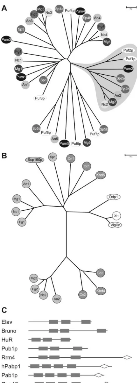
inter-FIG. 6. (A) PUM-type proteins in fungal model organisms. An unrooted dendrogram was created by the distance-based minimum-evolution method, based on 1,000 replicates (Materials and Methods). PUM-type protein sequences fromS.cerevisiae,S.pombe(Sp), and fungi were used, whose genomes were sequenced as part of the fungal genome initiative (A.nidulans, An;F.graminearum, Fg;M.grisea, Mg;
N.crassa, Nc; andU.maydis, Um). Proteins are either symbolized by ovals in the case ofS.cerevisiae(Puf1p to Puf7p) andU. maydis(Pum1 to Pum5) or by circles with different shading in case of all other fungi. Two proteins each fromC. neoformans(Cn) andC. cinereus(Cc) were omitted from the analysis because a reasonable gene prediction was not possible due to the presence of several introns. The area shaded in gray depicts PUM proteins with one or more RRMs. The accession numbers are as follows: An1, AN6587.2; An2, AN7474.2; An3, AN4285.2; An4, AN0320.2; An5, AN1235.2; Fg1, FG06710.1; Fg3, FG05203.1; Fg4, FG00841.1; Mg1, MG04985.4; Mg2, MG06318.4; Mg3, MG03158.4; Mg4, MG06982.4; Mg5, MG03872.4; Nc2, NCU06199.1; Nc3, NCU01760.1; Nc4, NCU01775.1; Puf3p (S. cerevi-siae), Q07807; Puf2p (S. cerevisiae), NP_015367; Puf1p/Jsn1p (S. cer-evisiae), P47135; Puf4p (S. cerevisiae), P25339; Puf5p/Mpt5p/Uth4p (S. cerevisiae), P39016; Puf6p (S. cerevisiae), Q04373; Puf7p (S. cerevisiae), P47077; Sp1, NP_593141; Sp2a, CAA18887; Sp2b, NP_595389; Sp3a, NP_593687; Sp3b, Q10238; Sp4a, Q09829; Sp4b, Q92359; Sp5a, NP_588564; Sp5b, Q92347; Pum2, UM00268.1; Pum3, UM05467.1; Pum4, UM03431.1; and Pum1 and Pum5, this study (Table 1). The scale bar denotes the number of substitutions per site. (B) Multi-KHD proteins in eukaryotes. An unrooted tree was derived as in panel A by aligning multi-KHD-type proteins from the same organisms as in panel A as well asD.melanogaster(Dm);Xenopus laevis, (Xl), andH. sapiens
(Hs). Proteins are either symbolized by ovals (Scp160p,S. cerevisiae; Ddp1,D. melanogaster; Vigilin,H. sapiens; and Khd4,U. maydis) or circles. Light gray, dark gray, or open symbols indicate the origin from ascomycetes, basidiomycetes, and higher eukaryotes, respectively. Ac-cession numbers: An1, AN2068.2; An2, AN6326.2; Ddp1 (D. melano-gaster), CAB52798; Fg1, FG09491.1; Fg2, FG06181.1; Vigilin (H. sa-piens), Q00341; Mg1, MG06496.4; Mg2, MG01781.4; Nc1, CAB91673; Nc2, XP_324623; Scp160p (S. cerevisiae), CAA46597; Sp1, NP_588106; Xl1, AAH44314; Khd4 and Khd5, this study (Table 1). The sequences of two proteins each from C. neoformans (Cn1 and Cn2) and C. cinereus(Cc1 and Cc2) were derived by conceptual translation (nucle-otide positions and predicted introns are given in Materials and Meth-ods). (C) Domain organization of ELAV-like and PABP proteins. RRM and PABC domains are given as filled rectangles and open diamonds, respectively. Accession numbers: ELAV (D. melanogaster), P16914; Bruno (D. melanogaster), AAB58464; HuR (H. sapiens), AAH03376; Pub1p (S. cerevisiae), NP014382; hPabp1 (H. sapiens), P11940; Pab1p, (S. cerevisiae), P04147; and Rrm4 and Rrm12, this study (Table 1).
on September 8, 2020 by guest
http://ec.asm.org/
esting to determine whether inU. maydismulti-KHD proteins such as Khd4 and Khd5 share functions in regulating morpho-genesis and pheromone signaling.
RNA-binding proteins of the RRM type in U. maydis. An
automated feature search predicted the presence of 43 RRM proteins in U. maydis (http://www.broad.mit.edu/cgi-bin /annotation/fungi/ustilago_maydis/findfeatures.cgi), a number that is close to the 53 RRM proteins fromS. cerevisiae(http: //smart.embl.de/). Phenotyping strains carrying a deletion in a subset of 11 RRM protein-encoding genes revealed that only the loss of Rrm4 resulted in discernible phenotypes. This was surprising since we would have expected more RRM proteins to be important for developmental processes inU. maydis. For example, Rrm8 shares the same domain organization as well as similar flanking sequences to the negative regulator of devel-opment Nrd1 from S. pombe. Loss of Nrd1 function causes cells to initiate sexual development in the absence of nutrient starvation (64). In contrast,rrm8⌬strains fromU. maydiswere not drastically disturbed in mating or pathogenicity.
In the case of Rrm4, we showed that neither pheromone secretion nor pheromone-responsive conjugation tube forma-tion was significantly disturbed. However, loss of Rrm4 nega-tively affects dikaryotic filaments as well as filamentation of a solopathogenic strain. This defect is likely to be the cause for the reduced disease symptoms, since it is conceivable that shorter filaments have difficulties in finding an appropriate entry site for plant penetration.
Rrm4 displays a novel domain organization that is so far unique in eukaryotes (Fig. 6C). At its C terminus, it contains a PABC domain that was initially found in poly(A)-binding pro-teins. However, this domain is also associated with a HECT domain (homologous to the E6-AP carboxyl terminus) found in ubiquitin-protein ligases and implicated as a protein-protein interaction interface (32, 39). The PABC domain in human poly(A)-binding protein contains four amino acids (F22, I25, A33, and K35) shown to form a deep hydrophobic pocket that could function as a peptide binding domain (32). Strikingly, all four residues are conserved in Rrm4. In its N terminus, Rrm4 contains three RRM domains with typical spacing known from ELAV-like proteins (3), such as Elav or Bruno fromD. mela-nogaster, HuR fromH.sapiens, and Pub1p fromS. cerevisiae. All of these proteins contain two RRM domains juxtaposed with each other and separated from a third by a hinge region of variable length (Fig. 6C). Members of this family of proteins have been described to function as sequence-specific RNA-binding proteins regulating mRNA translation or stability (3). For example, Bruno represses translation of Oskar, a determi-nant for posterior body patterning in embryos fromD. mela-nogaster, by binding to Bruno response elements in the 3⬘ -untranslated region of theoskmRNA (47, 68, 71). HuR from humans has been implicated in regulating AUUUA-mediated mRNA decay (16, 46). Pub1p from S. cerevisiae recognizes stabilizer elements and thereby protects mRNAs that contain upstream ORFs from degradation by the nonsense-mediated decay pathway (52).
According to sequence similarity, we hypothesize that Rrm4 is a sequence-specific RNA-binding protein that might be in-volved in regulation of mRNA splicing, transport, translation, or stability. For two reasons, we do not believe that Rrm4 functions as poly(A)-binding protein. Rrm4 does not contain
the typical four RRM domains in its N terminus, and in vitro binding experiments revealed that Rrm4 does not bind to poly(A) or poly(U) but recognizes specific sequences enriched by SELEX (C. Julius and M. Feldbru¨gge, unpublished obser-vation). A much better candidate for a poly(A)-binding protein in U. maydis is Rrm12, since it contains the characteristic domain organization (Fig. 6C) and exhibits high sequence sim-ilarity to other PABPs over its entire length (comparison to Pab1p from S. cerevisiaeresults in a BLAST expect value of e⫺132).
Conclusions.A great surprise from our study of
RNA-bind-ing proteins inU. maydiswas the fact that, despite our exten-sive phenotypic analysis, the majority of gene replacement mutants did not exhibit any discernible phenotypes. A possible explanation would be functional redundancy of related RNA-binding proteins. Alternatively, it could be possible that our assays were not sensitive enough or that we did not test the affected phenotype. Nevertheless, we were able to identify 2 out of 18 RNA-binding proteins implicated in pathogenic de-velopment ofU. maydis. One of them is a founding member of a new group of KHD proteins, and the other is a so far unique ELAV-like RRM protein. Investigation of the subcellular lo-calization of these candidates in combination with identifica-tion of specifically recognized target RNAs will allow us to unravel the connection between the mode of action of these RNA-binding proteins and pathogenic development. For ex-ample, nuclear localization would indicate that they function in mRNA maturation or export. We expect to find within the group of target RNAs important determinants of pathogenic development whose expression is manifested at the posttran-scriptional level. The involvement of RNA-binding proteins in regulation of pathogenic development supports our initial as-sumption predicting fundamental similarities to developmental programs of higher eukaryotes.
ACKNOWLEDGMENTS
We acknowledge A. Brachmann, P. Mu¨ller, R. Kahmann, and STaR laboratory members for valuable discussions and critical reading of the manuscript. We thank S. Hester and J. Hohenner for excellent tech-nical assistance. We are grateful to Julian Ko¨nig for generating gene replacement constructs.
This work was supported by Bayer CropScience.
REFERENCES
1.Abovich, N., and M. Rosbash.1997. Cross-intron bridging interactions in the yeast commitment complex are conserved in mammals. Cell89:403–412. 2.Altschul, S. F., W. Gish, W. Miller, E. W. Myers, and D. J. Lipman.1990.
Basic local alignment search tool. J. Mol. Biol.215:403–410.
3.Antic, D., and J. D. Keene.1997. Embryonic lethal abnormal visual RNA-binding proteins involved in growth, differentiation, and posttranscriptional gene expression. Am. J. Hum. Genet.61:273–278.
4.Banuett, F.1995. Genetics ofUstilago maydis, a fungal pathogen that induces tumors in maize. Annu. Rev. Genet.29:179–208.
5.Banuett, F., and I. Herskowitz.1989. Differentaalleles are necessary for maintenance of filamentous growth but not for meiosis. Proc. Natl. Acad. Sci. USA86:5878–5882.
6.Birney, E., S. Kumar, and A. R. Krainer. 1993. Analysis of the RNA-recognition motif and RS and RGG domains: conservation in metazoan pre-mRNA splicing factors. Nucleic Acids Res.21:5803–5816.
7.Bo¨lker, M.2001.Ustilago maydis—a valuable model system for the study of fungal dimorphism and virulence. Microbiology147:1395–1401.
8.Bo¨lker, M., S. Genin, C. Lehmler, and R. Kahmann.1995. Genetic regula-tion of mating, and dimorphism inUstilago maydis. Can. J. Bot.73:320–325. 9.Bo¨lker, M., M. Urban, and R. Kahmann.1992. Theamating type locus ofU.
maydisspecifies cell signaling components. Cell68:441–450.
10.Brachmann, A., J. Ko¨nig, C. Julius, and M. Feldbru¨gge.Reverse genetic approach for generating gene replacement mutants inUstilago maydis.Mol. Genet. Genomics272:216–226.