Disruption of Higher Order DNA Structures in
Friedreich’s Ataxia (GAA)
n
Repeats by PNA or
LNA Targeting
Helen Bergquist1,2, Cristina S. J. Rocha2, Rube´n A´ lvarez-Asencio3¤, Chi-Hung Nguyen4, Mark. W. Rutland3, C. I. Edvard Smith2, Liam Good5, Peter E. Nielsen6, Rula Zain2,7*
1 Department of Medical Biochemistry and Microbiology, Microbiology-Immunology, Uppsala University, Uppsala, Sweden, 2 Department of Laboratory Medicine, Clinical Research Center, Karolinska Institutet, SE-141 86, Huddinge, Sweden, 3 KTH Royal Institute of Technology, School of Chemical Science and Engineering, Department of Chemistry, Stockholm, Sweden, 4 Laboratoire de Pharmacochimie, Institut Curie, PSL Research University, UMR 9187 – U 1196 CNRS-Institut Curie, INSERM, Centre Universitaire, Orsay, France, 5 Department of Pathology and Infectious Diseases, Royal Veterinary College, University of London, United Kingdom, 6 Department of Cellular and Molecular Medicine, Faculty of Health and Medical Sciences, University of Copenhagen, The Panum Institute, Copenhagen, Denmark, 7 Department of Clinical Genetics, Centre for Rare Diseases, Karolinska University Hospital, SE-171 76, Stockholm, Sweden
¤ Current address: Fundacio´n IMDEA Nanociencia, Faraday, 9, Campus Universitario de Cantoblanco, Madrid, Spain.
*rula.zain@ki.se
Abstract
Expansion of (GAA)nrepeats in the first intron of the Frataxin gene is associated with
reduced mRNA and protein levels and the development of Friedreich’s ataxia. (GAA)n
expansions form non-canonical structures, including intramolecular triplex (H-DNA), and R-loops and are associated with epigenetic modifications. With the aim of interfering with higher order H-DNA (like) DNA structures within pathological (GAA)nexpansions, we
exam-ined sequence-specific interaction of peptide nucleic acid (PNA) with (GAA)nrepeats of
dif-ferent lengths (short: n=9, medium: n=75 or long: n=115) by chemical probing of triple helical and single stranded regions. We found that a triplex structure (H-DNA) forms at GAA repeats of different lengths; however, single stranded regions were not detected within the medium size pathological repeat, suggesting the presence of a more complex structure. Furthermore, (GAA)4-PNA binding of the repeat abolished all detectable triplex DNA
struc-tures, whereas (CTT)5-PNA did not. We present evidence that (GAA)4-PNA can invade the
DNA at the repeat region by binding the DNA CTT strand, thereby preventing non-canoni-cal-DNA formation, and that triplex invasion complexes by (CTT)5-PNA form at the GAA
repeats. Locked nucleic acid (LNA) oligonucleotides also inhibited triplex formation at GAA repeat expansions, and atomic force microscopy analysis showed significant relaxation of plasmid morphology in the presence of GAA-LNA. Thus, by inhibiting disease related higher order DNA structures in the Frataxin gene, such PNA and LNA oligomers may have poten-tial for discovery of drugs aiming at recovering Frataxin expression.
a11111
OPEN ACCESS
Citation: Bergquist H, Rocha CSJ, A´lvarez-Asencio R, Nguyen C-H, Rutland M.W, Smith CIE, et al. (2016) Disruption of Higher Order DNA Structures in Friedreich’s Ataxia (GAA)nRepeats by PNA or
LNA Targeting. PLoS ONE 11(11): e0165788. doi:10.1371/journal.pone.0165788
Editor: Rakesh N. Veedu, University of Queensland, AUSTRALIA
Received: June 9, 2016
Accepted: October 7, 2016
Published: November 15, 2016
Copyright:©2016 Bergquist et al. This is an open access article distributed under the terms of the Creative Commons Attribution License, which permits unrestricted use, distribution, and reproduction in any medium, provided the original author and source are credited.
Data Availability Statement: All relevant data are within the paper and its Supporting Information files.
Funding: This work was financially supported by The Knut och Alice Wallenbergs Stiftelse, Magnus Bergvalls Stiftelse, Carl Tryggers Stiftelse fo¨r Vetenskaplig Forskning, The Swedish Research Council and La¨ndells stiftelse fo¨r neurologisk forskning.
Introduction
Friedreich’s ataxia (FRDA) is the most common inherited autosomal recessive ataxia, and in >96% of cases the disease is correlated by expansion of (GAA)nrepeats in the first intron of
theFrataxingene (FXN) [1,2]. Disease-associated expanded alleles consist of approximately 70 to more than 1000 repeats [1–3]. The (GAA)nexpansions result in a substantial reduction in
FrataxinmRNA and protein levels [4], and non-symptomatic carriers (heterozygous for the expanded allele) show ~50% reduction [1,3–5]. Frataxin deficiency causes excessive free radi-cal production, dysfunction of Fe-S center containing enzymes, and progressive iron accumu-lation in mitochondria [6]. Several studies have focused on increasing the level ofFrataxin through targeting the epigenetic regulation of gene expression. Unfortunately, there are no FRDA therapeutics available, only symptomatic management strategies.
Expanded (GAA)nrepeats readily form non-canonical (non-B-DNA) structures including
intramolecular triplex structures (H-DNA) (Fig 1), and R-loops, which have also been pro-posed to be of importance in other triplex-repeat disorders [7]. Several models have been pro-posed for alternative structures formed at expanded (GAA)nrepeats [8–12]. Formation of a
higher order structure named “sticky DNA” has been reported in (GAA)ncontaining plasmids
and the structure was analyzed using electron microscopy [8,13–16]. It is believed that the structural properties of (GAA)nrepeats that lead to the formation of higher order structures
also affect the genomic stability of the repeat length as well as the expression ofFrataxin[17–
19]. Long (GAA)nrepeats stall replication inSaccharomyces cerevisiae[20] and inhibit
tran-scription bothin vitro[5,21] and in mammalian cells [13,22]. The observed effects on DNA replication and transcription are dependent on the length and orientation of the (GAA)n
repeats, which correlate with the predisposition of these repeats to form well-defined second-ary/tertiary DNA structures [7,23]. Finally, the expanded repeats are associated with silenced chromatin via DNA methylation and histone trimethylation and deacetylation in the adjacent regions [24,25].
An understanding of the DNA structural properties and chromatin modifications associ-ated with (GAA)nrepeats raises possibilities to reverseFrataxinsilencing. For example, histone
deacetylase inhibitors have been used to increaseFrataxinmRNA in FRDA mouse models and in patient cell lines [26–29]. Also, sequence-specific polyamides and low molecular weight minor groove binders enhanceFrataxinexpression [30,31]. Therefore, it is particularly inter-esting to develop specific (GAA)nrepeat targeting molecules to elucidate the possible
patho-logical structures formed at theFrataxin locus, which could facilitate the development of new therapeutic strategies.
The GAA expanded repeats in FRDA consist of large polypurine.polypyrimidine (R.Y) regions. In principle, these stretches can be specifically targeted by triplex forming oligonucle-otides (TFOs), which bind in the major groove of the dsDNA. This, so-called anti-gene strategy, can be used to modulate transcription at specificloci[32–34] and to induce recombi-nation [35,36] and repair [37–39]. For example, to enhance TFO binding of DNA under physi-ological conditions, modified nucleotides, such as morpholinophosphoroamidates (PMO), locked (LNA) or peptide nucleic acids (PNA) can be exploited [40].
Fig 1. Purine and pyrimidine H-DNA motifs formed at (GAA)nrepeats.
PNAs are DNA mimics having a peptide like backbone [41]. PNA binds to sequence comple-mentary DNA or RNA with high affinity and sequence specificity. More interestingly, PNA is able to invade dsDNA through binding of the purine (R) strand leaving the pyrimidine (Y) strand displaced. PNA was originally designed to bind dsDNA to form a triplex structure; however, it was soon discovered that an invasion mechanism is involved and several other PNA-DNA com-plexes can also be formed [41]. Binding of short homopyrimidine PNA to dsDNA leads mainly to formation of a triplex-invasion structure, of very high stability [42]. Formation of a triplex-invasion complex is slow and negatively affected by DNA duplex stabilizing conditions, such as physiological salt concentrations [43]. Formation of triplex-invasion structures requires two PNA molecules for Watson-Crick and Hoogsteen binding, respectively, of the target. To increase the rate of formation of a triplex-invasion complex, bis-PNAs, where two PNA molecules are linked together, have been developed [44]. Duplex invasion complexes have been reported for homo-purine PNAs [45] and also for backbone modified, high affinity PNAs [46] (e.g. gamma-PNAs [47]). Additionally, long homopyrimidine PNAs (>15 nucleotides) can form regular triplex structures (PNA-triplexes) at physiologically relevant salt concentrations [48], whereas stable PNA-triplex and triplex-invasion complexes have not been reported for homopurine PNAs.
Locked nucleic acid (LNA) is an RNA analogue having a 20-oxygen and 40 -carbon-methy-lene linkage. The presence of this bridge promotes a conformational restriction in LNA con-taining oligonucleotides, favoring duplex formation [49]. LNA modification has also been introduced in TFOs to increase triplex stability. Like PNA, LNA is also able to invade dsDNA through Watson-Crick hydrogen bond formation to the DNA complementary sequences or through combined Watson-Crick and Hoogsteen hydrogen bonds forming a bisLNA con-struct, analogous to bisPNA [50].
In a previous study on the formation of triplex structures at FRDA (GAA)nrepeats, we
showed that a low-molecular weight benzoquinoquinoxaline compound (BQQ), recognizes triplex structures formed at (GAA)nrepeats in plasmids. BQQ is a DNA intercalating
com-pound that specifically binds and stabilizes triplex structures of both purine and pyrimidine motifs [51–54]. Furthermore, BQQ is cell permeable, and we have shown that the compound binds and stabilizes H-DNA structures formed in plasmids in growingEscherichia colicells [55]. Additionally, we have converted BQQ to a triplex-specific cleaving agent (BQQ-OP) by conjugation to a 1,10-phenanthroline ligand [52]. In the presence of Cu2+and a reducing agent BQQ-OP causes dsDNA cleavage specifically at the site of formation of a triplex, and we have previously demonstrated the ability of BQQ-OP to probe triplex formation of both H-DNA and TFO-directed triplex structures in plasmidsin vitro[55,56].
Sequence-specific targeting of GAA repeats using synthetic single strand oligonucleotides (ONs) is an attractive approach to examine the molecular mechanisms of non-canonical DNA structure formation at FRDA repeat expansions. While modified ONs as mRNA targeting therapeutics have made major progress lately, also in relation to triplet-repeat diseases [57,58], successful DNA targeting of nucleotide repeats using ONs has not been reported. In this study we aimed to target the alternative DNA structures at FRDA (GAA)nrepeats using modified
ONs. We chose to examine PNA and LNA binding to these repeats owing to their ability to both invade dsDNA and to form triplex structure and thereby interfere with the formation of H-DNA and other higher order structures in FRDA expanded repeats. BQQ-OP mediated tri-plex-specific cleavage of dsDNA and chemical modification of ssDNA regions using chloroa-cetaldehyde were used to characterize the DNA structures and PNA-DNA complexes formed in short, medium and long (GAA)nrepeats. To assess global structural changes in plasmids
containing (GAA)nrepeats in the presence of LNA oligomers, we used Atomic Force
Materials and Methods
Plasmids
(GAA)n-containing plasmids, pMP179, pMP178 and pMP141, hereafter referred to as
pMP179(115 repeats), pMP178(75 repeats) and pMP141(9 repeats) to denote the number of repeats, were a kind gift from Prof. M. Pandolfo’s Laboratory. The plasmids are derived from pSPL3 and the inserts are all flanked by 352 and 256 bp of human genomic sequences 50and 30 of the (GAA)nrepeat, respectively [8,13].
PNA and LNA Oligomers
PNA oligomers were synthesized by solid phase Boc-chemistry, purified by RP-HPLC and characterized by MALDI-TOF mass spectrometry and HPLC [41]. LNA-PS oligomers were synthetized, purified by Reverse Phase - HPLC; quality control was accessed by MALDI-TOF mass spectrometry and bought from Eurogentec S.A.
BQQ-OP Triplex-Specific DNA Cleavage of DNA-PNA Complexes at
(GAA)
nRepeats
Plasmid pMP179(115 repeats) was linearized using ApaI, followed by DNA isolation using miniprep columns (Qiagen). 0.2μg linearized pM179(115 repeats) (2.2 nM) was incubated at 37˚C during 1 h in the presence of a 12-mer GAA-PNA (3320), a 15-mer CTT-PNA (3482) (10μM) or a 20-mer single strand CTT-DNA (4μM) (Table 1) in buffer (10 mM sodium caco-dylate, 100 mM NaCl and 0, or 2 mM MgCl2, pH 7.5). BQQ-OP (1.5μM) and CuSO4(2μM)
were premixed at room temperature for 15 min and then added to the plasmid. The mixture was left for 25 min at room temperature and mercaptopropionic acid (MPA, 2 mM, final vol-ume 20μl) was added to initiate the cleavage reaction, which was allowed to proceed for 2 h at 37˚C. Samples containing the crude reaction mixtures were then analyzed using 0.7% agarose gel electrophoresis (50 V, 1 h) and ethidium bromide staining. Gel-doc XR with Quantity One 4.5.2 software (Bio-Rad) was used for gel analysis and quantification of the gel bands. MassRu-ler (Fermentas) served as a molecular weight DNA ladder.
BQQ-OP Mediated Cleavage of H-DNA at (GAA)
115Repeats in the
Presence of PNA and LNA
Plasmid pMP179(115 repeats) (1μg, 11 nM) was incubated in buffer (10 mM sodium cacody-late, pH 7.5 and 100 mM NaCl and 2 mM MgCl2) at 37˚C during 2 h in the presence of 10μM
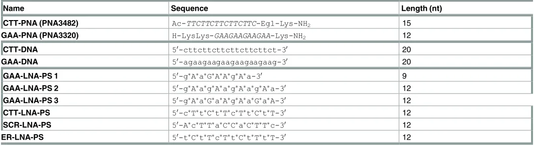
Table 1. PNA, DNA and LNA oligomers used in the study.
Name Sequence Length (nt)
CTT-PNA (PNA3482) Ac-TTCTTCTTCTTCTTC-Eg1-Lys-NH2 15
GAA-PNA (PNA3320) H-LysLys-GAAGAAGAAGAA-Lys-NH2 12
CTT-DNA 50-cttcttcttcttcttcttct-30 20
GAA-DNA 50-agaagaagaagaagaagaag-30 20
GAA-LNA-PS 1 50-gAaGAAgAa-30 9
GAA-LNA-PS 2 50-gAagAagAagAa-30 12
GAA-LNA-PS 3 50-gAaGaAgAaGaA-30 12
CTT-LNA-PS 50-cTtCtTcTtCtT-30 12
SCR-LNA-PS 50-AcTTaCCaCTTc-30 12
ER-LNA-PS 50-tCtTcTtCtTtT-30 12
of either a 12-mer GAA-PNA (PNA3320), a 15-mer CTT-PNA (PNA3482), a 9-mer GAA-L-NA-PS (GAA-LGAA-L-NA-PS 1), a 12-mer GAA-LGAA-L-NA-PS (GAA-LGAA-L-NA-PS 2 or 3), a 12-mer CTT-LNA-PS (CTT-CTT-LNA-PS), a 12-mer targeting the end of the repeat (ER-CTT-LNA-PS), a 20-mer sin-gle strand GAA-or a 20-mer CTT-DNA (Table 1). BQQ-OP (1μM) and CuSO4(1.5μM) were
premixed at room temperature (15 min) and then added to the plasmid solution. The mixture was left for 45 min at room temperature and mercaptopropionic acid (MPA, 2 mM, final vol-ume 20μl) was added to initiate the cleavage reaction. The reaction was allowed to proceed for 3 h at 37˚C, followed by isolation of DNA using miniprep columns (Qiagen). As a control, plasmid pMP179(115 repeats) was cleaved in the absence of ONs using BQQ-OP under similar experimental conditions. The isolated DNA was digested using ApaI (1 U, Promega) during 3 h at 37˚C and then analyzed using 0.7% agarose gel electrophoresis (50 V, 1 h) and ethidium bromide or SYBR-safe (Life Technologies) staining. Gel-doc XR with Quantity One 4.5.2 soft-ware (Bio-Rad) was used for gel analysis and quantification of the gel bands.
Analysis of DNA and DNA-PNA structure formation at short and long
(GAA)
nrepeats using BQQ-OP cleavage and CAA modification
In all probing reactions, the plasmid (pMP141(9 repeats): 1μg, 11.5 nM or pMP178(75 repeats): 11.2 nM) was first incubated in buffer (10 mM sodium cacodylate, 100 mM NaCl, 2 mM MgCl2, pH 7.5) at 37˚C, 2 h in the absence or presence of either 12-mer GAA-PNA or
15-mer CTT-PNA (10μM) (Table 1). PNA binding was followed by BQQ-OP mediated DNA cleavage or DNA chemical modification using chloroacetaldehyde (CAA).
BQQ-OP cleavage: BQQ-OP (1μM) and CuSO4(1.5μM) were premixed (15 min) at room
temperature and added to the PNA-plasmid solution. The mixture was left for 45 min at room temperature and mercaptopropionic acid (MPA, 2 mM, final volume 20μl) was added to initi-ate the cleavage reaction. The reaction was allowed to proceed for 3 h at 37˚C, followed by iso-lation of DNA using miniprep columns (Qiagen). The isolated DNA was digested using ApaI (Fermentas Fastdigest) and the enzyme was then inactivated, 5 min at 65˚C.
CAA chemical modification: CAA (2%) was added to the plasmid solution (final volume 20μl) and the reaction was allowed to proceed during 30 min at 37˚C, followed by isolation of the DNA using miniprep columns (Qiagen). Samples incubated under similar conditions in the absence of CAA were used as controls. The isolated DNA was digested as described in the previous section.
Primer extension: The primer pair pMP1764F (50 -CTCTGGAGTAGCTGGGATTACAG-30) and pMP1333R (50-CCAACATGGTGAAACCCAGTATCTAC-30) were 50-radioactively labeled using [γ-32P]ATP and T4 polynucleotide kinase (Fermentas) according to the manu-facturer’s protocol and subsequently purified using a QIAquick Nucleotide Removal Kit (QIA-GEN). Plasmids treated by BQQ-OP or CAA were used as templates. A primer extension (PE) mix (2 mM MgCl2, 1 U taq polymerase (Fermentas), 5 nM primer, 2 mM of each dNTP) was
AFM Analysis of pMP179(115 Repeats) in the Presence of LNA
Plasmid pMP179(115 repeats) (500 ng) was incubated in intranuclear buffer (final concentra-tion: 50 mM Tris-acetate, 120 mM KCl, 5 mM NaCl, 0.5 mM Mg-acetate and pH 7.4) at 37˚C, overnight and in the absence or presence of 10μM of either GAA-LNA-PS 3, CTT-LNA-PS-or a 12-mer scrambled phosphorothioate LNA (SCR-LNA-PS) (Table 1), after which the samples were immediately frozen until further use. Sample preparations prior the AFM measurements were identical for every treatment, where, after de-frost, 5μL of each plasmid preparation (2.5 ng/μL) was mixed with 25μL of 20 mM NiCl2.5H2O solution (Sigma-Aldrich) and 25μL
of 10 mM 4-(2-hydroxyethyl)-1-piperazineethanesulfonic acid buffer, pH 7.2 (HEPES, Sigma-Aldrich), in a similar way as described by Pyne etal. [60]. The mixture was deposited on a freshly cleaved mica surface (Sigma-Aldrich) and incubated during 30 minutes at room tem-perature. The sample was then diluted with 50μL of 10 mM HEPES buffer, pH 7.2, which reduces the NiCl2concentration to 5 mM. Subsequently, the mica surfaces were analyzed in
the liquid solution. PeakForce1tapping mode (PTM) measurements were performed on an atomic force microscope (Dimension Fast Scan, Nanoscope V, Bruker1) located in the Alba-Nova Nanofabrication Facility (Stockholm, Sweden). In these measurements the scanner vibrates at a low frequency (1–3 kHz), resulting in a tip-sample interaction with every oscilla-tion. The maximum force applied was kept constant with a feedback loop, which adjusts the overall extension of the piezo during scanning. This allows direct control of the force exerted, which is not the case for the traditional tapping mode (TM) [61,62]. In this work, the AFM images were obtained using silicon nitride cantilevers with silicon tips (ScanAsyst-Fluid+, Bru-ker1) with a tip nominal radius of 2 nm and spring constants ranging between 0.4 and 0.7 Nm-1. The images presented in this work were obtained with scan rates between 1 and 1.5 Hz, maximum force of 500 pN (lowest force possible to achieve due to optical interference), scan-ner oscillation amplitudes between 60–40 nm, scanscan-ner resonance frequency of 2 kHz, image resolution of 512 x 512 pixels and scan sizes around 5 x 5 and 1.3 x 1.3μm2. In all the images, the vertical limit was reduced to 1μm in order to enhance the resolution. Plasmid areas from all scans obtained were calculated by Image J software and GraphPad Prism 6 software was used for statistical analysis.
Statistical Analysis
Data is presented as means±S.D. or mean±S.E.M., when indicated. Values were tested for normality by the D’Agostino-Pearson normality test (omnibus K2). Statistical significance was determined by one-way ANOVA two-sided, followed by comparison of each treatment with the group control by Fisher’s Least Significance Difference (LSD) test (Graph Pad Prism 6 Soft-ware, Graph Pad SoftSoft-ware, Inc.). In all casesP<0.05 was considered significant.
Results and Discussion
Formation of PNA-Directed Triplex at (GAA)
nRepeats
DNA does not discriminate between H-DNA formed inherently by the FRDA (GAA)nrepeats
and a TFO-directed structure. Because H-DNA only readily forms in supercoiled plasmids, linearization of the GAA carrying plasmid by a unique site restriction enzyme is required before carrying out the BQQ-OP cleavage assay to ensure that only the intermolecular PNA-triplex (and not H-DNA) would be formed and detected.
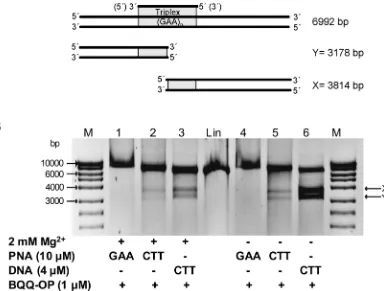
We used plasmid pMP179(115 repeats), which contains 115 GAA repeats and flanking sequences derived from the first intron of theFXNgene. After linearization with ApaI, the plasmid was incubated with CTT-PNA or GAA-PNA and cleaved by BQQ-OP. As control a triplex was also formed in the presence of a single strand CTT-DNA (Table 1). If dsDNA cleav-age by BQQ-OP would occur specifically at the site of triplex formation, the reaction should result in the formation of two DNA fragments. The size of these fragments is approximately 3814 and 3178 bp, assuming an average triplex cleavage to occur in the middle of the triplex forming (GAA)115repeat (Fig 2A). Binding of CTT-PNA or CTT-DNA followed by BQQ-OP
dsDNA cleavage resulted in the formation of two DNA fragments having the expected sizes
(Fig 2B, lanes 2 and 5 or 3 and 6, respectively). A stronger intensity of the bands corresponding
to the BQQ-OP cleavage was seen when CTT-DNA was used compared to conditions obtained by CTT-PNA binding, indicating either stronger binding of the DNA (although PNA-DNA2
Fig 2. Triplex-specific DNA cleavage of PNA triplex at (GAA)115repeats in linearized pMP179. A) Schematic
presentation of TFO-directed triplex formation and the two fragments generated after BQQ-OP cleavage indicated as X (3814 bp) and Y (3178 bp). B) Representative gel of linearized pMP179(115 repeats) (Apa1) incubated with GAA-PNA (10μM), CTT-PNA (10μM) or CTT-DNA (4μM) (Table 1) in buffer (10 mM sodium cacodylate, 100 mM NaCl and 0 or 2 mM MgCl2, pH 7.5), as indicated. BQQ-OP mediated cleavage of pMP179(115 repeats) was carried out in the presence of Cu2+and MPA. Reference linearized pMP179(115 repeats) (Lin) and a molecular weight DNA ladder (M) are also shown.
triplexes of identical length are much more stable than DNA triplexes [63]) or more efficient BQQ-OP cleavage of the DNA triplex versus the PNA-DNA2triplex. The same trend was
observed both in the presence as well as the absence of Mg2+ions. However, it is well known that an additional PNA-DNA complex, a PNA-triplex invasion, can be formed when homo-pyrimidine PNA are targeted to polyhomo-pyrimidine-polypurine dsDNA, and this is more stable than a PNA-triplex [64]. In the case of PNA-triplex invasion, only the GAA DNA strand is engaged in the triplex while the other strand is displaced. Therefore, BQQ-OP would only lead to a single strand cleavage of the plasmid. Hence, the resulting DNA fragment would be of the same size as the linearized plasmid (Lin), which can be seen inFig 2B(lanes 2 and 5).
Next, we examined the ability of GAA-PNA to form a purine motif intermolecular triplex using the BQQ-OP dsDNA cleavage assay. We detected only a single DNA fragment exhibiting slightly slower gel mobility than the linearized plasmid (Fig 2B, lanes 1 and 4), which could indicate stable PNA binding to the dsDNA plasmid, maybe due to formation of a duplex inva-sion complex. Nevertheless, triplex formation in the presence of GAA-PNA was not detected using the BQQ-OP assay. Interestingly, we did not observe a purine motif TFO-DNA triplex nor a purine motif H-DNA in earlier studies [56]. Based on these observations and that the tri-plex formed by CTT-PNA binding resulted in a clear BQQ-OP cleavage, we conclude that a purine motif triplex is not formed in the presence of GAA-PNA.
PNA Binding to H-DNA Forming FRDA (GAA)
115Repeats
H-DNA is found in two different motifs (parallel pyrimidine or antiparallel purine triplex) and both have been proposed to form at FRDA (GAA)nrepeats (Fig 1). We recently showed
that expanded (GAA)115repeats form a pyrimidine motif triplex in supercoiled plasmids by
using the BQQ-OP assay [56]. Also, binding of a single strand CTT oligonucleotide (identical in sequence and length to the currently used CTT-DNA,Table 1) to the H-DNA forming plas-mid enhanced triplex-formation significantly, whereas an analogous GAA oligonucleotide (corresponding to GAA-DNA,Table 1) had no such effect. These findings prompted us to examine whether binding of modified ON such as CTT-PNA or GAA-PNA (Table 1) to the (GAA)115repeats would have a comparable effect on H-DNA formation at this site. When
BQQ-OP mediated dsDNA cleavage occurs within the (GAA)115triplex-forming repeat, the
subsequent unique site enzymatic digestion is expected to generate two linear DNA fragments of approximately 3814 and 3178 bp (as shown inFig 3A). In the absence of DNA or PNA olig-omers, dsDNA cleavage by BQQ-OP, corresponding here only to inherent H-DNA formation, was estimated, by quantification of the gel bands (Fig 3B, lane 3), to be on average 35.3±3.5%
(Fig 3C). On the other hand, binding of CTT-PNA to the (GAA)115repeat leads to a
statisti-cally significant increase of the amount of triplex, as measured by the extent of BQQ-OP medi-ated DNA cleavage (50.3±2.1%,P=0.0068) (Fig 3B, lane 1 andFig 3C). As previously reported, the presence of CTT-DNA also leads to a significant increase in the level of BQQ-OP cleavage to 52.7±8.4%,P=0.0031 (Fig 3B, lane 4 andFig 3C). Taken together, our findings suggest that sequence-specific binding of CTT-PNA to FRDA expanded repeats enhances triplex formation under these conditions.
We envision three models to account for these results. Either the H-DNA structure is stabi-lized by PNA binding to the single stranded GAA loop formed as part of the H-DNA structure or the H-DNA is replaced by a PNA-DNA2triplex, or a PNA2-DNA triplex invasion structure
1 and 4), reflects mainly a stabilization of the H-DNA structure through binding of CTT-DNA or CTT-PNA (via a PNA2-DNA triplex structure) to the single stranded GAA repeat region of
the H-DNA.
Interestingly, when we targeted FRDA repeats in pMP179(115 repeats) using a GAA-PNA oligomer (Table 1) there was no detectable triplex-containing structure, including H-DNA,
(Fig 3B, lane 2). Only one major DNA fragment was observed upon BQQ-OP treatment,
cor-responding in size and gel mobility to the linearized plasmid. These results indicate a
completely different behavior of the GAA-PNA when interacting with theFXNtriplet repeats as compared to GAA-DNA (Fig 3B, lane 5). Apparently, GAA-DNA does not bind to the
Fig 3. BQQ-OP mediated DNA cleavage of H-DNA forming (GAA)115repeats in the presence of PNA. A)
Schematic presentation of pMP179 and the H-DNA forming site. The two DNA fragments generated by triplex-specific cleavage followed by enzymatic digestion are indicated as X (3814 bp) and Y (3178 bp). B) Representative gel for pMP179(115 repeats) incubated with 10μM PNA (CTT-PNA or GAA-PNA, lane 1 or 2, respectively), in the absence of ONs (lane 3) or with 10μM ssDNA (CTT-DNA or GAA-DNA, lane 4 or 5, respectively) in buffer (10 mM sodium cacodylate, 100 mM NaCl, 2 mM MgCl2, pH 7.5). Triplex-specific cleavage was carried out in the presence of Cu2+and MPA, followed by digestion with ApaI. Reference supercoiled (SC) and linearized (Lin) pMP179(115 repeats) and a molecular weight DNA ladder (M) are also shown. C) Graph represents the percentage of BQQ-OP mediated cleavage obtained for pMP179(115 repeats) in the absence (indicate in the graph as pMP179) or in the presence of PNA- or DNA oligomers. Values indicate the ratio between the intensity of DNA double strand cleavage to the total band intensity from the respective lane and are expressed as mean±S.D. (n=3). No cleavage was obtained in the presence of GAA-PNA and therefore not included in the graph.**P0.01 in relation to plasmid in the absence of oligonucleotide (pMP179).
repeat region and consequently H-DNA is still formed to a comparable extent as in the control sample (41.3±4.0%,Fig 3B, lane 3 andFig 3C). It is worth noting that a slower-mobility band is detected in the sample containing the (GAA)115repeat plasmid and GAA-PNA (Fig 3B, lane
2). The presence and intensity of this band indicates a stable interaction, which we believe can be attributed to a sequence-specific GAA-PNA binding to (GAA)115repeats in the plasmid.
However, this assay cannot unravel the nature of this interaction, which called for further detailed analysis as described in the following section. Nevertheless, our findings show that binding of a GAA-PNA oligomer to FRDA repeats has the unique ability to completely abolish all triplex structures, which are detected by BQQ-OP, including H-DNA, under these
conditions.
Structural Analysis of CTT-PNA and GAA-PNA Binding to (GAA)
nRepeats
To better understand the binding modes of CTT-PNA and GAA-PNA oligomers to the FRDA expanded (GAA)nrepeats and the effect of PNA binding on H-DNA, or other higher order
structure formation at these expansions, we examined PNA binding into two different super-coiled plasmids carrying short or medium (GAA)nrepeats (n=9 or 75, respectively) and
including FRDA flanking sequences. We employed structural probing analysis using either chemical modification of ssDNA regions by chloroacetaldehyde (CAA) or BQQ-OP mediated cleavage of DNA triplex structures. Chemical modifications of DNA and triplex-specific cleav-age by BQQ-OP were analyzed using primer extension (PE) reactions where each of the (GAA)nor (CTT)ncontaining strands, which are referred to as the R-strand and the Y-strand,
respectively, were used as template. Chloroacetaldehyde reacts with single strand adenosines and cytosines, which prevents PE by DNA polymerase. BQQ-OP cleavage, on the other hand, is used to map regions of triplex formation. Both probing reactions were carried out in the absence or presence of PNA (CTT-PNA or GAA-PNA). For all DNA reactions, we included a control sample, where the plasmid was incubated in the absence or presence of PNA but no DNA chemical modification or BQQ-OP cleavage reaction was carried out (control C1, C2, and C3). When analyzing DNA cleavage or chemical modification, the intensity of each band was compared to the corresponding band in the control sample. This is necessary to identify background signals, which are not related to any specific DNA modification or cleavage but result from DNA polymerase pausing/arrest of PE at PNA binding sites, or by formation of stable structures, in the DNA template.
H-DNA Formation at Short (GAA)
9Repeats
Chloroacetaldehyde probing clearly demonstrates the presence of a single strand region start-ing in the middle of the (GAA)9repeat and covering the 50-half of the repeat region (Fig 4,
R-strand, lane 2). Analysis of the corresponding Y-R-strand, only revealed a very short (1–3 nt) sin-gle strand region (Fig 4, Y-strand, lane 14). This region is localized in the middle of the mirror repeat (GAA)9sequence, which is consistent with formation of a short loop in a 503030
-pyrimi-dine motif H-DNA.Fig 5A(upper panel) shows the sites of chloroacetaldehyde modifications (indicated by blue arrows) and the corresponding H-DNA (lower panel, right side).
BQQ-OP probing of triplex regions of the R-strand showed that cleavage occurred mainly at the 30-end of the repeat and also extended to the shorter A-rich (A
5– A9) flanking region
(Fig 4, lane 10). Moreover, cleavage is also detected at the 50-end flanking nucleotides
corre-sponding to the longer A-rich region (A10– A23). The results suggest engagement of the
Fig 4. Structural- and chemical probing of DNA and DNA-PNA complex formation at (GAA)9repeats. Non-denaturing PAGE of DNA
compared to cleavage of the R-strand. We previously reported that intercalation of BQQ-OP in pyrimidine motif triplex DNA and the correspondingin situradical reaction results in a lower rate of DNA cleavage of the pyrimidine-rich strand (corresponding here to the Y-strand) and a more pronounced cleavage of the purine-rich strand (corresponding here to the R-strand) [52]. BQQ-OP mediated cleavage is illustrated inFig 5A(middle panel) and the site and intensity of cleavage is indicated by red arrows (higher level of cleavage) or circles (lower level). The combined chloroacetaldehyde and BQQ-OP probing results are in accordance with previous reports [9,56] and the proposed 503030H-DNA structure is based on these results (lower panel, right side).
However, triplex-directed dsDNA cleavage using BQQ-OP indicates the formation of two different H-DNA pyrimidine motif triplexes, but only one H-DNA isomer (Fig 5A, lower panel, right side), corresponding to a 503030H-DNA is also supported by the chloroacetalde-hyde probing results (Fig 4, lane 2), where a clear single strand formation is demonstrated. The different time kinetics of the chloroacetaldehyde reaction (30 min) and the BQQ-OP tri-plex-specific cleavage (3 h) could (partly) provide an explanation. It has been shown that the kinetics of H-DNA formation is different for each of the two isomers (503030and 305050). Rob-erts and Crothers reported, in a study using palindromic pyrimidine strands, which can fold into a triplex structure upon binding of a complementary purine strand, that the 503030-isomer of the intramolecular triplex folds 10–50 times faster than the 305050isomer even though both isomers are equally stable as final complexes [65]. Consequently, it is possible that chloroace-taldehyde probing reveals only formation of the kinetically favored 503030H-DNA isomer, whereas the much slower BQQ-OP cleavage detects both isomers. This suggestion is supported by the fact that the intercalating moiety in BQQ, BQQ-OP and other BQQ-conjugates has a higher affinity to T.AxT stretches, as compared to C.GxC triads [52–54], which is the case here including the 50-end flanking sequence of the R-strand. Therefore, the results of BQQ-OP cleavage could correspond to the formation of an additional H-DNA isomer (Fig 5A; lower panel, left side).
Probing PNA Binding at Short (GAA)
9Repeats
PNA binding to the (GAA)9repeats in pMP141(9 repeats) was analogously studied using
chloroacetaldehyde modification and BQQ-OP cleavage.
Binding of GAA-PNA. Chloroacetaldehyde reaction of the GAA-PNA treated pMP141(9
repeats) showed that a single strand region is formed throughout the entire repeat and flanking sequences of the R-strand (Fig 4, lane 6) as compared to control samples that were not sub-jected to chemical modification (Fig 4, lanes 1 and 5). In contrast, only a few single strand nucleotides were identified at the 50-end flanking sequence of the repeat of the Y-strand (Fig 4, lane 18, T15-T16) as compared to control lanes (Fig 4, lane 13 and 17). Only very weak bands
on both the R- and Y-strands were obtained from the BQQ-OP cleavage of the GAA-PNA treated plasmid, pMP141(9 repeats) (Fig 4, lane 12 and 24, respectively), as compared to con-trol lanes (Fig 4, lane 5 and 17, respectively). This together with a clear formation of single strand region throughout the R-strand is therefore ascribed to GAA-PNA duplex invasion of the repeat region, as illustrated in the proposed model structure inFig 5C(lower panel).
Binding of CTT-PNA. Chloroacetaldehyde treatment of the R-strand in the presence of
CTT-PNA indicated two short single strand sites at the 30- and 50-end flanking nucleotides within the A-rich regions (A5– A9and A10– A23) (Fig 4, lane 4). As mentioned previously, a
Fig 5. Models showing the most predominant DNA and DNA-PNA structures formed at pMP141(9 repeats) in the absence or the presence of CTT-PNA or GAA-PNA respectively. A) The H-DNA structure (lower panel, right side structure) corresponds to the predominant structure formed in the absence of PNA. The H-DNA structure (lower panel, left side) corresponds to the putative 305050
-isomer of the H-DNA. B) The triplex-invasion structure (lower panel) corresponds to the predominant structure formed in the presence of CTT-PNA. C) The duplex-invasion structure (lower panel) corresponds to the predominant structure formed under these conditions. In A, B and C) CAA modifications and BQQ-OP cleavage sites of pMP141(9 repeats) in the absence or presence of PNA are indicated in the DNA duplex as blue ( ) and red ( ), respectively and circles are used to mark a very low level of modification or cleavage. The height of the arrow represents the relative within-lane intensities of gel bands corresponding to CAA modifications and BQQ-OP cleavage. The nucleotides marked in blue or red indicate the sites of chloroacetaldehyde DNA modification or BQQ-OP mediated DNA cleavage, respectively. All indicated modifications, of samples treated with BQQ-OP or CAA, are compared to the controls.
(CTT)9repeat sequence (Fig 4, lane 16). The gel also shows that the band intensity in lane 16
gradually increases towards the 30-end of the repeat. Again, a control sample consisting of a CTT-PNA treated plasmid was included (Fig 4, lane 15). The results are compatible with the formation of a triplex invasion complex (Fig 5B, lower panel) of two CTT-PNA oligomers bind-ing to the R-strand at the (GAA)9repeats in pMP141(9 repeats). The prevalence of this structure
is also supported by BQQ-OP mediated triplex-specific cleavage (Fig 4, lane 11), which shows only very little additional cleavage as compared to the control sample (Fig 4, lanes 3), possibly indicating remaining H-DNA. Taken together, our results indicate that CTT-PNA binding to the repeat region in pMP141(9 repeats) favors formation of a triplex-invasion.
Formation of Stable H-DNA Structure(s) at Medium (GAA)
75Repeats
Pathological GAA repeat expansions in FRDA may vary in length and structural probing of long DNA sequences has proven challenging. Therefore, we chose to examine H-DNA and higher order structure formation in a medium GAA repeats using an FRDA derived DNA sequence that consists of 75 repeats and genomic flanking regions and we attempted to map formation of single- and triple strand regions using chloroacetaldehyde and BQQ-OP, respec-tively. Analysis of the R- and Y-strands that were subjected to BQQ-OP probing clearly indi-cated the presence of triplex structures as judged from the gel bands corresponding to DNA cleavage within the repeat region (Fig 6A, lanes 3 and 15). However, the band intensity in lane 3 shows that BQQ-OP mediated DNA cleavage is more pronounced at the 30-end of the R-strand indicating formation of a 503030- pyrimidine H-DNA (Fig 6B, structure 1). Reaction of the Y-strand also indicates some preference at the 30-end. In contrast to the clear single strand probing of the GAA strand within the short repeat (Fig 4), chloroacetaldehyde modification of the longer repeat failed to reveal bands that could indicate the presence of non base-paired nucleotides in either of the R- or Y-strand. These findings may reflect the presence of more complex higher order DNA structures at the medium GAA repeats indicating a difference between the pathological and normal repeat lengths.
The Effect of GAA-PNA Binding on H-DNA Formation at (GAA)
75Repeats
Binding of GAA-PNA to the (GAA)75repeats in supercoiled plasmid was examined using
chloroacetaldehyde and BQQ-OP reactions. Binding of GAA-PNA resulted in a clear and strong chloroacetaldehyde reaction with the R-strand providing evidence for a single strand formation in particular at the 30-end of the GAA repeat (Fig 6A, lane 8), whereas reaction above background could not be detected within the Y-strand (Fig 6A, lane 20) under similar conditions. Most interestingly, no BQQ-OP mediated triplex-specific cleavage of the R- or Y-strand was seen when the plasmid was incubated with GAA-PNA (Fig 6A, lanes 9 and 21 com-pared to the control lanes 7 and 19, respectively).
To the best of our knowledge, this is the first study that demonstrates formation of a duplex invasion complex, which forms through binding of GAA-PNA to expanded FRDA repeats, thereby dissolving a triplex containing higher order structure.
The Effect of CTT-PNA Binding on H-DNA Formation at (GAA)
75Repeats
Based on the results of H-DNA probing and the outcome of PNA binding of pMP179(115 repeats) (Fig 3), we concluded that the presence of CTT-PNA enhances triplex formation within the repeat sequence. Therefore, we examined how this PNA oligomer affects the dsDNA medium repeat region by using the single strand and triplex region probing assays.
Targeting the (GAA)75repeats in pMP178(75 repeats) with CTT-PNA resulted in an
enhanced triplex-directed cleavage by BQQ-OP of the R-strand (Fig 6A, lane 6) as compared to the control (lane 3). This result quantitatively agrees with triplex-specific cleavage at (GAA)115repeats, analyzed using agarose gel electrophoresis (Fig 3B, lane 1). In addition, an
analogous enhancement was seen for the Y-strand (Fig 6A, lane 18 compared to lane 15). Chemical modification using chloroacetaldehyde of the pMP178(75 repeats) in the pres-ence of CTT-PNA did not detect any major single-strand regions in the R-strand (Fig 6A, lane 5 as compared to control lane 4), as also observed in the absence of PNA (Fig 6A, lane 2 as compared to control lane 1). However, chloroacetaldehyde probing of the Y-strand in pMP178(75 repeats) in the presence of CTT-PNA strongly indicates the formation a single strand region that extends to nearly half the length of the mirror repeat sequence (towards the 30-end) (Fig 6A, lane 17). Since homopyrimidine PNA has the ability to invade dsDNA poly-purine-polypyrimidine duplexes, we attribute the single strand in the Y-strand to formation of a PNA2-DNA triplex-invasion complex (Fig 6B, structure 3). This result is in full accordance
with the data obtained for the pMP141(9 repeats) as shown inFig 4andFig 5, although it can-not, on its own, explain the pronounced BQQ-OP cleavage in the presence of this PNA. Thus, a triplex-invasion complex of the dsDNA may co-exist with H-DNA under these conditions
(Fig 6B, structure 2). CTT-PNA may also bind to the GAA repeat forming a traditional
PNA-DNA2triplex (Fig 6B, structure 4) analogous to that formed by CTT-DNA
oligonucleo-tides. Such a triplex is cleaved by BQQ-OP (Fig 2, lanes 2 and 5), but much less efficiently than pure DNA-triplexes, and thus could not account for the increased BQQ-OP sensitivity. There-fore the present data do not allow for a detailed quantitative structural description of the inter-action of CTT-PNA with medium GAA repeats, but a combination of PNA triplex invasion as well as H-DNA (like) structures must be involved. The difficulties in interpreting this data may also reflect different kinetics of formation of the different DNA structures (B-DNA,
Fig 6. Structural- and chemical probing of DNA and DNA-PNA complex formation in pMP178(75 repeats). A) Non-denaturing PAGE of DNA fragments mapped by BQQ-OP cleavage and chloroacetaldehyde (CAA) modification followed by primer extension (PE). Plasmid pMP178(75 repeats), was incubated in the absence (No PNA) or the presence of 10μM of PNA (GAA-PNA or CTT-PNA) in buffer (10 mM sodium cacodylate, 100 mM NaCl, 2 mM MgCl2, pH 7.5). The plasmid was then chemically modified using 2% CAA or cleaved using 1μM BQQ-OP. Non-treated plasmid was used in a set of four different controls (C) of the PE reactions C1= plasmid incubated in the absence of PNA. C2= plasmid incubated in the presence of GAA-PNA. C3= plasmid incubated in the presence of CTT-PNA. C4= plasmid not incubated. All samples were linearized by ApaI then subjected as templates for the PE reaction. Sequence ladders using dideoxynucleotides (C=ddCTP, T=ddTTP, G=ddGTP and A=ddATP), linearized pMP178(75 repeats) and the PE reaction control (C4) are shown as references. The DNA fragments detected in a PE reaction using the GAA (R)-strand as template, and the right gel panel shows the DNA fragments detected in a PE reaction using the CTT (Y)-strand as template. The A1to A9nucleotides flanking the repeats of the R-strand and T1to T12flanking in the Y-strand and the middle point of the repeat sequence are indicated (M). B) Models showing the most predominant DNA and DNA-PNA structures formed at pMP178(75 repeats) in the absence or the presence of CTT-PNA or GAA-PNA respectively. They correspond to: 1. 503030
-pyrimidine motif H-DNA, 2. CTT-PNA stabilized 503030-pyrimidine motif H-DNA, 3. Triplex-invasion, 4. Intermolecular triplex and 5.
Duplex-invasion structure. Thin line= purine strand, thick line= pyrimidine strand, grey line= PNA. Regions modified by CAA (π) or cleaved by BQQ-OP (o) are indicated.
H-DNA and triplex-invasion structures), which can transiently co-exist and hence conse-quently be detected by chloroacetaldehyde or BQQ-OP reactions.
LNA Binding to H-DNA Forming FRDA (GAA)
115Repeats
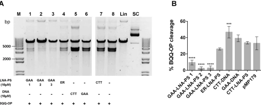
To examine whether another type of modified oligomer, with comparable dsDNA invasion properties as PNAs, could mediate results similar to those previously observed, the BQQ-OP cleavage assay was performed in the presence of CTT- or GAA-LNA-PS oligomers.Fig 7A
shows, in an analogous way as inFig 3B, that indeed in the presence of a 9-mer GAA-LNA-PS (lane 1) or 12-mer GAA-LNA-PS (lane 2 and 3) the BQQ-OP cleavage was almost abolished, presumably due to the destabilization of the H-DNA structure. When compared with the plas-mid alone, a statistic significance (P0.0001) was determined for all GAA-LNA-PS ONs, how-ever different mean±S.D. percentages of BQQ-OP cleavage were found depending of the ONs size, decreasing with increase of length (GAA-LNA-PS 1: 9.5±2.9%, while GAA-LNA-PS 2 and 3: 2.4±1.8% and 2.7±2.5%, respectively) (Fig 7B). Interestingly, is the lack of significance for the ER-LNA-PS ON (26.1±1.4%), showing that indeed the ON effect is sequence specific. Regarding CTT-LNA-PS, no significant increase in the BQQ-OP cleavage percentage was found (33.7±1.5%). These results support the hypothesis that modified GAA ONs can be used to destabilize triplex structures formed at (GAA)nrepeats.
Monitoring the Effect of LNA Binding to (GAA)
115Containing Plasmids
Using AFM
DNA topology is affected by formation of higher order DNA structures not the least at expanded triplet repeats. Atomic force microscopy (AFM) has been used to examine cruciforms, H-DNA and higher order structures at CAG and GAA repeats, respectively. In a similar way, we reasoned
Fig 7. BQQ-OP mediated DNA cleavage of H-DNA forming (GAA)115repeats in the presence of LNA-PS. A) Representative gel
from pMP179(115 repeats) incubated with 10μM LNA-PS (GAA-LNA-PS 1, 2 or 3 or ER-LNA-PS or CTT-LNA-PS, lane 1, 2, 3, 4 or 7, respectively), in the absence of oligonucleotide (lane 8) or with 10μM ssDNA (CTT-DNA or GAA-DNA, lane 5 or 6, respectively) in buffer (10 mM sodium cacodylate, 100 mM NaCl, 2 mM MgCl2, pH 7.5). Triplex-specific cleavage was carried out in the presence of Cu2+and MPA, and the plasmid was subsequently digested using ApaI. The two obtained DNA fragments have approximately 3814 and 3178 bp. Reference supercoiled (SC) and linearized (Lin) pMP179(115 repeats) and a molecular weight DNA ladder (M) are also shown. B) Graph represents the percentage of BQQ-OP mediated cleavage for pMP179(115 repeats) in the absence (indicate in the graph as pMP179) or in the presence of LNA- or DNA oligomers. Values indicate the ratio between the intensity of DNA double strand cleavage to the total band intensity from the respective lane and are expressed as mean±S.D. (n=3).***P0.001,****P<0.0001 in relation to plasmid in the absence of oligonucleotide (pMP179).
that oligomer binding to structure forming GAA expansions should be reflected by changes in DNA topology (and thereby morphology of circular DNA) and could be detectable by AFM. To examine this hypothesis, we monitored the effects of LNA binding to pMP179(115 repeats) using CTT-, GAA- or a scrambled-LNA (Table 1) and the binding conditions described above. We assumed that physiological buffers, containing Mg2+and Na+, which are both required for DNA structure stabilization, would be sufficient for visualization of DNA [66,67]. However, while Mg2+binds very weakly to mica surfaces, the presence of Na+promotes the release of DNA molecules from the surface. This can be overcome by the addition of Ni2+ions to the buffer. Thus, the Ni2+concentration used in this work permits suitable attachment of DNA to the mica surface, avoiding Ni2+precipitation and poor DNA binding, occurring at higher and lower NiCl2concentrations, respectively [60,68]. Studies by Billingsley et al. (2010) demonstrate
that the hydration of the sample during AFM measurements is crucial to determine morphology of supercoiled plasmids. Specifically, they show that supercoiled samples prepared with Ni2+ with high hydration are more condensed, with a large number of crossovers, local conformation changes and more turns effects that seem to be topologically driven. They also postulate that under these conditions, the configuration is closer to the 3D solution form [69]. DNA supercoil-ing is also dependent on the plasmid integrity, and PeakForce1tapping (PTM) mode, where the applied force can be controlled and decreased below the nano-Newton range, is a suitable tech-nique for this type of measurements where the preservation of the sample is crucial [61].
In view of these considerations, we performed AFM measurements by PTM in liquid in the presence of Ni2+ions using pMP179(115 repeats) incubated in the presence or absence of LNA ONs. All analyses were performed under the same conditions. As shown inFig 8, pMP179(115 repeats) the supercoiled state (Fig 8A) presents a very condensed morphology, resembling a compact sphere structure and this morphology is maintained in the presence of scrambled-LNA (Fig 8B) or CTT-LNA (Fig 8C) oligomers. However, in the presence of GAA-LNA (Fig 8D) a clearly more relaxed morphology was observed. As control, we per-formed similar AFM analysis using the parent plasmid pSPL3, lacking the GAA repeat sequence, in the absence or presence of the same set of oligomers, and in all cases we did not observe any change of the plasmid morphology (S1 Fig). This confirms that oligomer mediated change of plasmid structure is dependent on the presence of the GAA expansions.
To provide a quantitative analysis of the oligomer binding effect, detected by AFM, we mea-sured the distribution (mean±S.E.M. values) of all the areas determined under each condition tested as shown in the graph ofFig 8E. On average, pMP179(115 repeats) occupies an area of 0.053±0.0016μm2(pMP179 SCR-LNA-PS: 0.050±0.0019μm2and pMP179 CTT-LNA-PS 0.059±0.0061μm2) while in the presence of GAA-LNA-PS 3 an area of 0.076±0.0047μm2is observed, significantly increasing the area by 43.4%. Statistical analysis shows that GAA-L-NA-PS 3 binding of the repeat-containing plasmid clearly has a significant effect (P0.0001), whereas CTT-LNA-PS shows no statistically significant difference as compared to the control (P=0.3740), consistent with the BQQ-OP cleavage data (Fig 7). In other words, GAA-LNA-PS 3 significantly changed the conformation of the GAA-repeat plasmid, both as compared to the control plasmid, and as compared to the CTT-LNA-PS treated plasmid (P=0.0071).
These results support the conclusion that GAA-LNA (but not CTT-LNA) in analogy to GAA-PNA can resolve triplex containing higher order structures thereby promoting a more relaxed structure that is topologically similar to duplex DNA.
Conclusions
Furthermore, we find that PNA and LNA sequence-specific targeting of Friedreich’s ataxia GAA repeat expansions can alter and resolve higher order DNA structures. BQQ-OP mediated triplex-specific cleavage of double strand DNA and chloroacetaldehyde chemical modification of single strand DNA regions at (GAA)nrepeats demonstrate that GAA-PNA binding result in a duplex
invasion complex, that completely dissociates all detectable triplex containing higher order struc-tures at this site, whereas this is not the case for CTT-PNA. Additionally, we obtained a similar pattern using LNA based ONs. Furthermore, a significant change in plasmid morphology in the presence of GAA-LNA was detected using atomic force microscopy. Our results suggest that DNA targeting by modified GAA-oligomers at expanded (GAA)nrepeats can be employed to
examine the possible role of non-canonical DNA structures inFXNgene silencing and potentially applied to develop new nucleic acids-based therapeutic strategies in Friedreich’s ataxia disease.
Fig 8. Topography analysis of pMP179 (115 repeats) in the absence or presence of LNA-PS ONs. Representative AFM topography images of 12.5 ng of supercoiled pMP179(115 repeats): A) in the absence of LNA; B) in the presence of SCR-LNA-PS oligomer; C) in the presence of CTT-LNA-PS or D) in the presence of GAA-LNA-PS 3. Sample preparation in a 10 mM HEPES supplemented with 5 mM Ni2+solution, pH 7.4 was deposited onto fresh clean mica. Colour scales: 2.5 nm. Images were acquired in liquid with the Dimension FastScan, Nanoscope V operating in Peakforce®tapping mode. E) Plasmid area distribution from different
AFM overview fields of pMP179(115 repeats) supercoiled alone or in the presence of the different LNA ONs. The graph shows the mean±S.E.M. determined for each condition (pMP179 n=312; pMP179+SCR-LNA-PS n=185; pMP179+CTT-LNA-PS n=63; and pMP179+GAA-LNA-PS 3 n=216). A highly significant increase in the area occupied by the plasmids is observed in the presence of the GAA-LNA-PS 3, representing a more relaxed morphology.**P0.01,****P0.0001 in relation to plasmid in the absence of oligonucleotide (pMP179).
Supporting Information
S1 Dataset. BQQ-OP cleavage raw data and statistical analysis. File contains all the data
analysis regarding the BQQ-OP cleavage experiments in the presence or absence of or PNA or LNA oligomers.
(XLSX)
S2 Dataset. AFM raw data and statistical analysis. File contains all the data analysis
regard-ing the AFM experiments with pMP179 or pSPL3 in the presence or absence of LNA oligo-mers.
(XLSX)
S1 Fig. Topography analysis of pSPL3 in the absence or presence of LNA-PS ONs.
Repre-sentative AFM topography images of 12.5 ng of supercoiled pSPL3: A) in the absence of LNA; B) in the presence of SCR-LNA-PS oligomer; C) in the presence of CTT-LNA-PS or; D) in the presence of GAA-LNA-PS 3. Sample preparation in a 10 mM HEPES supplemented with 5 mM Ni2+solution, pH 7.4 was deposited onto fresh clean mica. Colour scales: 2.0 nm. Images were acquired in liquid with the Dimension FastScan, Nanoscope V operating in Peakforce1 tap-ping mode. E) Plasmid area distribution from different AFM overview fields of pSPL3 super-coiled alone or in the presence of the different LNA ONs. The graph shows the mean±S.E.M. determined for each condition (pSPL3 n=159; pSPL3+SCR-LNA-PS n=152; pSPL3+CTT-LNAPS n=127; and pSPL3+GAA-LNA-PS 3 n=154). No statistic significance was found among all the treatments when compared to plasmid (pSPL3) in the absence of oligonucleotide. (TIF)
Acknowledgments
The authors thank Prof. M. Pandolfo for the kind gift of plasmids pMP141, pMP178 and pMP179. The authors also thank Dr Matthew Fielden at the Albanova Nanofabrication Facility for his skillful technical support.
Author Contributions
Conceptualization: RZ LG CIES HB CSJR.
Formal analysis: HB CSJR RAA.
Funding acquisition: RZ CIES HB.
Investigation: HB CSJR RAA.
Methodology: HB CSJR CIES LG RZ.
Resources: RZ CIES MWR PEN CHN.
Supervision: RZ.
Writing – original draft: HB CSJR RZ.
Writing – review & editing: HB CSJR RAA CHN MWR CIES LG PEN RZ.
References
2. Cossee M, Schmitt M, Campuzano V, Reutenauer L, Moutou C, Mandel J-L, et al. Evolution of the Frie-dreich’s ataxia trinucleotide repeat expansion: Founder effect and premutations. Proc Natl Acad Sci. 1997; 94: 7452–7457. doi:10.1073/pnas.94.14.7452PMID:9207112
3. Du¨rr A, Cossee M, Agid Y, Campuzano V, Mignard C, Penet C, et al. Clinical and Genetic Abnormalities in Patients with Friedreich’s Ataxia. N Engl J Med. 1996; 335: 1169–1175. doi:10.1056/
NEJM199610173351601PMID:8815938
4. Campuzano V, Montermini L, Lutz Y, Cova L, Hindelang C, Jiralerspong S, et al. Frataxin is Reduced in Friedreich Ataxia Patients and is Associated with Mitochondrial Membranes. Hum Mol Genet. 1997; 6: 1771–1780. doi:10.1093/hmg/6.11.1771PMID:9302253
5. Bidichandani SI, Ashizawa T, Patel PI. The GAA Triplet-Repeat Expansion in Friedreich Ataxia Inter-feres with Transcription and May Be Associated with an Unusual DNA Structure. Am J Hum Genet. 1998; 62: 111–121. doi:10.1086/301680PMID:9443873
6. Pandolfo M. Frataxin deficiency and mitochondrial dysfunction. Mitochondrion. 2002; 2: 87–93. doi:10. 1016/S1567-7249(02)00039-9PMID:16120311
7. Groh M, Lufino MMP, Wade-Martins R, Gromak N. R-loops Associated with Triplet Repeat Expansions Promote Gene Silencing in Friedreich Ataxia and Fragile X Syndrome. PLoS Genet. 2014; 10: e1004318. doi:10.1371/journal.pgen.1004318PMID:24787137
8. Sakamoto N, Chastain PD, Parniewski P, Ohshima K, Pandolfo M, Griffith JD, et al. Sticky DNA. Mol Cell. 1999; 3: 465–475. doi:10.1016/S1097-2765(00)80474-8PMID:10230399
9. Potaman VN. Length-dependent structure formation in Friedreich ataxia (GAA)n•(TTC)n repeats at neutral pH. Nucleic Acids Res. 2004; 32: 1224–1231. doi:10.1093/nar/gkh274PMID:14978261 10. Hanvey JC, Shimizu M, Wells RD. Intramolecular DNA triplexes in supercoiled plasmids. Proc Natl
Acad Sci. 1988; 85: 6292–6296. doi:10.1073/pnas.85.17.6292PMID:3413097
11. Mariappan SVS, Catasti P, Silks LA, Bradbury EM, Gupta G. The high-resolution structure of the triplex formed by the GAA/TTC triplet repeat associated with Friedreich’s ataxia. J Mol Biol. 1999; 285: 2035– 2052. doi:10.1006/jmbi.1998.2435PMID:9925783
12. Jain A, Rajeswari MR, Ahmed F. Formation and Thermodynamic Stability of Intermodular (R*R•Y) DNA Triplex In GAA/TTC Repeats Associated with Freidreich’s Ataxia. J Biomol Struct Dyn. 2002; 19: 691–699. doi:10.1080/07391102.2002.10506775PMID:11843630
13. Ohshima K, Montermini L, Wells RD, Pandolfo M. Inhibitory Effects of Expanded GAA•TTC Triplet Repeats from Intron I of the Friedreich Ataxia Gene on Transcription and Replicationin Vivo. J Biol Chem. 1998; 273: 14588–14595. doi:10.1074/jbc.273.23.14588PMID:9603975
14. Son LS, Bacolla A, Wells RD. Sticky DNA: in Vivo Formation in E. coli and in Vitro Association of Long GAA•TTC Tracts to Generate Two Independent Supercoiled Domains. J Mol Biol. 2006; 360: 267–284. doi:10.1016/j.jmb.2006.05.025PMID:16764889
15. Vetcher A a., Napierala M, Iyer RR, Chastain PD, Griffith JD, Wells RD. Sticky DNA, a long
GAAGAATTC triplex that is formed intramolecularly, in the sequence of intron 1 of the frataxin gene. J Biol Chem. 2002; 277: 39217–39227. doi:10.1074/jbc.M205209200PMID:12161437
16. Vetcher AA, Napierala M, Wells RD. Sticky DNA: Effect of the Polypurine•Polypyrimidine Sequence. J Biol Chem. 2002; 277: 39228–39234. doi:10.1074/jbc.M205210200PMID:12161438
17. Napierala M. Structure-dependent Recombination Hot Spot Activity of GAA•TTC Sequences from Intron 1 of the Friedreich’s Ataxia Gene. J Biol Chem. 2004; 279: 6444–6454. doi:10.1074/jbc. M309596200PMID:14625270
18. Wojciechowska M, Napierala M, Larson JE, Wells RD. Non-B DNA Conformations Formed by Long Repeating Tracts of Myotonic Dystrophy Type 1, Myotonic Dystrophy Type 2, and Friedreich’s Ataxia Genes, Not the Sequences per se, Promote Mutagenesis in Flanking Regions. J Biol Chem. 2006; 281: 24531–24543. doi:10.1074/jbc.M603888200PMID:16793772
19. Gacy AM, Goellner GM, Spiro C, Chen X, Gupta G, Bradbury EM, et al. GAA Instability in Friedreich’s Ataxia Shares a Common, DNA-Directed and Intraallelic Mechanism with Other Trinucleotide Diseases. Mol Cell. 1998; 1: 583–593. doi:10.1016/S1097-2765(00)80058-1PMID:9660942
20. Krasilnikova MM, Mirkin SM. Replication Stalling at Friedreich’s Ataxia (GAA)n Repeats In Vivo. Mol Cell Biol. 2004; 24: 2286–2295. doi:10.1128/MCB.24.6.2286-2295.2004PMID:14993268
21. Grabczyk E. The GAATTC triplet repeat expanded in Friedreich’s ataxia impedes transcription elonga-tion by T7 RNA polymerase in a length and supercoil dependent manner. Nucleic Acids Res. 2000; 28: 2815–2822. doi:10.1093/nar/28.14.2815PMID:10908340
23. Follonier C, Oehler J, Herrador R, Lopes M. Friedreich’s ataxia–associated GAA repeats induce replica-tion-fork reversal and unusual molecular junctions. Nat Struct Mol Biol. 2013; 20: 486–494. doi:10. 1038/nsmb.2520PMID:23454978
24. Greene E, Mahishi L, Entezam A, Kumari D, Usdin K. Repeat-induced epigenetic changes in intron 1 of the frataxin gene and its consequences in Friedreich ataxia. Nucleic Acids Res. 2007; 35: 3383–3390. doi:10.1093/nar/gkm271PMID:17478498
25. Al-Mahdawi S, Pinto RM, Ismail O, Varshney D, Lymperi S, Sandi C, et al. The Friedreich ataxia GAA repeat expansion mutation induces comparable epigenetic changes in human and transgenic mouse brain and heart tissues. Hum Mol Genet. 2007; 17: 735–746. doi:10.1093/hmg/ddm346PMID: 18045775
26. Rai M, Soragni E, Jenssen K, Burnett R, Herman D, Coppola G, et al. HDAC Inhibitors Correct Frataxin Deficiency in a Friedreich Ataxia Mouse Model. PLoS One. 2008; 3: e1958. doi:10.1371/journal.pone. 0001958PMID:18463734
27. Herman D, Jenssen K, Burnett R, Soragni E, Perlman SL, Gottesfeld JM. Histone deacetylase inhibitors reverse gene silencing in Friedreich’s ataxia. Nat Chem Biol. 2006; 2: 551–558. doi:10.1038/
nchembio815PMID:16921367
28. Rai M, Soragni E, Chou CJ, Barnes G, Jones S, Rusche JR, et al. Two new pimelic diphenylamide HDAC inhibitors induce sustained frataxin upregulation in cells from Friedreich’s ataxia patients and in a mouse model. PLoS One. 2010; 5: e8825. doi:10.1371/journal.pone.0008825PMID:20098685 29. Xu C, Soragni E, Chou CJ, Herman D, Plasterer HL, Rusche JR, et al. Chemical Probes Identify a Role
for Histone Deacetylase 3 in Friedreich’s Ataxia Gene Silencing. Chem Biol. 2009; 16: 980–989. doi: 10.1016/j.chembiol.2009.07.010PMID:19778726
30. Burnett R, Melander C, Puckett JW, Son LS, Wells RD, Dervan PB, et al. DNA sequence-specific poly-amides alleviate transcription inhibition associated with long GAA•TTC repeats in Friedreich’s ataxia. Proc Natl Acad Sci. 2006; 103: 11497–11502. doi:10.1073/pnas.0604939103PMID:16857735 31. Grant L, Sun J, Xu H, Subramony SH, Chaires JB, Hebert MD. Rational selection of small molecules
that increase transcription through the GAA repeats found in Friedreich’s ataxia. FEBS Lett. 2006; 580: 5399–5405. doi:10.1016/j.febslet.2006.09.006PMID:16989817
32. Wu Q, Gaddis SS, MacLeod MC, Walborg EF, Thames HD, DiGiovanni J, et al. High-affinity triplex-forming oligonucleotide target sequences in mammalian genomes. Mol Carcinog. 2007; 46: 15–23. doi: 10.1002/mc.20261PMID:17013831
33. Jain A, Magistri M, Napoli S, Carbone GM, Catapano C V. Mechanisms of triplex DNA-mediated inhibi-tion of transcripinhibi-tion initiainhibi-tion in cells. Biochimie. 2010; 92: 317–320. doi:10.1016/j.biochi.2009.12.012 PMID:20045441
34. Carbone GM, Napoli S, Valentini A, Cavalli F, Watson DK, Catapano C V. Triplex DNA-mediated down-regulation of Ets2 expression results in growth inhibition and apoptosis in human prostate cancer cells. Nucleic Acids Res. 2004; 32: 4358–4367. doi:10.1093/nar/gkh744PMID:15314206
35. Datta HJ. Triplex-induced Recombination in Human Cell-free Extracts. Dependence on XPA and HsRad51. J Biol Chem. 2001; 276: 18018–18023. doi:10.1074/jbc.M011646200PMID:11278954 36. Liu Y, Nairn RS, Vasquez KM. Processing of triplex-directed psoralen DNA interstrand crosslinks by
recombination mechanisms. Nucleic Acids Res. 2008; 36: 4680–4688. doi:10.1093/nar/gkn438PMID: 18628293
37. Wang G, Seidman MM, Glazer PM. Mutagenesis in mammalian cells induced by triple helix formation and transcription-coupled repair. Science. 1996; 271: 802–805. doi:10.1126/science.271.5250.802 PMID:8628995
38. Vasquez KM, Christensen J, Li L, Finch R a, Glazer PM. Human XPA and RPA DNA repair proteins par-ticipate in specific recognition of triplex-induced helical distortions. Proc Natl Acad Sci U S A. 2002; 99: 5848–5853. doi:10.1073/pnas.082193799PMID:11972036
39. Zhao J, Jain A, Iyer RR, Modrich PL, Vasquez KM. Mismatch repair and nucleotide excision repair pro-teins cooperate in the recognition of DNA interstrand crosslinks. Nucleic Acids Res. 2009; 37: 4420– 4429. doi:10.1093/nar/gkp399PMID:19468048
40. Karkare S, Bhatnagar D. Promising nucleic acid analogs and mimics: Characteristic features and appli-cations of PNA, LNA, and morpholino. Appl Microbiol Biotechnol. 2006; 71: 575–586. doi:10.1007/ s00253-006-0434-2PMID:16683135
42. Demidov V V, Yavnilovich M V, Belotserkovskii BP, Frank-Kamenetskii MD, Nielsen PE. Kinetics and mechanism of polyamide (“peptide”) nucleic acid binding to duplex DNA. Proc Natl Acad Sci. 1995; 92: 2637–2641. doi:10.1073/pnas.92.7.2637PMID:7708697
43. Kosaganov YN, Stetsenko D a, Lubyako EN, Kvitko NP, Lazurkin YS, Nielsen PE. Effect of Tempera-ture and Ionic Strength on the Dissociation Kinetics and Lifetime of PNA−DNA Triplexes †. Biochemis-try. 2000; 39: 11742–11747. doi:10.1021/bi0006417PMID:10995242
44. Egholm M, Christensen L, Dueholm KL, Buchardt O, Coull J, Nielsen PE. Efficient pH-independent sequence-specific DNA binding by pseudoisocytosine-containing bis-PNA. Nucleic Acids Res. 1995; 23: 217–222. Available:http://www.ncbi.nlm.nih.gov/pmc/articles/PMC306657/. PMID:7862524 45. Nielsen PE, Christensen L. Strand Displacement Binding of a Duplex-Forming Homopurine PNA to a
Homopyrimidine Duplex DNA Target. J Am Chem Soc. 1996; 118: 2287–2288. doi:10.1021/ja953125q 46. Zhang X, Ishihara T, Corey DR. Strand invasion by mixed base PNAs and a PNA-peptide chimera.
Nucleic Acids Res. 2000; 28: 3332–3338. doi:10.1093/nar/28.17.3332PMID:10954602
47. Rapireddy S, He G, Roy S, Armitage BA, Ly DH. Strand Invasion of Mixed-Sequence B-DNA by Acri-dine-Linked,γ-Peptide Nucleic Acid (γ-PNA). J Am Chem Soc. 2007; 129: 15596–15600. doi:10.1021/ ja074886jPMID:18027941
48. Bentin T, Hansen GI, Nielsen PE. Structural diversity of target-specific homopyrimidine peptide nucleic acid-dsDNA complexes. Nucleic Acids Res. 2006; 34: 5790–5799. doi:10.1093/nar/gkl736PMID: 17053099
49. Lundin KE, Højland T, Hansen BR, Persson R, Bramsen JB, Kjems J, et al. Biological Activity and Bio-technological Aspects of Locked Nucleic Acids. Advances in genetics. 2013. pp. 47–107. doi:10.1016/ B978-0-12-407676-1.00002-0PMID:23721720
50. Moreno PMD, Geny S, Pabon YV, Bergquist H, Zaghloul EM, Rocha CSJ, et al. Development of bis-locked nucleic acid (bisLNA) oligonucleotides for efficient invasion of supercoiled duplex DNA. Nucleic Acids Res. 2013; 41: 3257–3273. doi:10.1093/nar/gkt007PMID:23345620
51. Escude´ C, Nguyen CH, Kukreti S, Janin Y, Sun J-SS, Bisagni E, et al. Rational design of a triple helix-specific intercalating ligand. Proc Natl Acad Sci U S A. 1998; 95: 3591–3596. doi:10.1073/pnas.95.7. 3591PMID:9520410
52. Zaid A, Sun J-S, Nguyen C-H, Bisagni E, Garestier T, Grierson DS, et al. Triple-Helix Directed Cleavage of Double-Stranded DNA by Benzoquinoquinoxaline-1,10-phenanthroline Conjugates. ChemBioChem. 2004; 5: 1550–1557. doi:10.1002/cbic.200400074PMID:15515089
53. Zain R, Marchand C, Sun JS, Nguyen CH, Bisagni E, Garestier T, et al. Design of a triple-helix-specific cleaving reagent. Chem Biol. 1999; 6: 771–777. doi:10.1016/S1074-5521(99)80124-0PMID: 10574778
54. Zain R, Polverari D, Nguyen C- HH, Blouquit Y, Bisagni E, Garestier T, et al. Optimization of Triple-Helix-Directed DNA Cleavage by Benzoquinoquinoxaline-Ethylenediaminetetraacetic Acid Conjugates. ChemBioChem. 2003; 4: 856–862. doi:10.1002/cbic.200300621PMID:12964160
55. Amiri H, Nekhotiaeva N, Sun J-S, Nguyen C-H, Grierson DS, Good L, et al. Benzoquinoquinoxaline Derivatives Stabilize and Cleave H-DNA and Repress Transcription Downstream of a Triplex-forming Sequence. J Mol Biol. 2005; 351: 776–783. doi:10.1016/j.jmb.2005.03.044PMID:16045927 56. Bergquist H, Nikravesh A, Ferna´ndez RD, Larsson V, Nguyen C- H, Good L, et al. Structure-Specific
Recognition of Friedreich’s Ataxia (GAA) n Repeats by Benzoquinoquinoxaline Derivatives. ChemBio-Chem. 2009; 10: 2629–2637. doi:10.1002/cbic.200900263PMID:19746387
57. Hu J, Liu J, Yu D, Aiba Y, Lee S, Pendergraff H, et al. Exploring the Effect of Sequence Length and Composition on Allele-Selective Inhibition of Human Huntingtin Expression by Single-Stranded Silenc-ing RNAs. Nucleic Acid Ther. 2014; 24: 199–209. doi:10.1089/nat.2013.0476PMID:24694346 58. Li L, Matsui M, Corey DR. Activating frataxin expression by repeat-targeted nucleic acids. Nat Commun.
2016; 7: 10606. doi:10.1038/ncomms10606PMID:26842135
59. Potaman VN, Bissler JJ. Overcoming a barrier for DNA polymerization in triplex-forming sequences. Nucleic Acids Res. 1999; 27: e5. doi:10.1093/nar/27.15.e5-iPMID:10454624
60. Pyne A, Thompson R, Leung C, Roy D, Hoogenboom BW. Single-Molecule Reconstruction of Oligonu-cleotide Secondary Structure by Atomic Force Microscopy. Small. 2014; 10: 3257–3261. doi:10.1002/ smll.201400265PMID:24740866
61. Pittenger BB, Erina N, Su C. Quantitative Mechanical Property Mapping at the Nanoscale with Peak-Force QNM. Burker Appl Note. 2009; 128: 1–12. doi:10.13140/RG.2.1.4463.8246
63. Hansen ME, Bentin T, Nielsen PE. High-affinity triplex targeting of double stranded DNA using chemi-cally modified peptide nucleic acid oligomers. Nucleic Acids Res. 2009; 37: 4498–4507. doi:10.1093/ nar/gkp437PMID:19474349
64. Nielsen PE, Egholm M, Buchardt O. Evidence for (PNA)2/DNA triplex structure upon binding of PNA to dsDNA by strand displacement. J Mol Recognit. 1994; 7: 165–170. doi:10.1002/jmr.300070303PMID: 7880540
65. Roberts RW, Crothers DM. Kinetic Discrimination in the Folding of Intramolecular Triple Helices. J Mol Biol. 1996; 260: 135–46. doi:10.1006/jmbi.1996.0388PMID:8764396
66. Suzuki Y, Yoshikawa Y, Yoshimura SH, Yoshikawa K, Takeyasu K. Unraveling DNA dynamics using atomic force microscopy. Wiley Interdiscip Rev Nanomedicine Nanobiotechnology. 2011; 3: 574–588. doi:10.1002/wnan.150PMID:21618449
67. Lyubchenko YL, Shlyakhtenko LS, Ando T. Imaging of nucleic acids with atomic force microscopy. Methods. 2011; 54: 274–283. doi:10.1016/j.ymeth.2011.02.001PMID:21310240
68. Pie´trement O, Pastre´ D, Fusil S, Jeusset J, David M- O, Landousy F, et al. Reversible binding of DNA on NiCl2 -Treated mica by varying the ionic strength. Langmuir. 2003; 19: 2536–2539. doi:10.1021/ la026942u