of complex regional pain syndrome:
a case report
Heather M Shearer,
BA(Hon), DC*†
Astrid Trim,
BSc, DC, FCCSS(C)**
A 44 year-old woman presented to a chiropractic clinic with swelling and point tenderness over the right metacarpals and right shoulder and elbow pain of insidious onset. Examination revealed right wrist and hand swelling, diminished grip strength, and reduced wrist and cervical ranges of motion. A bone scan, radiographs, and clinical examination led to the diagnosis of complex regional pain syndrome (CRPS). Following chiropractic care, the patient had improved grip strength, functional abilities, and pain reduction. The primary characteristics of CRPS include motor, trophic and sensory changes, usually in a peripheral limb following some form of trauma. Due to the varied symptom presentation, it may be unclear which conservative therapies will be most beneficial in the treatment of CRPS. A multidisciplinary approach to treatment should be pursued with these patients. More investigation of therapies such as chiropractic care as it relates to the pathophysiology of CRPS is needed. (JCCA 2006; 50(1):20–26)
k e y wo r d s: pain, neuropathy, chiropractic.
Une femme, âgée de 44 ans, s’est présentée dans une clinique de chiropratique avec une enflure et une sensibilité, localisée sur le dessus du métarcapien droit et l’épaule droite, ainsi qu’une douleur au coude d’une apparence insidieuse. Un examen a signalé une enflure du poignet et de la main droite, une diminution de la force de préhension, de même qu’une diminution de l’amplitude des mouvements du poignet droit et du cervical. Une scintigraphie osseuse, des radiographies et un examen clinique ont mené au diagnostic du syndrome douloureux régional complexe (SRDC). Suite à des soins en chiropraxie, la patiente a amélioré sa force de préhension, ses capacités fonctionnelles et la douleur a diminué. Les caractéristiques principales du SDRC incluent des changements moteur, trophiques et sensoriels qui apparaissent habituellement sur un membre inférieur, suite à une forme quelconque de traumatisme. Étant donné, la variété de symptômes qui se présentent, il n’est pas toujours évident de déterminer parmi les thérapies conventionnelles, laquelle est la plus favorable pour traiter le SDRC. Une approche multidisciplinaire du traitement devrait être envisagée pour de tels patients. Une analyse plus approfondie des thérapies, telles que, les soins en chiropratique, reliés à la pathophysiologie est nécessaire.
(JACC 2006; 50(1):20–26)
m o t s c l é s : douleur, neuropathie, chiropratique.
** Clinical Sciences Resident, Graduate Education and Research, Canadian Memorial Chiropractic College, 6100 Leslie Street, Toronto, Ontario M2H 3J1. Phone 416-482-2340 ext: 287.
*† Author responsible for negotiations concerning the manuscript.
Correspondence may be sent to Dr. Heather M. Shearer at the above address or by e-mail: [email protected] ** Faculty, Canadian Memorial Chiropractic College, Private practice, Mississauga, Ontario.
Research procedures conformed to the ethical standards of the Canadian Memorial Chiropractic College Review Board. The research was approved February 9, 2005.
Introduction
Complex Regional Pain Syndrome (CRPS) represents a revised taxonomic system for two forms of neuropathies. CRPS Type I describes regional sympathetic dystrophy (RSD) and may occur without a definite nerve lesion. Type II represents causalgia, which involves cases in which a definite nerve lesion occurred.1 The taxonomy for the syn-dromes was developed in 1994 by the International Asso-ciation for the Study of Pain (IASP) and was introduced based on patient history, symptoms, and examination find-ings.2 The word complex was chosen to represent the varied clinical presentation of the syndromes.
The primary characteristics of the syndrome include motor, trophic and sensory changes, usually in a periph-eral limb following some form of trauma to the region. Some features include persistent pain at disproportionate levels to the injury, evidence of edema, and alterations in blood flow and motor activity.1
The clinical picture of CRPS Type I has been de-scribed as occurring following a range of trauma, from sprains, bruises, soft tissue trauma to fractures or surgery of the affected limb.3 Additional inciting events may in-clude stroke, myocardial infarction, and shingles.4 Al-though CRPS is usually associated with trauma, it has been reported that no precipitant could be identified in ten percent of diagnosed cases.5 Sensory changes most commonly appear early. Physical examination may reveal a hypersensitivity to light touch and pinprick testing. Au-tonomic abnormalities of swelling, hyperhydrosis, and skin blood flow are reported as common diagnostic cri-teria.6 Temperature differences have often been used to describe the temporal aspect of the syndrome. Acute presentations have been described as having warmer symptomatic limbs, while chronic patients are described as being in a cold end-phase.4 Others who have not found the temperature changes to be a reliable indicator of dis-ease stage have disputed this temporal classification.5 Other commonly reported findings include muscle weak-ness, joint stiffweak-ness, impaired accurate movements, and severity of symptoms disproportionate to trauma, and re-gional osteoporosis in chronic patients.4
As noted above, CRPS Type II occurs following a trau-matic peripheral nerve lesion. All symptoms are very similar to those of CRPS Type I including extreme hyper-sensitivity, distal extremity swelling, and symptoms spreading beyond peripheral nerve distribution.3
Case Study
History
A 44 year-old woman presented to a chiropractic clinic with swelling over the dorsum of the right hand, point tenderness over the 4th and 5th right metacarpals, and right shoulder and elbow pain. According to the patient, the symptoms had been present for several weeks and were of insidious onset. During the subsequent month, the patient developed progressive weakness of the right hand and wrist and was having difficulty performing dai-ly activities such as brushing her hair and teeth, and writ-ing. She also reported occasional paresthesia in the right hand and arm. The swelling persisted and the patient not-ed that the affectnot-ed hand had decreasnot-ed range of motion and increased temperature in comparison with the asymptomatic hand. At the time of the initial examina-tion, the patient rated her pain as 9/10. Her medical histo-ry was unremarkable aside from being post-menopausal since the age of 39.
Findings
At the initial examination, there was visible swelling in the right wrist and hand. All right wrist ranges of motion were reduced by 50%. Grip strength was 4kg and 23kg in the right and left wrists, respectively. An upper limb neu-rological examination was unremarkable. There was sig-nificantly decreased cervical flexion, left rotation, and right lateral flexion. Right shoulder abduction was dimin-ished by 10–20%. Adson’s test was positive and re-strictions were noted in motion palpation of the right first rib, clavicle, sternoclavicular joint, and cervicothoracic joints.
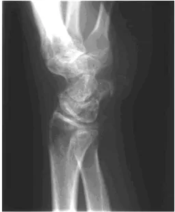
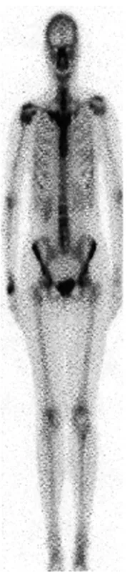
increased activity in either the metacarpals or carpal bones at the base of the right first, second, and third met-acarpals (See Figures 3–4). There was milder non-specif-ic diffuse increased uptake in the remaining right wrist region and medial half of the right clavicle. Based on these findings, a medical diagnosis of shoulder-hand syn-drome was made by the rheumatologist.
Treatment
The patient was treated by a chiropractor two to three times weekly for the initial three weeks, then one to two times per week for another five weeks. The goal of the treatment was to restore first rib, clavicular, sternoclavic-ular, and lower cervical joint biomechanics using spinal manipulative therapy. Soft tissue therapy was also per-formed on the scalenes, trapezius and levator scapulae musculature. Initially, no hand or wrist treatment was provided due to the level of pain. After one week, the
Acupuncture was the chosen modality for pain control. In accordance with Traditional Chinese Medicine patterns, the two common wrist points used were “heart 7” and “triple energizer 5”. Both acupuncture and chiropractic care continued for several months.
Results
A follow-up appointment with the rheumatologist oc-curred at five months. He re-confirmed the diagnosis, al-though he did remark that it was an unusual presentation of the shoulder-hand syndrome. Due to the improvement in both the hand and shoulder symptoms he recommend-ed the patient continue with her current therapy and exer-cises. He also suggested the patient take Actonel due to her moderate osteopenia, and noted that it may aid in her CRPS symptoms. The patient chose not to take the medi-cation.
An evaluation performed at nine months post-onset re-vealed that pain intensity levels had decreased to 1–2/10. The pain was described as a dull ache in the right wrist and shoulder only. Cervical spine and shoulder ranges of motion were unremarkable, as were all orthopaedic tests. Palpation revealed mild hypertonicity of the right sca-lene, and tenderness of the right clavicle and first rib. At this time, the wrist symptoms had greatly improved since the initial examination. Active and passive wrist flexion was full, while there was 30% decrease in active
exten-sion. Tenderness was reported at the base of the right metacarpals and the carpals. Motion palpation of the wrist was tender, with restrictions noted at the radiocar-pal joint and midcarradiocar-pal region. Grip strength was equal bilaterally. Right wrist mobilizations were initiated by the chiropractor at this point.
At the one-year follow-up, the patient had full cervical and shoulder ranges of motion. Right wrist ranges of mo-tion were full with pain at end-ranges and pain with pas-sive wrist extension. Grip strength was equal bilaterally. Figure 3 Wrist bone scan with increased uptake in right
wrist.
At two-year follow-up, the patient reported some mild residual difficulty with prolonged writing, leading to pain in the right thumb and forearm. Cervical ranges of mo-tion and orthopaedic testing were unremarkable. Mild tenderness was noted over the right first rib and trapezius musculature. Right wrist ranges of motion were full, with mild pain at the end range of extension. Grip strength was equal bilaterally with normal colour and muscle appear-ance in comparison to the left hand.
Discussion
Although the new taxonomy for CRPS has been in place for several years, it is evident that many clinicians contin-ue to be unfamiliar with the new terminology. A literature search revealed the use of various synonymous terminol-ogy including Sudek’s atrophy, reflex sympathetic dys-trophy, shoulder-hand syndrome, alygodysdys-trophy, and CRPS. In fact, the taxonomy used often varies depending on the country concerned, the specialty treating the pa-tient, or the personally favored terminology. Part of the confusion stems from the variety of applied diagnostic criteria and until further clarity is gained with respect to the clinical presentation of CRPS, it is important that cli-nicians continue to be very specific and complete when describing the clinical presentation of CRPS.
The pathophysiological mechanism for CRPS remains poorly understood. Early theories focused on hyperactive sympathetic responses causing the symptoms. Propo-nents of this theory cite the salient response to sympa-thetic blockades as evidence of the pathogenesis.6 Others have argued that because blockades are not effective in all patients, other possible pathological mechanisms need to be explored.
Another proposed theory gaining support is the possi-bility of an exaggerated sensitivity to sympathetic nervous system neurotransmitters. A denervation hypersensitivity of the peripheral B-adrenergic receptor is thought to occur following nerve injury. The lowered adrenergic outflow from sympathetic neurons is compensated for by latent re-ceptor up-regulation. Increased rere-ceptor function, synap-tic efficacy and exaggerated response to otherwise normal neurotransmitter loads is the result. The end result is symptoms which are attributed to an over-active sympa-thetic nervous system.4
Recent investigations have reported that an exaggerat-ed inflammatory response may also be a possible disease
mechanism.5 Localized neurogenic inflammation may be related to increased vasodilation, acute edema, and sweating. Clinical findings which indicate that there may be an inflammatory process in the pathogenesis of early CRPS include joint effusions measured with MRIs, in-creased synovial protein concentration, hypervascularity, and neutrophil infiltration.3
The lack of a gold standard test for the diagnosis of CRPS may lead to over diagnosis of this condition. As such, CRPS can be a difficult condition for both patients and clinicians. Other neuropathic disorders, undifferenti-ated arthritis, rheumatism, inflammatory arthropathies, and unilateral vascular occlusive disorders should all be considered as possible differentials. In order to discern between various diagnoses, the clinical and imaging find-ings must not be examined in isolation. Taken together, the findings will quickly help to narrow down the dif-ferential diagnoses. For example, the inflammatory ar-thropathies which affect the hand and wrist include rheumatoid arthritis, psoriatic arthritis, and gout. Infec-tion should also be considered another differential. Some of the radiographic findings of CRPS include normal joint spaces and margins. This assists in differentiating CRPS from infection or rheumatoid arthritis. In addition, the clinical pictures would be notably different for all three diagnoses. Although the clinical findings between CRPS and gout do have several similarities, the radio-graphic findings for both this and psoriatic arthritis are not consistent with those of CRPS.
Several authors cite the misdiagnosis of dysfunctional postures as CRPS Type I, and that a multidisciplinary ap-proach for the diagnosis and treatment of both conditions is needed.8 Currently we must rely on a detailed history and physical examination in order to make the appropri-ate diagnosis. Certain key information must be ascer-tained. This includes any history of inciting trauma, and history of sensory, autonomic, and motor changes. The physical examination should reveal any neurological changes, distribution of pain, swelling, sweating, and trophic changes.
cold forms. Those with colder limbs were more inclined to have been suffering from CRPS for a longer period of time.5 Due to the inconsistencies of presenting symptoms among acute and chronic sufferers, it has also been sug-gested that the syndrome be classified as either mild, moderate, or severe.7 The current diagnostic criteria re-quire that the following four criteria be satisfied:2
1. The presence of an initiating noxious event or a cause of immobilization.
2. Continuing pain, allodynia, or hyperalgesia with which the pain is disproportionate to any inciting event. 3. Evidence at some time of edema, changes in skin
blood flow, or abnormal sudomotor activity in the re-gion of the pain.
4. This diagnosis is excluded by the existence of condi-tions that otherwise would account for the degree of pain and dysfunction.
In the IASP classification, Merskey and Bogduk noted that only criteria 2 and 4 had to be satisfied.
Paraclinical testing is important to provide information regarding autonomic, sensory, and motor changes. Bone scintigraphy is useful in providing information about vas-cular bone changes, especially in later disease stages. Plain radiographs need only be taken at the chronic stage, when bone mineralization status can be evaluated. Quan-titative sensory testing (QST) and autonomic function testing for these patients has been reviewed in the litera-ture, although it is beyond the scope of this paper to dis-cuss the details of the testing.3,7
Not only has the taxonomy of this syndrome changed, the therapeutic approach has also been greatly modified. Sympathetic blockades were a common diagnostic tool and form of therapy. Studies have shown that there is in-adequate evidence to unequivocally report the benefit of sympathetic blockades on pain levels and motor function. It was reported that approximately 85% of CRPS patients report a positive acute effect following sympathetic blocks.3 In contrast, others have reported that sympathet-ic blockade or sympathectomy had minimal effect with only 7% reporting lasting success.5
More recently, there has been a shift towards restored functional abilities instead of pain control as the primary goal of treatment. Exercise therapy is considered one of the key elements of this process.7 Physical therapy is an
important component to treatment although aggressive therapy can be detrimental in early stages when pain lev-els are high. Although one functional goal is to improve joint motion and strength, passive movements are fre-quently too painful and should be avoided in acute pa-tients.4 Passive therapy, followed by active isometric and then active isotonic training can be pursued when pain levels diminish.3
A multidisciplinary approach to treatment should be pursued with these patients. Various modes of therapy may be considered. The primary goal of pharmacological intervention is pain relief.7 In the early onset, under the assumption that neurogenic inflammation is the patho-logical mechanism, NSAIDs and steroids may be admin-istered as a form of pain control. Even so, Vacariu reported that few clinical trials have shown benefits.7 There are no long-term studies on the use of opioids in treating CRPS or other neuropathic pain although this is thought to be an effective initial course of therapy in or-der to manage pain levels. Tri-cyclic antidepressants have been shown to have an analgesic effect and are used in doses smaller than those needed to produce anti-depres-sant effects (75–150mg/day).3 The use of TCAs may be related to interrupting the pain cycle by improving sleep, mood, and anxiety. Physical therapy, and to a lesser de-gree occupational therapy, has been effective in reducing pain and improving active mobility in patients with CRPS I of less than a one year duration.9
These two CRPS cases are encouraging with respect to the results obtained using a non-pharmacological multi-disciplinary approach to care. CRPS can be a severe, de-bilitating condition of which the effects may be long lasting. A key factor in the prognosis and recovery of CRPS patients is early intervention focusing on restora-tion of funcrestora-tional abilities. Manipulative therapy serves to restore joint function and may be effective in improv-ing somatic dysfunction associated with CRPS and other similar conditions.4 Home exercises and a return to activ-ities of daily living may limit the overall disability and shorten recovery time.
Although the diagnostic criteria of CRPS were met in the current case, the true pathophysiology of the disease is yet to be completely understood. Due to the varied symptom presentation and extent of affliction, it may be unclear which conservative therapies will be most benefi-cial in the treatment of CRPS. In order for clinicians to make truly accurate diagnoses and treatment protocols for this group of patients, more investigation of therapies such as chiropractic care, acupuncture, massage, and ex-ercise as they relate to the pathophysiology are needed.
Acknowledgements
The authors would like to thank Dr. Paula Stern for her assistance in reviewing the manuscript.
References
1 Cepeda S, Carr D. Local anaesthetic sympathetic blockade for complex regional pain syndrome. The Cochrane Database of Systematic Reviews 2004, Issue 1. Art. No.:CD004598.
2 Stanton-Hicks M, Janig W, Hassenbusch S, Haddox JD, Boas R, Wilson P. Reflex sympathetic dystrophy: changing concepts and taxonomy. Pain 1995; 63:127–133.
3 Wasner G, Schattschneider J, Binder A, Baron R. Complex regional pain syndrome – diagnostic, mechanisms, CNS involvement and therapy. Spinal Cord 2003; 41:61–75 4 Muir JM, Vernon H. Complex regional pain syndrome and
chiropractic. JMPT 2000; 23(7):490–497.
5 Veldman PHJM, Reynan HM, Arntz IE, Goris RJA. Signs and symptoms of reflex sympathetic dystrophy:
prospective study of 829 patients. The Lancet 1993; 342:1012–1016.
6 Turner-Stokes L. Reflex sympathetic dystrophy – a complex regional pain syndrome. Disability and Rehabilitation 2002; 24:939–947.
7 Vacariu G. Complex regional pain syndrome. Disability and Rehabilitation 2002; 24:435–442.
8 Stutts JT, Kasdan ML, Hickey SE, Bruner A. Reflex sympathetic dystrophy: misdiagnosis in patients with dysfunctional postures of the upper extremity. The Journal of Hand Surgery 2000; 25A:1152–1156.
9 Oerlemans HM, Oostendorp RAB, de Boo, Goris RJA. Pain and reduced mobility in complex regional pain syndrome I: outcome of a prospective randomized controlled clinical trial of adjuvant physical therapy versus occupational therapy. Pain 1999; 83:77–83.