Rehabilitation protocol for undisplaced
Colles’ fractures following cast removal
Stephen Balsky,
BSc(Hons), DC*
Richard J Goldford,
BSc, DC, FCCSS(C), FCCRS(C)* Austin Rehabilitation & Treatment Clinic, 4 – 2930 Islington Avenue, North York, Ontario M9L 2K5. Phone: (416) 742-5952 Fax: (416) 742-7591.
Reprint information can be directed to Dr. Stephen Balsky at the above address. © JCCA 2000.
Colles’ fracture is a relatively uncommon presentation to a chiropractic office. A case of a 74-year-old woman complaining of pain, loss of strength and diminished range of motion in her left wrist is presented. These complaints were the result of a slip and fall causing a Colles’ fracture that occurred four weeks prior to presentation. Dynamometer and goniometric testing revealed significant losses of strength and range of motion when compared to the unaffected wrist. Initial therapy consisted of ice, wax bath application and gentle range of motion mobilizations for two weeks followed by entry into a supervised active rehabilitation program for a further three weeks. After thirteen visits, the patient demonstrated objective improvement in both range of motion and grip strength as well as subjective improvement in pain intensity. A rehabilitation protocol is proposed for clinicians with patients suffering from Colles’ fractures. Appropriate management may begin passively and ultimately leads to a supervised active program for optimal results.
(JCCA 2000; 44(1):29–33)
K E Y W O R D S: fracture, wrist, rehabilitation.
Les cas fracture de Colles sont relativement peu
fréquents en chiropratique. Voici l’histoire d’une femme de 74 ans qui se plaint de douleur, d’une diminution de la force de préhension et de l’amplitude des mouvements du poignet gauche à la suite d’une chute ayant causé une fracture de Colles quatre semaines auparavant. Les épreuves au dynamomètre et au goniomètre révèlent en effet une diminution importante de la force de préhension et de l’amplitude des mouvements du poignet gauche par rapport au poignet droit. La première phase de traitement a consisté en l’application de glace et de bains de cire et en la mobilisation de faible amplitude de l’articulation; cette première phase a duré deux semaines et a été suivie d’un programme supervisé de réadaptation active pendant trois autres semaines. Au bout de treize visites, on a noté une amélioration objective de la force de préhension et de l’amplitude des mouvements du poignet, et la patiente a fait état d’une diminution subjective de la douleur. Le présent article propose donc aux cliniciens un protocole de réadaptation pour les patients ayant subi une fracture de Colles. Le traitement peut commencer par des exercices passifs et finir par un programme supervisé d’exercices actifs pour l’obtention de résultats optimaux.
(JACC 2000; 44(1):29–33)
Introduction
Colles’ fracture is defined as a linear transverse fracture of the distal radius approximately 20–35 mm proximal to the articular surface with dorsal angulation of the distal frag-ment.1 Females are predelected more than males for this
type of injury and there is often a precedent history of osteoporosis.1 Stable Colles’ fractures present with
mini-mal comminution. Unstable fractures are distinctly com-minuted often with corresponding avulsions of the ulnar or radial styloid that have the potential to cause compression neuropathies, especially of the median nerve.1 Other
com-plications that have been reported include reflex sympa-thetic dystrophy and degenerative joint disease.1
A case report of a 74-year-old female who presented one month post injury to her left wrist is described. A treatment protocol is presented to restore patients with this type of injury to their pre-accident activities.
Case report
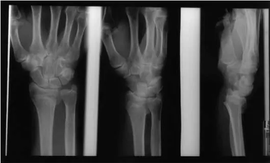
A 74-year-old woman reported an acute onset of left wrist pain following a slip and fall on the ground and landing on an outstretched, extended hand. She was immediately taken to a hospital facility where a routine series of plain film radiographs revealed a non-comminuted Colles’ frac-ture. Figure 1 displays the radiograph of a similar injury. She was placed in a plaster cast for 4 weeks at which time she was referred to the clinic for assessment and rehabilita-tion.
The patient complained of a persistent pain and loss of motion accompanied by moderate effusion of the left distal radius. The pain was reported to be worse upon wak-ing in the mornwak-ing and was marginally relieved by medica-tion.
Goniometric evaluation of the left wrist revealed a loss of active range of motion in extension completely, in
flexion by 50o and in radial and ulnar deviation by 10o and
20o respectively. Dynamometer testing revealed a grip
strength of 5 lbs in the left extremity and 35 lbs in the right extremity. Neurological testing of the cervical spine and upper extremity were unremarkable bilaterally. Active range of motion of the unaffected upper extremity joints were full and pain free bilaterally. Orthopaedic testing of the cervical spine, shoulder joints, elbow joints and for carpal tunnel syndrome were unremarkable. Palpatory evaluation revealed moderate atrophy of the left wrist ex-tensor muscles with severe pain to mild pressure and mod-erate effusion 1 cm proximal to the left distal radius.
Initial treatment consisted of the application of ice to reduce swelling, followed by gentle, passive range of mo-tion exercise to patient tolerance. Once the swelling had abated, application of heat using a paraffin wax bath was introduced to increase circulation and mobility. At this point she entered a supervised active program for a further three weeks. The program focused on increasing mobility and strength to the injured extremity.
At the end of the five week treatment period, swelling of the distal radius had reduced considerably and goniometric assessment of the active range of motion had improved by 25o in flexion, 50o in extension and 5o in both radial and
ulnar deviation. Subjectively, the patient still reported a dull pain with wrist extension, however the severity was considerably reduced from initial presentation using a 10 point numerical rating scale.
Discussion
The literature surrounding conservative management of Colles’ fractures reveals conflicting results. Dias et al.2
report that early wrist mobilization resulted in rapid recov-ery of both strength and movement without adversely in-fluencing the progression of residual deformity. Poorer prognoses were associated with the use of plaster casting over a crepe bandage and displacement of fracture lines. McAuliffe and colleagues3 report that early mobilization
demonstrated distinct improvement in strength and pain, however there was no significant improvement in the final range of movement of the healed wrist. In their study of post-fracture weakness and diminished range of motion, Kaufman et al.4 reported significant recovery from a
unique treatment regimen employing manipulation of the intercarpal and radiocarpal joints in flexion and extension. Despite this variability in outcome, it is generally
ac-cepted that early rehabilitation of acute injuries can main-tain mobility of the joint capsule and ligaments, prevents adherence of soft tissues, provides increased circulation to the healing bone and assists in the reduction of edema.5
The case report outlines a rehabilitation protocol utilized to improve range of motion and grip strength in an undisplaced, stable Colles’ fracture.
Following presentation and evaluation, our patient be-gan a treatment regimen that consisted initially of passive interventions designed to improve circulation and prevent immobilization adhesion formation. These treatments in-cluded application of an ice pack to reduce edema fol-lowed by application of a wax bath on the affected wrist. Gentle range of motion mobilizations were then intro-duced that could only be performed in flexion and exten-sion to the patient’s pain tolerance. The mobilizations performed were similar to those described by Collins.5
Three sets of 5 flexion/extension repetitions were per-formed on the affected wrist. In addition, the joint was mobilized in circumduction, ulnar flexion and radial flexion to the patient’s level of tolerance.
Following six treatments in this fashion, the patient then entered a supervised active rehabilitation program. The program focused on restoring active range of motion and strength using a variety of different techniques. Table 1 outlines the stepwise progression of exercises employed in the program.
After nine visits under this regimen, the patient was re-evaluated to monitor progress. Range of motion was as-sessed using a goniometer and strength was measured using a grip dynamometer. Effusion and sensitivity to pal-pation was compared to the initial assessment findings. In addition, the patient was asked to subjectively rate her current status using a 10 point scale. She was then edu-cated to perform the same active protocol at home at the same frequency and intensity. In addition she was encour-aged to resume functional activities that involve the wrist and hand such as writing, cooking and sewing. These ac-tivities give the patient a tangible outcome measure be-yond the clinical setting. In this way the patient is better able to grade their progression.
the joints and musculature both above and below the site of injury. A review of our protocol demonstrates that in addi-tion to exercises for the wrist, routines were also devel-oped for the finger intrinsics, elbow and shoulder. Patients who present post Colles’ fracture tend to guard the entire upper extremity as a means of protecting the wrist. By introducing exercise for the entire limb, disuse atrophy and stiffness due to immobilization will be avoided.6 In
addi-tion, muscle balancing for strength and endurance in the entire upper/lower extremity will be attained.
As a patient progresses through their program, the clini-cian should be regularly monitoring patient progress and noting any changes in range of motion, strength, degree of effusion and level of pain and disability. Should the symptoms not abate or regress with intervention, the cli-nician should consider referral to an appropriate
special-Table 1
Rehabilitation protocol for Colles’ Fracture
A) ISOMETRIC EXERCISE 1) Wrist flexors and extensors
B) ACTIVE RANGE OF MOTION EXERCISE
1) Assisted stretch to forearm flexors and extensor musculature and radial/ulnar deviation
2) Weight bearing wrist extension exercise (hands on the table with the patient leaning forward on them) to patient tolerance
3) Active stretch to shoulder girdle and rotator cuff musculature 4) Active stretch to elbow flexor and extensor musculature C) INTRINSIC HAND MUSCLE EXERCISE
1) Thumb/digit opposition
2) Repetitive squeezing of theraputty 3) Repetitive towel wringing exercise D) STRENGTHENING ROUTINE
1) Biceps curls with 1½ – 2 pound weights bilaterally
2) Shoulder abduction, flexion and extension reps with 2 pound weights bilaterally 3) Repetitive squeezing of rubber ball in affected wrist
4) Flexion and extension of wrist using 1½ pound weights increasing as tolerated E) FUNCTIONAL ACTIVITIES
1) Patient is encouraged to resume pre-accident activities that involve the affected extremity (eg. writing, typing, cooking, etc.)
ist for further investigation. The complications resulting from Colles’ fractures have been well described in the literature.4,5,7,8 The reported sequelae include carpal
tun-nel syndrome, reflex sympathetic dystrophy, tenosynovi-tis of the extensor carpi ulnaris, avulsion of the ulnar styloid process and rupture of the triangular fibrocarti-lage, to name a few. The clinician should also be aware of the potential for malunion of the fractured fragments that is often the result of impaired circulation, inadequate early immobilization or excessive distraction of the wrist.1
Noteworthy to the clinician are studies by Dias2 and
McAuliffe3 which indicate that early mobilization does not
increase the magnitude or the rate of deterioration of the bony deformity. The radiographic results obtained indi-cated that there were no significant changes in the meas-urement of dorsal angulation, radial deviation and radial shortening when comparing those patients that received early mobilization from those that received prolonged im-mobilization. Given these findings, clinicians should therefore endeavor to begin an active protocol as quickly as possible.
.
Summary
Colles’ fractures are transverse linear breaks in the distal radius, often accompanied by dorsal angulation of the ulna following a fall on an outstretched, extended wrist. A case of a 74-year-old woman who slipped and fell and suffered a Colles’ fracture of the left wrist is presented. Therapy was initially passive consisting of ice application to decrease swelling and control pain, wax application to improve circulation and passive range of motion mobilizations to prevent adhesion formation. The patient then entered an active rehabilitation program to restore strength and range of motion to the injured and adjacent extremities. The protocol utilized is outlined along with suggestions for re-evaluation and a delineation of possible complications of such a fracture.
Acknowledgements
The authors would like to thank the Canadian Memorial Chiropractic College Radiology Department for their ef-forts in providing the radiographs included in this report.
References
1 Yochum T, Rowe L. Essentials of skeletal radiology. 2nd edition Vol. 1. Maryland: Williams & Wilkins, 1996; 664–665, 756–757.
2 Dias JJ, Wray CC, Jones JM, Gregg PJ. The value of early mobilization in the treatment of Colles’ fractures. J Bone Joint Surg 1987; 69(3):463–467.
3 McAuliffe TB, Hilliar KM, Coates CJ, Grange WJ. Early mobilization of Colles’ fractures. J Bone Joint Surg 1987; 69(5):727–730.
4 Kaufman Rod L, Bird Joel. Manipulative management of post-Colles’ fracture weakness and diminished active range of motion. J Manipulative Physiol Ther 1999; 22(2):105–107.
5 Collins DC. Management and rehabilitation of distal radius fractures. Ortho Clin North America 1993; 24(2):36–77. 6 Liebenson C. Rehabilitation of the spine – a practitioner’s
manual. Pennsylvania: Williams & Wilkins, 1996: 14–16. 7 Tsukazaki Tomoo, Iwasaki Katsuro. Ulnar wrist pain after
Colles’ fracture. Acta Orthop Scand 1993; 64(4):465–468. 8 Altissimi Maurizio, Antenucci Renato, Fiacca Claudio,