University of Pennsylvania
ScholarlyCommons
Publicly Accessible Penn Dissertations
2016
Temporal Dynamics Of The Skin Microbiome In
Disease
Michael Austin Loesche
University of Pennsylvania, loesche@upenn.edu
Follow this and additional works at:
https://repository.upenn.edu/edissertations
Part of the
Microbiology Commons
This paper is posted at ScholarlyCommons.https://repository.upenn.edu/edissertations/2444
For more information, please contactrepository@pobox.upenn.edu.
Recommended Citation
Loesche, Michael Austin, "Temporal Dynamics Of The Skin Microbiome In Disease" (2016).Publicly Accessible Penn Dissertations. 2444.
Temporal Dynamics Of The Skin Microbiome In Disease
Abstract
The skin is colonized by communities of bacteria, fungi, and viruses, collectively referred to as the skin microbiome. These microbial communities are shaped by the topology and diseases of the skin. Dysbiosis of the cutaneous microbiome has been associated with several ailments of the skin including atopic dermatitis, acne, rosacea, psoriasis, and chronic wounds. However, our understandings of the processes by which these microbes initiate, maintain, or modulate skin diseases is lacking. Moreover, previous research on the topic has largely been limited by cross-sectional study designs, neglecting the natural dynamism of microbial
communities. Here we present a comprehensive analysis of the temporal dynamics of the skin microbiome in various diseases. In the first section, we characterize the diversity and dynamics of both bacterial and fungal communities colonizing chronic wounds and its associations with clinical outcomes. In a study of 100 subjects with diabetic foot ulcers, we sampled the wound microbiota in 2-week intervals until healing, amputation of 26 weeks of follow-up. We demonstrate the high levels of community instability in chronic wounds and expose the positive association between wound healing community instability. We also reveal the effect of antibiotic perturbation on the microbiota. The fungal component was found to have associations with various bacteria and clinical outcomes. Our results should inform the design of future studies and provides evidence that microbial dynamics may be an effective biomarker for identifying high-risk ulcers. The second section investigates the body-site specific effects of psoriasis on the skin microbiome and how it responds to therapy. We reveal these patterns in a study of 114 subjects, across 6 body sites, and over 112 weeks of follow-up. The effect of psoriatic lesions was found to be mild and body-site specific. In contrast, ustekinumab treatment was found to induce moderate shifts in microbial composition, including an increase in atypical skin bacteria and inter-individual heterogeneity. These results suggest that the effect of psoriasis lesions is secondary to the effect the broad effects of the immune environment. Together the work presented in this thesis represents a significant advancement in our understanding of the microbial dynamics of the skin and their associations with human health.
Degree Type
Dissertation
Degree Name
Doctor of Philosophy (PhD)
Graduate Group
Cell & Molecular Biology
First Advisor
Elizabeth A. Grice
Second Advisor
Frederic D. Bushman
Keywords
Chronic Wounds, Foot Ulcer, Microbiology, Microbiome, Psoriasis, Skin
Subject Categories
Microbiology
TEMPORAL DYNAMICS OF THE SKIN MICROBIOME IN DISEASE
Michael Austin Loesche
A DISSERTATION
in
Cell and Molecular Biology
Presented to the Faculties of the University of Pennsylvania
in
Partial Fulfillment of the Requirements for the
Degree of Doctor of Philosophy
2016
Supervisor of Dissertation Co-Supervisor of Dissertation
_______________________ ________________________
Elizabeth A. Grice, PhD Frederic D. Bushman, PhD
Assistant Professor in Dermatology William Maul Measey Professor in Microbiology
Graduate Group Chairperson
________________________ Daniel S. Kessler
Associate Professor of Cell and Developmental Biology
Dissertation Committee
Aimee Payne, MD, PhD Phillip Scott, PhD
Albert M. Kligman Associate Professor of Microbiology and
Professor of Dermatology Immunulogy
David Margolis, MD, PhD Hongzhe Li, PhD
Professor of Dermatology Professor of Biostatistics and
ii
DEDICATION
iii
ACKNOWLEDGMENT
I have been the recipient of incredible support and had the privilege to work with and
learn from amazing individuals. I am deeply thankful for having been the beneficiary of the
advising, support, and generosity of my mentor, Elizabeth Grice. She has always been my
advocate and encouraged me to purse my interests, in and out of the lab. Elizabeth has had a
tremendous influence on my development as a physician-scientist and as an individual. I have
benefitted immensely from her tutelage and friendship, and for that I am eternally grateful.
For their guidance and support, I thank my co-advisor, Frederic Bushman, and the
members of my thesis committee, Aimee Payne, Hongzhe Li, David Margolis, and Phil Scott. My
development as an independent physician-scientist was significantly advanced by their thoughtful
feedback and critical analysis of my work. I would particularly like to thank my thesis chair,
Aimee Payne. She is an incredible student advocate, leader in her field, and accomplished mentor,
and I am indebted for her support and guidance. My committee pushed me to succeed as a
scientist, but never forgot my commitment to being a physician; for that I am eternally grateful.
I am incredibly fortunate to have been a part of Club Grice, the most amazing lab in the
world. I have learned so much from my lab mates, past and present, and cherish their friendship.
Every one of them has provided intellectual and emotional support during my thesis work, and
listened to me ramble for more hours than anyone deserves. For this I need to thank: Geoffrey
Hannigan, Amanda Tyldsley, Jackie Meisel, Adam SanMiguel, Brendan Hodkinson,Joseph
iv
I am thankful for the continued support of Skip Brass and the rest of the combined-degree
office. I especially need to thank Maggie Krall and Maureen Kirsch. I would not be where I am
today without their amazing generosity and exceptional patience.
None of this would have been possible had it not been for the incredible support of my
friends and family. My family has always been a source of unquestioning love and support, which
has allowed me to pursue my highest aspirations without fear or hesitation. My parents always
enabled my passions and supported me every step of the way. I am incredibly lucky to have six of
the most amazing people I know, as my brothers and sisters. Finally, I must thank my friends for
helping keep me sane throughout my training. I am eternally grateful to have been surrounded by
v
ABSTRACT
TEMPORAL DYNAMICS OF THE SKIN MICROBIOME IN DISEASE
Michael A Loesche
Elizabeth A Grice, PhD
The skin is colonized by communities of bacteria, fungi, and viruses, collectively referred to as
the skin microbiome. These microbial communities are shaped by the topology and diseases of
the skin. Dysbiosis of the cutaneous microbiome has been associated with several ailments of the
skin including atopic dermatitis, acne, rosacea, psoriasis, and chronic wounds. However, our
understandings of the processes by which these microbes initiate, maintain, or modulate skin
diseases is lacking. Moreover, previous research on the topic has largely been limited by
cross-sectional study designs, neglecting the natural dynamism of microbial communities. Here we
present a comprehensive analysis of the temporal dynamics of the skin microbiome in various
diseases. In the first section, we characterize the diversity and dynamics of both bacterial and
fungal communities colonizing chronic wounds and its associations with clinical outcomes. In a
study of 100 subjects with diabetic foot ulcers, we sampled the wound microbiota in 2-week
intervals until healing, amputation of 26 weeks of follow-up. We demonstrate the high levels of
community instability in chronic wounds and expose the positive association between wound
healing community instability. We also reveal the effect of antibiotic perturbation on the
microbiota. The fungal component was found to have associations with various bacteria and
clinical outcomes. Our results should inform the design of future studies and provides evidence
vi
second section investigates the body-site specific effects of psoriasis on the skin microbiome and
how it responds to therapy. We reveal these patterns in a study of 114 subjects, across 6 body
sites, and over 112 weeks of follow-up. The effect of psoriatic lesions was found to be mild and
body-site specific. In contrast, ustekinumab treatment was found to induce moderate shifts in
microbial composition, including an increase in atypical skin bacteria and inter-individual
heterogeneity. These results suggest that the effect of psoriasis lesions is secondary to the effect
the broad effects of the immune environment. Together the work presented in this thesis
represents a significant advancement in our understanding of the microbial dynamics of the skin
vii
TABLE OF CONTENTS
LIST OF TABLES ... X
LIST OF FIGURES ... X
CHAPTER 1 – INTRODUCTION TO THE SKIN MICROBIOME ... 1
1.1 From Microscopes to Microbiomes ... 1
1.2 The Human Microbiome ... 2
1.3 Microbiome W orkflow ... 4
1.4 The Structure and Function of Human Skin ... 6
1.5 The Healthy Skin Microbiome ... 8
1.6 W ound Healing and Chronic W ounds ... 11
1.7 A Note on Terminology ... 14
1.8 References ... 14
CHAPTER 2 – TEMPORAL STABILITY IN CHRONIC WOUND
MICROBIOTA IS ASSOCIATED WITH POOR HEALING ... 22
2.1 Abstract ... 22
2.2 Introduction ... 23
2.3 Results ... 25
2.3.1 Characterization of the DFU microbiota at baseline ... 25
2.3.2 DFU microbiota can be partitioned into four community types ... 26
2.3.3 The frequency of Community Type transitions in DFU are associated with clinical outcomes ... 27
2.3.4 DFU with more dynamic microbiota heal faster than those with less dynamic microbiota ... 28
2.3.5 Effect of antibiotics on temporal stability in DFU microbiota ... 29
2.4 Discussion ... 30
2.5 Materials and Methods ... 32
2.5.1 Study Design ... 32
2.5.2 Setting and Sample ... 32
2.5.3 Study Variables ... 33
2.5.4 Data Analyses ... 35
3.6 Author Contributions ... 36
3.7 Acknowledgements ... 36
viii
3.9 Tables ... 45
2.10 References ... 49
CHAPTER 3 – REDEFINING THE CHRONIC WOUND MICROBIOME:
FUNGAL COMMUNITIES ARE PREVALENT, DYNAMIC, AND
ASSOCIATED WITH DELAYED HEALING ... 54
3.1 Contributions ... 54
3.2 Abstract ... 54
3.3 Importance ... 55
3.4 Introduction ... 56
3.5 Results ... 58
3.5.1 Study overview ... 58
3.5.2 Characterization of the DFU Fungal Mycobiome ... 59
3.5.3 The DFU Mycobiome Has High Interpersonal and Intrapersonal Variation ... 61
3.5.4 The DFU Mycobiome is Associated with Clinical Outcomes ... 62
3.5.5 Pathogens versus allergens in the DFU mycobiome ... 64
3.5.6 The DFU Mycobiome Forms Multi-Species Biofilms with Bacteria ... 66
3.6 Discussion ... 67
3.7 Materials and Methods ... 70
3.7.1 Study Design ... 70
3.7.2 Study Variables ... 70
3.7.3 Fungal and Bacterial Manipulation ... 74
3.7.4 Data Analyses ... 76
3.8 Data Availability ... 76
3.9 Funding Information ... 76
3.10 Acknowledgements ... 77
3.11 Figures ... 78
3.12 Tables ... 93
3.13 References ... 97
CHAPTER 4 – LONGITUDINAL STUDY OF THE
PSORIASIS-ASSOCIATED SKIN MICROBIOME DURING THERAPY WITH
USTEKINUMAB ... 107
4.1 Abstract ... 107
4.2 Background ... 108
4.3 Results: Phase I – Response to Ustekinumab ... 111
ix
4.3.2 Skin microbiome differences between lesion and non-lesion skin are mild and site
specific. ... 112
4.3.3 Lesion and non-lesion skin microbiota respond similarly to ustekinumab therapy ... 113
4.3.4 Microbiota of lesion and non-lesion skin diverges with treatment ... 115
4.3.5 Greater heterogeneity within psoriatic lesions than non-lesion skin ... 116
4.4 Results: Phase II – Duration of Ustekinumab Response ... 117
4.4.1 The skin microbiome is not predictive of the duration of therapeutic response to ustekinumab ... 117
4.4.2 Recurrent lesions do not resemble original lesions ... 118
4.4.3 No difference between standard and tailored dosing on effect of skin microbiome ... 118
4.5 Discussion ... 119
4.6 Conclusions ... 123
4.7 Methods ... 125
4.7.1 Study Design ... 125
4.7.2 Sample Sequencing and Processing ... 126
4.7.3 Data Analysis ... 127
4.9 Figures ... 129
4.10 Tables ... 140
CHAPTER 5 – CONCLUSIONS AND FUTURE DIRECTIONS ... 147
5.1 Conclusions and Future Directions ... 147
x
LIST OF TABLES
Table 2-S1 Patient and ulcer characteristics for the total sample and by complication
status 45
Table 2-S2 Summary of microbial communities in DFU samples. 46
Table 2-S3 Summaries of taxonomic composition by community type 47
Table 2-S4 Estimated Markov chain parameters of DFU CT transitions 48
Table 3-1 Subject Demographics and wound characteristics 93
Table 3-2 Distribution of the top 1% identified fungal taxa 94
Table 3-S1 Summary of Spearman correlation coefficients and p-values for
supplementary figure 1 95
Table 3-S2 Differentially abundant taxa between forefoot and hindfoot wounds 96
Table 4-1 Characterization of subjects’ demographics and results of randomization
process 140
Table 4-2 Psoriatic lesion classification accuracy by body site at the baseline visit 141
LIST OF FIGURES
Figure 2-1 The DFU microbiome clusters into four Community Types 38
Figure 2-2 DFU Community Types are dynamic 39
Figure 2-3 Inter-visit Weighted UniFrac distances associations with healing time for
subjects that healed within 24 weeks 40
Figure 2-4 Effects of antibiotics on microbial communities in DFUs 42
Figure 2-S1 Laplace approximation predicts 4 clusters as optimal. 43
Figure 3-1 The DFU mycobiome is diverse and highly heterogeneous 78
Figure 3-2 The DFU mycobiome is temporally unstable 80
Figure 3-3 The DFU mycobiome is associated with clinical outcomes 81
Figure 3-4 Pathogens are associated with necrotic tissue and poor outcomes 82
Figure 3-5 Pathogens form inter-kingdom biofilms 84
Figure 3-S1 Heatmap illustrating positive and negative correlations between microbiome
factors and clinical factors at baseline 85
Figure 3-S2 Subjects with positive yeast culture result 86
xi
Figure 3-S4 Shannon Diversity Index for subjects administered an antibiotic during the
course of the study or experienced a complication 88
Figure 3-S5 Dendogram and heatmap illustrating positive and negative correlations between the taxa found in >1% abundance across the entire sample set and the
pathogens and allergens groups 89
Figure 3-S6 Dendogram and heatmap illustrating positive and negative correlations between the fungal and bacterial taxa found in >1% abundance across the entire sample
set 90
Figure 3-S7 Quantitative culture data 91
Figure 4-1 Clinical trial design diagram 129
Figure 4-2 The effect of the psoriatic lesions on the skin microbiota at baseline 130
Figure 4-3 Longitudinal changes in taxonomic composition following ustekinumab
treatment 139
Figure 4-4 Lesion and non-lesion sites exhibit similar amounts of change due to
ustekinumab therapy 133
Figure 4-5 Divergence of lesion and non-lesion skin following treatment 134
Figure 4-6 Body sites variance and distinctness increases with ustekinumab therapy135
Figure 4-7 Recurrent lesions do not resemble prior lesions 136
Figure 4-8 Ustekinumab dosing frequency does not impact the skin microbiota 137
Figure 4-S1 Line plot showing the mean PGA score for each treatment group 138
1
CHAPTER 1 – Introduction to the Skin Microbiome
1.1 From Microscopes to Microbiomes
It was advancements in the field of microscopy that led to the discovery of bacteria and
other microbes inhabiting our world in 1676 by Antonie van Leeuwenhoek (Dobell 1960),
ushering in an era of awe and fascination with the microscopic world that persists to this day. It
did not take long before many noticed large discrepancies between numbers of bacteria visible
under a microscope and the number of colonies they could culture on a plate. In some cases the
difference between the two was several orders of magnitude, a phenomenon known as the great
plate count anomaly (Staley & Konopka 1985). Bacteria, such as E. coli, that are easily cultured
have fueled decades of discovery in genetics, molecular biology, and modern medicine. However,
the fastidious and oligotrophic bacteria that have eluded being cultured greatly outnumber those
that have. It is now estimated that 95-99% of bacteria are not readily culturable, though focused
efforts to improve culturing surveys are changing this number (Browne et al. 2016).
It was the development of next-generation sequencing technology that facilitated the
study of entire microbial communities or microbiomes, overcoming the biases and limitations of
culturing techniques. The term microbiome refers to the community of bacteria, fungi, viruses
and other microbes occupying an ecological niche. Microbiome studies have revealed rich and
complex communities inhabiting nearly all surfaces of the environment, including the human
body (Gevers et al. 2012). They have led to significant increases in our understanding of
microbial ecology, host-microbe interactions, and microbe-microbe interactions. To manage the
massive amounts of data generated by microbiome studies, the field of microbiology has become
2
have also attracted inter-disciplinary collaborations resulting in the adoption of theories and
methodologies from many fields including ecology, multivariate statistical modeling,
machine-learning, evolution, phylogenetics, and network analysis.
The study of the world’s microbiomes is still in its infancy, though rapid progress is
being made. Studies characterizing microbial communities and documenting ecological
phenomenon are being published at an ever-increasing rate. Standards for sample collection,
processing, and analysis are beginning to become formalized; and tools to manipulate the
microbiome, experimentally and medically, are being developed. After more than a century of
mechanistic and hypothesis driven work, microbiology has returned to the stage of observation.
Like the naturalists of the past, we have been give a new lens to discover a world previously
unknown.
1.2 The Human Microbiome
The human microbiome is the collection of ecological communities of bacteria, fungi,
viruses, and other microbes that colonize our bodies. The commensal bacteria perform a variety
of beneficial roles including immune system education, vitamin production, and protection form
invading pathogens. They inhabit nearly every surface of the human body exposed to the external
environment including the skin, gut, vagina, oral cavity, and even the upper airways of the lung
(Grice & Segre 2012; Charlson et al. 2012). While some constituents are conserved, the structure,
composition, and biomass of the human microbiome vary greatly between body sites and are
3
reflects the health of its host, and in many cases may be a driving factor in the initiation or
persistence of disease (Cho & Blaser 2012).
The complexity of human-microbe interactions is staggering in its breath and depth. Even
the bacterial component alone, by far the best studied, is unfathomable in its scale. The average
human body is composed of 30 trillion cells, but it is colonized by approximately 39 trillion
bacterial cells (Sender et al. 2016). These commensal bacteria interact with their human host via a
wealth of secreted compounds, metabolites, antigens, and occasionally toxins. They also interact
with each other by competing for nutrients, producing antibiotics and bacteriocins, facilitating
horizontal gene transfer, and myriad other antagonistic and mutualistic interactions. Nowhere is
this more apparent than in the gut, where the microbiome has been shown to contribute to disease,
modulate physiologic responses to diet, and provide protection from invading pathogens
(Shreiner et al. 2015). There is mounting evidence for the role of the gut microbiome in
inflammatory bowel diseases (Morgan et al. 2012), coronary artery disease (Koeth et al. 2013),
obesity (Turnbaugh et al. 2006), insulin resistance (Suez et al. 2014), depression (Foster &
McVey Neufeld 2013), hepatic encephalopathy (Bajaj et al. 2012), and differential drug
absorption (Clayton et al. 2009). Moreover, it is well established that antibiotic perturbations of
the gut microbiota leads to dysbiotic states (Modi et al. 2014), which increase the risk of
developing Clostridium difficile infections (Buffie et al. 2014), which can be successfully treated
with fecal microbiota transplantation (Kassam et al. 2013).
The centrality of the gut microbiome in many ailments has raised the possibility that the
microbiome of other body sites may hold similar promise as driving determinants of health and
4
Alterations in the vaginal microbiome have been linked to bacterial vaginosis (Mayer et al. 2015),
chronic yeast infections (Liu et al. 2013), and preterm delivery (DiGiulio et al. 2015). The oral
microbiome may contribute to periodontal disease and dental caries (Wade 2013). Even the lung,
which is generally thought to be a sterile site, has been shown to have a microbiome that can
reflect the immunity status of its host (Charlson et al. 2012; Wang et al. 2016; Young et al. 2014;
Quinn et al. 2014).
Seminal work put forth by Grice et al characterized the healthy skin microbiome and how
it varies spatially across the body(Grice et al. 2009). Subsequent studies have since linked
dysbiosis of the skin microbiome with psoriasis (Takemoto et al. 2014; Alekseyenko et al. 2013;
Ganju et al. 2016), chronic wounds (Grice et al. 2010; Gardner et al. 2013), acne (Fitz-Gibbon et
al. 2013), and atopic dermatitis (Kong et al. 2012); however, whether the skin microbiome plays a
causative or reactive role remains to be established. The majority of these studies implemented
cross-sectional study designs, and those with longitudinal analyses have been limited by sample
number. Consequently, the temporal dynamics of the skin microbiome in disease are poorly
understood and may yield better understanding and potential therapeutic targets for diseases of
the skin. The work presented here endeavors to advance our understanding in this regard by
characterizing the microbial dynamics of chronic wounds and psoriasis and their associations
with clinically meaningful outcomes.
1.3 Microbiome Workflow
Microbiome studies can be broadly classified into metataxonomic and metagenomic
5
conserved marker genes, which serve as a proxy for taxonomic classification. The 16S rRNA
gene has been extensively used to characterize prokaryotic communities, whereas the 18S rRNA
gene and the internal transcribed spacer (ITS) regions are used for fungal communities.
Bacteriophage and other viruses do not possess universally conserved genes, making
metataxonomic analyses impossible to perform. These marker genes contain conserved and
hypervariable regions, which allow for universal primer annealing and taxonomic classification
respectively. The 16S rRNA gene is approximately 1.5 kilobases in length and contains 9
hypervariable regions, denoted V1 through V9. Because of the current limitations in sequence
lengths generated by next-generation sequencing technologies, specific sub-regions must be
chosen for amplification and sequencing. The most commonly used sub-regions are V1-V3 and
V4, however, resident evidence suggests that the V1-V3 region introduces less bias for skin
communities (Meisel et al. 2016). In contrast, metagenomic studies involve shotgun sequencing
the combined genomic content of the entire community. Metagenomic studies provide both a
taxonomic and functional perspective of the community, but require much greater sequencing
depth, computational resources, and controlling for human contamination.
Metaxonomic studies begin with the aggregation of similar sequences into clusters,
termed operational taxonomic units (OTU), which serve as proxies for species (Kopylova et al.
2016). OTUs are taxonomically identified and are then used to estimate alpha and beta diversity
by a variety of metrics. Alpha metrics measure the diversity intrinsic to a sample by quantifying
the richness (number of observed OTUs) and evenness of the community. Beta metrics measure
the amount of shared diversity between samples. Diversity metrics differ in how abundance or
phylogenetic relationships are weighted, which reveal different components of the community
6
popular adoption of software packages such as QIIME and MOTHUR has partially standardized
these analyses (Caporaso et al. 2010; Schloss et al. 2009).
1.4 The Structure and Function of Human Skin
The skin is one of the most exposed organs of the body and serves a critical role as the
primary interface between the external environment and the underlying tissues. The skin provides
a barrier against constant physical, chemical, and immunological insults. It also performs key
homeostatic functions in regulating body temperature, fluid balance, and production of vitamin D
(Telofski et al. 2012). The skin covers approximately 1.8 m2 with multiple microenvironments
created by variation in exposure (folds, invaginations, and clothing), sweat and sebum production,
and hair distribution. The diversity of skin microenvironments is reflected by the rich and
complex communities of microbes that colonize the skin. Thus, to fully understand the skin
microbiome and its interactions with the skin, we must be familiar with the biology and topology
of the skin.
The skin is composed of two layers, the dermis and epidermis, which rest above a layer
of subcutaneous fat (Simpson et al. 2011). The deeper of the two, the dermis is composed of
dense, irregular connective tissue and contains a variety of receptors, vessels, and glands involved
in maintenance of epidermal integrity. Superficial to the dermis is the epidermis, which itself is
composed of four layers of keratinocytes various stages of development. The stratum basale, as
the name implies, is the basal layer, which is separated from the dermis by the epidermal
basement membrane (Fuchs & Raghavan 2002). This layer contains the undifferentiated stem
7
immature keratinocytes begin to flatten and develop lamellar bodies and keratin fibrils. As
keratinocytes continue to mature, they develop into the stratum granulosum, characterized by
abundant keratohyalin granules. The granules contain filagrin, keratin, loricin, and involucrin,
which are critical for the proper barrier function of the epidermis. To complete the differentiation
process, the keratinocytes then anucleate and fully flatten forming the stratum corneum. Cells at
this stage are termed corneocytes for their highly cornified envelope (Candi et al. 2005).
The orderly layers of the epidermis are regularly interrupted by hair follicles and glands
that extend into the dermis. Sebaceous glands specialized in the secretion of a lipid-rich substance
termed sebum, which contributes to the waterproofing of the skin (Zouboulis & Boschnakow
2001). The glands are often associated with hair follicles forming pilosebacious units, particularly
prevalent on the face and scalp. These units provide an ideal environment for anaerobic bacteria
that metabolize the secreted lipids into free fatty acids, contributing to the relatively acidic pH of
the skin (Puhvel et al. 1975). Sweat glands in contrast are distributed throughout the body,
though they are particularly concentrated in the axillae, palms, soles, and forehead (Lu & Fuchs
2014). The eccrine sweat glands produce a salty solution primarily composed of sodium-chloride
and water, which is critical to their role in thermoregulation. The apocrine sweat glands secrete a
more specialized solution including steroids, proteins, and lipids, which are thought to be
involved in pheromone production and are particularly active during puberty. The apocrine
secretions, initially odorless, are metabolized by microbes into volatile compounds associated
with body odor.
Critical to the barrier function of the skin is its role in protection again invading
8
and innate immune system (Pasparakis et al. 2014). As part of its immunological role,
keratinocytes produce a variety of antimicrobial peptides, which function as broad antibiotics.
Keratinocytes express Toll- and Nod-like receptors, which are activated by conserved molecules
produced by commensals and pathogens alike (Heath & Carbone 2013). When activated,
keratinocytes are prolific producers of proinflammatory cytokines and chemokines, which recruit
nearby cells of the immune system. Langerhans cells, a dendritic cell subset, monitor the dermis
and epidermis for microbial antigens, though they are also involved in promoting tolerance to
self-antigens. When activated by foreign antigen, Langerhans cells will migrate to the
skin-draining lymph nodes and present the offending antigen to naïve T-cells. Differentiated effector
and memory T-cells then migrate to the skin to address the potential pathogen.
1.5 The Healthy Skin Microbiome
The skin features rich and complex communities of microbes, which reflect the diversity
of microenvironments of the body. Body sites may be sebaceous, moist, or dry; haired or
glabrous; exposed or occluded. They vary in humidity, pH, temperature, and level of
antimicrobial peptides. These features define the microenvironments that microbes interact with
and are major determinants of the community composition and structure. In general, the skin is
dominated by four major taxa – Propionibacterium, Staphylococcus, Corynebacterium, and
Proteobacteria – though the relative abundances of each vary with body site (Grice & Segre
2011). Foundational work by Grice et al revealed the topographical diversity of the bacterial
microbiome at 20 distinct body sites, characterized as moist, sebaceous, or dry (Grice et al. 2009).
Sebaceous sites were dominated by Propionibacterium species, with some contributions of
9
β-Proteobacteria, and Staphylococcus species, though the distribution between these three varied
considerably. The dry sites in contrast are more diverse, with no taxa being particularly dominant,
however, increased prevalence of β-Proteobacteria and Flavobacteriaceae species were apparent.
Sebaceous sites exhibited low levels of community diversity, whereas the dry sites exhibited the
highest levels. This was found to be true for both the number of observed OTUs (richness) and
Shannon Diversity Index (evenness).
Body sites have been shown to be the greatest determinants of community structure in
healthy individuals. Community differences between body sites of the same individual are
significantly larger than those between individuals of the same body site (Costello et al. 2009).
Even so, differences between individuals at the same body site are greater than those between
contralateral samples of the same subject and body site. Similarly, individuals are more similar to
themselves over time (1-3 months), than between individuals at the same time point (Grice et al.
2009; Costello et al. 2009). Importantly, skin microbiome was shown to be less stable than
communities of the gut or mouth (Costello et al. 2009), however, a recent metagenomic analysis
found the skin to be more stable than previously reported (Oh et al. 2016).
Body sites also vary in the permissiveness of accepting microbiome transplants from
other body sites. Costello et al found that the microbiome of the forehead, a sebaceous site, was
significantly more resistant to microbiome transplants than the forearm, a dry site (Costello et al.
2009). The forehead also regained its original community structure faster than the forearm. This
10
Fungal communities colonizing the skin have not been as extensively studied, but are
beginning to emerge (Findley et al. 2013). Malassezia species were found to dominate fungal
communities at most body sites, though species level signatures could be detected. In contrast, the
fungal communities of the foot exhibited much greater levels of diversity and had significant
contributions from Aspergillus and Epicoccum. Interestingly, community types clustered by body
site geography rather than physiologic niche (sebaceous, moist, dry). This suggests that the fungal
communities are less sensitive to these selective pressures.
The microbiota of the skin develops with age, beginning with birth and normalizing with
the completion of puberty. Neonatal skin is markedly different from that of adults – the stratum
corneum is thinner and composed of smaller corneocytes (Stamatas et al. 2010), the epidermal
barrier is more permeable as a result of lower lipid content (Nikolovski et al. 2008), and the skin
pH is more alkaline (Giusti et al. 2001). The cutaneous microbiome of neonates is marked by
high levels of Staphylococcus and Streptococcus, but body-site specific patterns begin to emerge
by six months of age (Capone et al. 2011). Using Tanner stages to distinguish between children
and adults, a study found that subjects in late puberty had developed the adult-like dominances of
Propionibacterium and Corynebacterium (Oh et al. 2012). Even whether a neonate is born
vaginally or via Caesarean section has profound differences on the composition of the skin
microbiota (Dominguez-Bello et al. 2010); however, it is unclear what impact this has on health
outcomes. In contrast to age, the effect of gender on the skin microbiome is less clear and likely
dwarfed by individual-specific forces (SanMiguel & Grice 2014).
The interactions between the immune system and the cutaneous microbiome are of
11
(Belkaid & Tamoutounour 2016). There is a growing appreciation for the role of the skin
microbiota in educating the immune system. One study compared the cutaneous immunity
profiles of germ-free (GF) and specific pathogen-free (SPF) mice (Naik et al. 2012). This study
documented significant decreases in the production of IFN-γ and IL-17A and increased presence
of Foxp3+ regulatory T-cells in GF mice. When mono-colonized by the human commensal
Staphylococcus epidermids, the IL-17A deficiency was abrogated. When applied to GF mice
infected with Leishmania major, S. epidermidis mono-colonization was enough to correct the
defective immune response normally mounted by GF mice. S. epidermidis has also been shown to
increase the production of antimicrobial peptides and proinflammatory cytokines, through its
activation of TLR2, leading to improved responses to infection with the bacteria Group A
Streptococcus and human papilloma virus (HPV) (Lai et al. 2010; Wanke et al. 2011; Percoco et
al. 2013). Lipoteichoic acid, a component of gram-positive cell walls, may also modulate
TLR3-mediated inflammation in keratinocytes during acute injury (Lai et al. 2009). The immune system
also shapes the cutaneous microbiome as demonstrated by studies in humans with primary
immunodeficiencies (Oh et al. 2013). Immunocompromised subjects’ skin was marked by
increased permissiveness to the opportunistic pathogen, Serratia marcesens, and other atypical
bacteria. Longitudinal stability and site specificity were also noted to be less pronounced in these
subjects, suggesting that the immune system plays an active role in defining the cutaneous
microbiome.
1.6 Wound Healing and Chronic Wounds
On occasion the integrity of the skin may be compromised by traumatic injury or some
12
organized and well-characterized processes collectively referred to as wound healing. Wound
healing can be divided into four phases with considerable overlap: hemostasis, inflammation,
proliferation, and remodeling; however, some include hemostasis as a part of the inflammation
phase (Velnar et al. 2009). The process begins with coagulation and the formation of a fibrin-rich
clot, ensuring hemostasis and additionally providing a matrix for tissue regeneration. Platelets,
embedded in the clot, degranulate, releasing chemokines and growth factors critical for
facilitating keratinocyte and leukocyte migration to the wound and cellular proliferation (Gurtner
et al. 2008).
Soon after, the early inflammation phase commences with the infiltration of neutrophils
into the wounded tissue (Hart 2002). Their primary purpose is to prevent infection by
phagocytosing bacteria and cellular debris. Neutrophils also produce elastases and collagenases
that assist in degrading the extracellular matrix, facilitating migration of other cells. The late
inflammatory phase begins 48-72 hours after injury and is characterized by the migration of
macrophages to the wound, which continue the process of phagocytosis and crucially, produce
copious amounts of additional growth factors promoting wound healing.
The proliferative phase begins at approximately the third day following injury and may
persist for two weeks or more depending on the extent of injury (Velnar et al. 2009). Fibroblasts
migrate into the wound and produce hyaluronan, proteoglycans, fibronectin, and pro-collagen,
which replace the makeshift fibrin-matrix created during the coagulation phase. After the first
week, fibroblasts mature into myofibroblasts, adhere to the extracellular matrix and contract,
pulling the edges of the wound closer together. During this phase, new blood vessels develop to
13
and migrate into the wound following chemotactic and proliferative signals. Together these
processes lead to the creation of vasculature, connective tissue, and extracellular matrix that
collective is termed granulation tissue. Re-epithelialization, the migration and proliferation of the
epithelial keratinocytes, begins as early as the first day of wounding but is most pronounced
during the proliferative phase.
Finally, the wound undergoes the remodeling phase, during which the healing tissue is
remodeled into mature, healthy skin. This includes thickening, organizing, and cross-linking of
collagen bundles and degradation of hyaluronic acid and fibronectin fibers. This process develops
the tensile strength of the wound and may take weeks to years to finish, depending on the size and
location of the injury.
In chronic wounds, this process is delayed or halted in the inflammatory phase. In the
case of diabetic foot ulcers, a common and costly complication of diabetes, these wounds may
persist for months to years before healing or in many cases terminating in amputation (Wolcott
2015). Chronic diabetes results in significant impairments of upwards of 100 physiologic factors,
dramatically increasing the risk of foot ulceration and impeding nearly all aspects of wound
healing (Brem & Tomic-Canic 2007). The hyperglycemic state of subjects with diabetes results in
the development of distal neuropathies and peripheral vascular disease. The severity of these
defects is a function of distance, which is why the distal extremity of the foot is the first and most
affected.
Diabetic distal neuropathy is manifested in autonomic, motor, and sensory deficits
14
in dry skin prone cracking, injury, and infection. Foot deformities may develop as a consequence
of motor defects, creating bony protuberances and other sites vulnerable to mechanical stress.
Sensory deficits exacerbate the situation, by increasing the risk of injury due to repetitive or acute
trauma. More importantly, diabetics with distal neuropathy are often unaware their injury, thus
foot ulcers continue to be exposed to trauma and infection before receiving any medical
intervention. All of this is compounded by deficiencies in phagocytic activity, growth factor
production, cellular migration, angiogenesis, and extracellular matrix accumulation, which
impede effective wound healing (Brem & Tomic-Canic 2007).
1.7 A Note on Terminology
The study of the human microbiome is relatively new and its vocabulary is rapidly
evolving and often ambiguous. The word microbiome is used generally to describe all microbes
and their genomes inhabiting a niche, however, it is can also be used to describe the bacterial
component specifically, with mycome and virome referring to the fungal and viral components
respectively. For the remainder of the text, I will use distinguish between the bacterial, fungal,
and viral components using these terms and adopt the convention of distinguishing between the
actual cells, microbiota/mycobiota, and the collective genetic content, microbiome/mycome.
1.8 References
Alekseyenko, A.V. et al., 2013. Community differentiation of the cutaneous microbiota in
psoriasis. Microbiome, 1(1), p.31.
Bajaj, J.S., et al., 2012. Linkage of gut microbiome with cognition in hepatic encephalopathy.
15
Belkaid, Y. & Tamoutounour, S., 2016. The influence of skin microorganisms on cutaneous
immunity. Nature Reviews Immunology, 16(6), pp.353–366.
Brem, H. & Tomic-Canic, M., 2007. Cellular and molecular basis of wound healing in diabetes.
The Journal of clinical investigation, 117(5), pp.1219–1222.
Browne, H.P. et al., 2016. Culturing of “unculturable” human microbiota reveals novel taxa and
extensive sporulation. Nature, 533(7604), pp.543–546.
Buffie, C.G. et al., 2014. Precision microbiome reconstitution restores bile acid mediated
resistance to Clostridium difficile. Nature, 517(7533), pp.205–208.
Candi, E., Schmidt, R. & Melino, G., 2005. The cornified envelope: a model of cell death in the
skin. Nature Reviews Molecular Cell Biology, 6(4), pp.328–340.
Capone, K.A. et al., 2011. Diversity of the Human Skin Microbiome Early in Life. Journal of
Investigative Dermatology, 131(10), pp.2026–2032.
Caporaso, J.G. et al., 2010. QIIME allows analysis of high-throughput community sequencing
data. Nature Methods, 7(5), pp.335–336.
Charlson, E.S. et al., 2012. Lung-enriched Organisms and Aberrant Bacterial and Fungal
Respiratory Microbiota after Lung Transplant. American Journal of Respiratory and Critical
Care Medicine, 186(6), pp.536–545.
Cho, I. & Blaser, M.J., 2012. The human microbiome: at the interface of health and disease.
Nature Reviews Genetics, 13(4), pp.260–270.
Clayton, T.A. et al., 2009. Pharmacometabonomic identification of a significant host-microbiome
metabolic interaction affecting human drug metabolism. Proceedings of the National
Academy of Sciences, 106(34), pp.14728–14733.
Costello, E.K. et al., 2009. Bacterial community variation in human body habitats across space
16
DiGiulio, D.B. et al., 2015. Temporal and spatial variation of the human microbiota during
pregnancy. Proceedings of the National Academy of Sciences, 112(35), pp.201502875–
11065.
Dobell, C., 1960. Antony Van Leeuwenhoek and His “Little Animals,” New York: Harcourt,
Brace, and Co.
Dominguez-Bello, M.G. et al., 2010. Delivery mode shapes the acquisition and structure of the
initial microbiota across multiple body habitats in newborns. Proceedings of the National
Academy of Sciences, 107(26), pp.11971–11975.
Falanga, V., 2005. Wound healing and its impairment in the diabetic foot. The Lancet, 366(9498),
pp.1736–1743.
Findley, K. et al., 2013. Topographic diversity of fungal and bacterial communities in human
skin. Nature, 498(7454), pp.367–370.
Fitz-Gibbon, S. et al., 2013. Propionibacterium acnes Strain Populations in the Human Skin
Microbiome Associated with Acne. Journal of Investigative Dermatology, 133(9), pp.2152–
2160.
Foster, J.A. & McVey Neufeld, K.-A., 2013. Gut–brain axis: how the microbiome influences
anxiety and depression. Trends in neurosciences, 36(5), pp.305–312.
Fuchs, E. & Raghavan, S., 2002. Getting under the skin of epidermal morphogenesis. Nature
Reviews Genetics, 3(3), pp.199–209.
Ganju, P. et al., 2016. Microbial community profiling shows dysbiosis in the lesional skin of
Vitiligo subjects. Scientific reports, 6, p.18761.
Gardner, S.E. et al., 2013. The neuropathic diabetic foot ulcer microbiome is associated with
clinical factors. Diabetes, 62(3), pp.923–930.
17
human microbiome. PLoS biology, 10(8), p.e1001377.
Giusti, F. et al., 2001. Skin barrier, hydration, and pH of the skin of infants under 2 years of age.
Pediatric dermatology, 18(2), pp.93-96.
Grice, E.A. & Segre, J.A., 2012. The human microbiome: our second genome. Annual review of
genomics and human genetics, 13(1), pp.151–170.
Grice, E.A. & Segre, J.A., 2011. The skin microbiome. Nature reviews. Microbiology, 9(4),
pp.244–253.
Grice, E.A. et al., 2010. Longitudinal shift in diabetic wound microbiota correlates with
prolonged skin defense response. Proceedings of the National Academy of Sciences, 107(33),
pp.14799–14804.
Grice, E.A. et al., 2009. Topographical and Temporal Diversity of the Human Skin Microbiome.
Science (New York, N.Y.), 324(5931), pp.1190–1192.
Gurtner, G.C. et al., 2008. Wound repair and regeneration. Nature, 453(7193), pp.314–321.
Hart, J., 2002. Inflammation. 1: Its role in the healing of acute wounds. Journal of Wound Care,
11(6), pp.205–209.
Heath, W.R. & Carbone, F.R., 2013. The skin-resident and migratory immune system in steady
state and memory: innate lymphocytes, dendritic cells and T cells. Nature immunology,
14(10), pp.978–985.
Kassam, Z. et al., 2013. Fecal Microbiota Transplantation for Clostridium difficile Infection:
Systematic Review and Meta-Analysis. The American journal of gastroenterology, 108(4),
pp.500–508.
Koeth, R.A. et al., 2013. Intestinal microbiota metabolism of l-carnitine, a nutrient in red meat,
promotes atherosclerosis. Nature Medicine, 19(5), pp.576–585.
18
and treatment in children with atopic dermatitis. Genome research, 22(5), pp.850–859.
Kopylova, E. et al., 2016. Open-Source Sequence Clustering Methods Improve the State Of the
Art N. Segata, ed. mSystems, 1(1), pp.e00003–15.
Lai, Y. et al., 2010. Activation of TLR2 by a small molecule produced by Staphylococcus
epidermidis increases antimicrobial defense against bacterial skin infections. The Journal of
investigative dermatology, 130(9), pp.2211–2221.
Lai, Y. et al., 2009. Commensal bacteria regulate Toll-like receptor 3-dependent inflammation
after skin injury. Nature Medicine, 15(12), pp.1377–1382.
Liu, M.-B. et al., 2013. Diverse Vaginal Microbiomes in Reproductive-Age Women with
Vulvovaginal Candidiasis J. Ravel, ed. PLoS ONE, 8(11), p.e79812.
Lu, C. & Fuchs, E., 2014. Sweat Gland Progenitors in Development, Homeostasis, and Wound
Repair. Cold Spring Harbor perspectives in medicine, 4(2), pp.a015222–a015222.
Mayer, B.T. et al., 2015. Rapid and Profound Shifts in the Vaginal Microbiota Following
Antibiotic Treatment for Bacterial Vaginosis. Journal of Infectious Diseases, 212(5),
pp.793–802.
Meisel, J.S. et al., 2016. Skin Microbiome Surveys Are Strongly Influenced by Experimental
Design. The Journal of investigative dermatology, 136(5), pp.947–956.
Modi, S.R., Collins, J.J. & Relman, D.A., 2014. Antibiotics and the gut microbiota. The Journal
of clinical investigation, 124(10), pp.4212–4218.
Morgan, X.C. et al., 2012. Dysfunction of the intestinal microbiome in inflammatory bowel
disease and treatment. Genome biology, 13(9), p.1.
Naik, S. et al., 2012. Compartmentalized Control of Skin Immunity by Resident Commensals.
Science (New York, N.Y.), 337(6098), pp.1115–1119.
19
transport properties of infant stratum corneum are different from adult and continue to
develop through the first year of life. Journal of Investigative ….
Oh, J. et al., 2012. Shifts in human skin and nares microbiota of healthy children and adults.
Genome Medicine, 4(10), p.77.
Oh, J. et al., 2016. Temporal Stability of the Human Skin Microbiome. Cell, pp.1–14.
Oh, J. et al., 2013. The altered landscape of the human skin microbiome in patients with primary
immunodeficiencies. Genome research, 23(12), pp.2103–2114.
Pasparakis, M., Haase, I. & Nestle, F.O., 2014. Mechanisms regulating skin immunity and
inflammation. Nature Reviews Immunology, 14(5), pp.289–301.
Percoco, G. et al., 2013. Antimicrobial peptides and pro‐inflammatory cytokines are differentially
regulated across epidermal layers following bacterial stimuli. Experimental Dermatology,
22(12), pp.800–806.
Puhvel, S.M., Reisner, R.M. & Sakamoto, M., 1975. Analysis of Lipid Composition of Isolated
Human Sebaceous Gland Homogenates After Incubation with Cutaneous Bacteria.
Thin-Layer Chromatography. Journal of Investigative Dermatology, 64(6), pp.406–411.
Quinn, R.A. et al., 2014. Biogeochemical Forces Shape the Composition and Physiology of
Polymicrobial Communities in the Cystic Fibrosis Lung. mBio, 5(2), pp.e00956–13–e00956–
13.
SanMiguel, A. & Grice, E.A., 2014. Interactions between host factors and the skin microbiome.
Cellular and Molecular Life Sciences, 72(8), pp.1499–1515.
Schloss, P.D., Westcott, S.L. & Ryabin, T., 2009. Introducing mothur: open-source,
platform-independent, community-supported software for describing and comparing microbial
communities. Applied and ….
20
cells in the body,
Shreiner, A.B., Kao, J.Y. & Young, V.B., 2015. The gut microbiome in health and in disease.
Current opinion in gastroenterology, 31(1), pp.69–75.
Simpson, C.L., Patel, D.M. & Green, K.J., 2011. Deconstructing the skin: cytoarchitectural
determinants of epidermal morphogenesis. Nature Reviews Molecular Cell Biology, 12(9),
pp.565–580.
Staley, J.T. & Konopka, A., 1985. Measurement of in Situ Activities of Nonphotosynthetic
Microorganisms in Aquatic and Terrestrial Habitats. Annual Review of Microbiology, 39(1),
pp.321–346.
Stamatas, G.N., Nikolovski, J. & Luedtke, M.A., 2010. Infant skin microstructure assessed in
vivo differs from adult skin in organization and at the cellular level. Pediatric ….
Suez, J. et al., 2014. Artificial sweeteners induce glucose intolerance by altering the gut
microbiota. Nature.
Takemoto, A. et al., 2014. Molecular characterization of the skin fungal microbiome in patients
with psoriasis. The Journal of Dermatology, 42(2), pp.166–170.
Telofski, L.S. et al., 2012. The Infant Skin Barrier: Can We Preserve, Protect, and Enhance the
Barrier? Dermatology Research and Practice, 2012(1, part 1), pp.1–18.
Turnbaugh, P.J. et al., 2006. An obesity-associated gut microbiome with increased capacity for
energy harvest. Nature, 444(7122), pp.1027–131.
Velnar, T., Bailey, T. & Smrkolj, V., 2009. The Wound Healing Process: An Overview of the
Cellular and Molecular Mechanisms. Journal of International Medical Research, 37(5),
pp.1528–1542.
Wade, W.G., 2013. The oral microbiome in health and disease. Pharmacological research, 69(1),
21
Wang, Z. et al., 2016. Lung microbiome dynamics in chronic obstructive pulmonary disease
exacerbations. pp.1–11.
Wanke, I. et al., 2011. Skin commensals amplify the innate immune response to pathogens by
activation of distinct signaling pathways. The Journal of investigative dermatology, 131(2),
pp.382–390.
Wolcott, R., 2015. Economic aspects of biofilm-based wound care in diabetic foot ulcers. Journal
of Wound Care, 24(5), pp.189–90– 192–4.
Young, J.C. et al., 2014. Viral Metagenomics Reveal Blooms of Anelloviruses in the Respiratory
Tract of Lung Transplant Recipients. American Journal of Transplantation, 15(1), pp.200–
209.
Zouboulis, C.C. & Boschnakow, A., 2001. Chronological ageing and photoageing of the human
22
CHAPTER 2 – Temporal Stability In Chronic Wound Microbiota Is Associated
With Poor Healing
The contents of this chapter are accepted for publication as:
Michael Loesche*, Sue E. Gardner*, Lindsay Kalan, Joseph Horwinski, Qi Zheng,
Brendan P. Hodkinson, Amanda S. Tyldsley, Carrie L. Franciscus, Stephen L. Hillis,
Samir Mehta, David J. Margolis, Elizabeth A. Grice. Temporal Stability In Chronic
Wound Microbiota Is Associated With Poor Healing. J. Inv. Dermatol. (In Press)
2.1 Abstract
Microbial burden of chronic wounds is believed to play an important role in impaired
healing and development of infection-related complications. However, clinical cultures have little
predictive value of wound outcomes, and culture-independent studies have been limited by
cross-sectional design and small cohort size. We systematically evaluated the temporal dynamics of the
microbiota colonizing diabetic foot ulcers, a common and costly complication of diabetes, and its
association with healing and clinical complications. Dirichlet multinomial mixture modeling,
Markov chain analysis, and mixed-effect models were used to investigate shifts in the microbiota
over time and its associations with healing. Here we show for the first time the temporal
dynamics of the chronic wound microbiome. Microbiota community instability was associated
with faster healing and improved outcomes. DFU microbiota were found to exist in one of four
community types that experienced frequent and non-random transitions, which corresponded to
the healing time. Exposure to systemic antibiotics destabilized the wound microbiota, rather than
23
evidence that the dynamic wound microbiome is indicative of clinical outcomes and may be a
valuable guide for personalized management and treatment of chronic wounds.
2.2 Introduction
Chronic, non-healing wounds affect 6.5 million patients annually in the US and are an
increasing public health and economic threat, exceeding estimated annual treatment costs of $9.7
billion (Bickers et al. 2006). Chronic wounds almost always affect individuals with an underlying
predisposition (e.g. obesity, advanced age, diabetes) and are often disguised as a comorbid
condition. A major type of chronic wound is the diabetic foot ulcer (DFU), a common
complication of diabetes that results from neuropathy coupled with mechanical stress and tissue
breakdown. Those with diabetes have a 15-25% lifetime incidence of DFU (Valensi et al. 2005)
and result in amputation in 15.6% of cases (Ramsey et al. 1999). Projections estimate that
diabetes will continue to increase in prevalence (Guariguata et al. 2014); thus addressing
management and treatment strategies for this complication is critical.
Microbial bioburden is believed to contribute to impaired healing of chronic wounds and
it is estimated that over 50% of DFUs are infected upon presentation (Prompers et al. 2007);
however, infections are difficult to diagnose due to the diminished or absent clinical signs in
DFUs (Glaudemans et al. 2015). Without clinical suspicion, wound cultures provide little
diagnostic value, as bacteria colonize all open wounds. Our previous work demonstrated that
clinical cultures underestimate bacterial diversity and load when compared to culture-independent
techniques, based on the prokaryote-specific 16S ribosomal RNA (rRNA) gene. Multiple
24
and abundance of potential pathogens (Gardner and Frantz 2008). Although other studies have
used culture-independent methods to examine DFUs and other chronic wound microbiomes, these
studies employed cross-sectional designs (Dowd et al. 2008; Price et al. 2009; Gontcharova 2010;
Gardner et al. 2013; Wittebole et al. 2014; Wolcott et al. 2015) and the relationship between the
wound microbiome and outcomes has not been rigorously examined.
Microbial communities exhibit a wide range of stabilities across the human body (Ding
and Schloss 2014; Flores et al. 2014); however, what these differing stabilities mean for the
health of the community or the host remain poorly understood. Very little is known about the
dynamics of the wound microbiota during healing, deterioration, or exposure to antibiotics. To
date, no study has investigated the microbial dynamics of chronic wounds. These dynamics may
contain information about the vulnerability of the wound to opportunistic infections or provide
insight as to the origin of stalled wound healing. It is critical to study these dynamics to enhance
our understanding of chronic wounds and improve our ability to effectively treat them.
We address several important limitations of previous studies by performing a study
designed to capture the longitudinal dynamics of microbiota colonizing DFUs and examining the
association between the DFU microbiome and clinical outcomes. Microbiota were sampled from
DFUs every two weeks for 26 weeks or until healed. We employed high throughput sequencing
of the 16S rRNA gene to define multiple metrics of the microbiome, including diversity, stability,
and relative abundance of potential pathogens and identified microbiomic features associated
with DFU clinical outcomes. Though our study was focused on the microbiota in DFU, many of
these findings may be true of other chronic wounds and should be considered in future studies
25
2.3 Results
We enrolled 100 subjects into a prospective, longitudinal cohort study to analyze
temporal dynamics of DFU microbiota and association with outcomes using culture-independent
approaches. DFU microbiota was collected at initial presentation (baseline) and resampled every
two weeks until: 1) DFU healed; 2) lower extremity amputation; or 3) the conclusion of 26 weeks
of follow up. All subject received standardized treatment of surgical debridement and offloading.
Of the 100 enrolled subjects, 31 experienced an infection-related complication, defined as: 1)
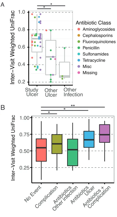
amputation; 2) wound deterioration, or 3) development of osteomyelitis. Table S1 summarizes
clinical factors by complication status.
2.3.1 Characterization of the DFU microbiota at baseline
DFU microbiomes were determined by sequencing of hypervariable regions V1 through
V3 of the 16S ribosomal RNA (rRNA) gene. The most abundant genus identified was
Staphylococcus, present in 345 of the 349 samples, with an average relative abundance of
22.77%. The second, third, and fourth most abundant genera were Streptococcus (11.98%; 318 of
349 samples), Corynebacterium (11.46%; 346 of 349 samples), and Anaerococcus (7%; 300 of
349 samples), respectively. All other genera represented <5% of bacterial relative abundance in
this dataset. A more detailed characterization can be found in Table S2. We further classified
Staphylococcus operational taxonomic units (OTUs) to species level for 79.5% of the OTUs. Of
the 22.77% attributed to Staphylococcus, 13.3% was classified as S. aureus, 5.3% was S.
pettenkoferi, and 4% was not further classified. While S. aureus is a common DFU isolate, the
26
in 2007 (Trülzsch et al. 2007), though it was identified as the cause of osteomyelitis in patient
with a chronic DFU in France (Loïez et al. 2007).
2.3.2 DFU microbiota can be partitioned into four community types
We assigned DFUs to community types with the Dirichlet multinomial mixture (DMM)
model-based approach (Holmes et al. 2012). The DMM model supposes a more biologically
relevant distribution of data, which overcomes limitations of alternative methods such as k-means
(Holmes et al. 2012) and PAM clustering (Ding and Schloss 2014). The DFU microbiomes were
clustered into 4 groups, or Community Types (CT), by minimizing the Laplace approximation
(Fig. S1). The top five differentiating taxa contributed 48.9% of the total difference between a
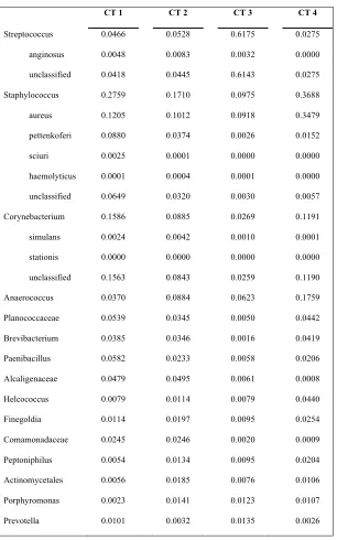
one and four component model, though the major distinguishing taxa were Streptococcus (25.6%)
and S. aureus (11.8%) (Fig. 1A). CT3 DFUs were characterized by high relative abundances of
Streptococcus (median = 64.0%). CT4 DFUs were comprised of relatively high levels of S.
aureus (median=23.8%). CT1 and CT2 were highly heterogeneous with no dominant taxa
contributing more than a median of 5% of total relative abundance. This was also reflected by
Theta values, a measure of cluster variability with smaller values corresponding to highly variable
communities, which were 3.7 and 6.9, for the CT1 and CT2 compared to 16.4 and 10.5 for CT3
and CT4, respectively. Community type summaries are described in greater detail in
Supplemental Table 3.
To better visualize how CTs were associated with microbiota composition and clinical
features, we generated a biplot depicting these relationships (Fig. 1B). As would be expected, the
taxa vectors for Streptococcus and S. aureus are closely associated with the CT3 and CT4,
27
in CT4, demonstrating the importance of the whole community in distinguishing clusters.
Streptococcus was closely associatedwith HbA1C levels and anaerobe levels with ulcer depth.
Serum C-reactive protein levels (CRP) and white blood cell counts (WBC), both measures of
inflammation used to inform the diagnosis of infections, localized separately with CT4 and CT3,
respectively. Subject outcomes also contributed to data separation, with amputation localizing
with CT1 and CT2, and unhealed subjects localizing with CT4.
2.3.3 The frequency of Community Type transitions in DFU are associated with
clinical outcomes
We next investigated the stability of the CTs by exploring the frequency and type of CT
transitions. The DFU microbiota was highly dynamic with CT transitions occurring every 1.76
study visits (approximately 3.52 weeks) on average (Fig. 2A). Transition frequencies were
significantly associated with subject outcomes (healed = 1.60, unhealed = 2.04, amputation =
3.08 study visits/CT-transition). We further subdivided healed subjects into those whose ulcers
closed in <12 weeks and those closed in >12 weeks. Consistent with our analysis, the faster
healing subjects experienced greater transition frequencies (<12 weeks = 1.45, >12 weeks 2.11
study visits/CT-transition, Wilcoxon p-value = 0.011).
We then questioned whether transition patterns between CTs were related to ulcer
outcomes. By quantifying transitions between CTs we could represent the data as a Markov
chain, with nodes representing CTs and edges representing transition frequencies by their weight
(Fig. 2B). The transition patterns between those that healed in <12 weeks and those that healed in
>12 weeks were significantly different (p-value < 0.0001). In those who healed in <12 weeks,
28
and 0.53, respectively. In contrast CT3 and CT4 experienced lower self-transition rates of 0.23
and 0.29, and had a predilection for transitioning to CT2. For subjects that took >12 weeks to
heal, there is a marked increase in self-transitions, with ulcers stalling in CT3 and CT4 at rates of
0.45 and 0.84, respectively, indicating that the stability of these CTs may be detrimental to wound
healing. Analysis of the stationary distribution and expected recurrence time revealed similar
trends (Table S4). The presence or absence of transitions between CT3 and CT4 also
differentiated the two groups, with no recorded instances in wounds healing in <12 weeks.
Together these findings suggest that community stability reflects a delayed healing phenotype.
2.3.4 DFU with more dynamic microbiota heal faster than those with less
dynamic microbiota
To address more subtle patterns of variation, which may not be apparent when examining
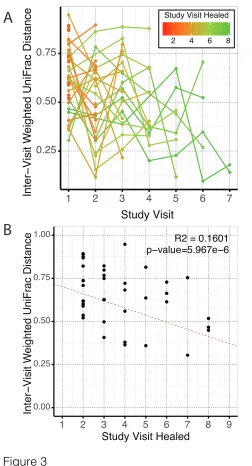
broad community types, we used the inter-visit weighted UniFrac (WUF) distance as a proxy of
stability. The weighted UniFrac metric measures the proportion of shared OTUs, their
phylogenetic relationships, and their relative distributions on a scale of 0 to 1, with higher values
indicating greater instability. We generated mixed-effect linear regressions to model the
relationship between microbiota instability and time required to heal in those that healed within
24 weeks. This model suggests that all ulcers are slowly stabilizing at a rate of -0.024/visit;
however, slow healing ulcers begin in a more stable state (-0.036 per visit required to heal) (Fig.
3A). Because mixed-effect models do not allow generation of a traditional R2 value, we
calculated marginal and conditional pseudo-R2 values, which reveals an estimate of the variance
due to the fixed effects alone and the combined model of fixed and random effects respectively.
The marginal R2 was estimated to be 0.201 and the conditional to be 0.280, indicating that our
29
The first inter-visit distance, between the baseline study visit and following visit, includes
the effect of the initial surgical debridement. Thus it was possible that the high instability in faster
healing wounds was an artifact of the first study visit being weighted more. To address this
concern, we investigated the relationship between healing time and the amount of change
between baseline and the following visit (2 weeks’ time) using a traditional linear model. We
found the same negative association between healing time and the inter-visit distance (R2 = 0.16,
p<0.0001) (Fig. 3B), suggesting the effect is independent of debridement.
2.3.5 Effect of antibiotics on temporal stability in DFU microbiota
During the course of the study, 32 subjects required the administration of antibiotics,
which afforded us the opportunity to glean the effects of antibiotics on ulcer microbiomes.
Antibiotic exposure did not drive microbiota variation in our samples (Fig 1B). Furthermore, we
did not detect any significant changes in community diversity as measured by the Shannon index
or OTU richness, perhaps due to unique interactions between specific antibiotic classes and
personal microbial communities. We assessed the potential for antibiotics to disrupt microbial
communities using the inter-visit WUF distances as before and by binning antibiotics into distinct
categories based on their class and mechanism of action. We did not detect significant differences
in microbial stability due to antibiotic class. However, in half of the cases, the antibiotics were
prescribed to treat infections not involving the studied ulcer (e.g. other ulcers, urinary tract
infection, upper respiratory infection, sinus infection). When we examined the subjects treated
specifically for the study ulcer, we found that antibiotics administered produced significantly