University of Pennsylvania
ScholarlyCommons
Publicly Accessible Penn Dissertations
1-1-2014
Augmenting Anti-Tumor Immunity by Targeting
Macrophage Cox-2 in Breast Cancer
Edward Po-Hwa Chen
University of Pennsylvania, edpchen@gmail.com
Follow this and additional works at:http://repository.upenn.edu/edissertations
Part of thePharmacology Commons
This paper is posted at ScholarlyCommons.http://repository.upenn.edu/edissertations/1232
For more information, please contactlibraryrepository@pobox.upenn.edu.
Recommended Citation
Chen, Edward Po-Hwa, "Augmenting Anti-Tumor Immunity by Targeting Macrophage Cox-2 in Breast Cancer" (2014).Publicly Accessible Penn Dissertations. 1232.
Augmenting Anti-Tumor Immunity by Targeting Macrophage Cox-2 in
Breast Cancer
Abstract
Cyclooxygenase-2 (COX-2) expression is associated with poor prognosis across a range of human cancers, including breast. While the contributions of tumor cell-derived COX-2 are well studied, those of the stroma remain ill-defined. Macrophages, an essential component of the tumor microenvironment, exist within a range of two polar phenotypes, influenced by signals in their local environment: anti-tumorigenic M1 or pro-tumorigenic M2. M2-like tumor-associated macrophages (TAM) are positively associated with tumorigenesis. This thesis investigates the contribution of macrophage COX-2 in two models of HER2/neu-induced
mammary tumorigenesis utilizing mice selectively lacking macrophage COX-2 (COX-2MÃ?KO) and the contribution of COX-2 derived products in modifying macrophage phenotype in vitro. Finally, a targeted macrophage COX-2 inhibitor is investigated in vitro and in vivo as a potential cancer therapeutic. Deletion of macrophage COX-2 led to reduced mammary tumorigenesis coincident with fewer TAMs and reduction in M2 characteristics of TAM. Further, depletion of CD8+cytotoxic T lymphocytes (CTLs), but not CD4+T helper and regulatory cells, restored tumor growth in COX-2MÃ?KO mice, suggesting enhanced CTL function caused by reduction in total and M2-like TAM. Investigation of COX-2-mediated polarization of bone marrow-derived macrophages (BMDM) in vitro revealed paracrine influences of prostaglandin (PG) E2
in modifying polarized macrophage phenotype to more closely resemble TAM. Interestingly, interference with macrophage COX-2 did not significantly modify BMDM polarization. This suggested that autocrine COX-2 minimally affects BMDM phenotype, and that polarization of COX-2MÃ?KO BMDM does not recapitulate reduced M2 characteristics observed in COX-2MÃ?KO TAM. Reconstituted high-density lipoprotein (rHDL) nanoparticles were utilized as a method to target macrophages in vitro and in vivo. rHDL conjugated to fluorescent dye DiR revealed efficient incorporation of rHDL nanoparticles with TAM. In preliminary experiments utilizing rHDL-celecoxib as a targeted macrophage COX-2 inhibitor, marked suppression of PGD2and PGE2generation was evident in lipopolysaccharide (LPS)-stimulated J774A.1 cells.
Importantly, urinary prostaglandin levels were not altered in mice treated with rHDL-celecoxib, suggesting no systemic inhibition of COX-2 with this targeted approach. These studies provide rationale for targeting macrophage COX-2 in mammary tumorigenesis and provide essential preliminary experiments in translating these findings into a potential chemopreventative or chemotherapeutic agent.
Degree Type
Dissertation
Degree Name
Doctor of Philosophy (PhD)
Graduate Group
Pharmacology
First Advisor
Emer M. Smyth
Keywords
Breast Cancer, COX-2, Macrophages, Tumor Microenvironment
Subject Categories
Pharmacology
AUGMENTING ANTI-TUMOR IMMUNITY BY TARGETING MACROPHAGE COX-2 IN BREAST CANCER
Edward P. Chen
A DISSERTATION In
Pharmacology
Presented to the Faculties of the University of Pennsylvania In
Partial Fulfillment of the Requirements for the Degree of Doctor of Philosophy
2014
Supervisor of Dissertation:
_________________________________________________________ Emer M. Smyth, Ph.D., Research Associate Professor of Pharmacology
Graduate Group Chairperson:
_________________________________________________________ Julie A. Blendy, Ph.D., Professor of Pharmacology
Dissertation Committee:
Marcelo Kazanietz, Ph.D., Professor of Pharmacology
AUGMENTING ANTI-TUMOR IMMUNITY BY
TARGETING MACROPHAGE COX-2 IN BREAST
CANCER
COPYRIGHT
2014
Edward P. Chen
This work is licensed under the Creative Commons Attribution-NonCommercial-ShareAlike 4.0
International License. To view a copy of this license, visit
http://creativecommons.org/licenses/by-nc-sa/4.0/deed.en_US.
Dedicated to my parents and sisters, and all of the people who have shaped their lives and mine.
And to my wife, who has never doubted me for second.
ACKNOWLEDGMENTS
The work described in this thesis was made possible through the concerted efforts of multiple individuals.
To my advisor, Dr. Emer Smyth: Thank you for your mentorship, guidance, and support over the course of my thesis. Without your encouragement and optimism, I would never have gotten this far. You have always been willing to spend your weekends and nights revising my work, and have always been accessible and approachable. I wish you the best in all of your future endeavors.
To Dr. Nune Markosyan, Ms. Victoire Ndong, and Ms. Emma Connolly: Thank you for your help in my work, both technically and intellectually. To Dr. Salam Ibrahim: thank you for valuable discussions. Finally, to Dr. Alex Frey: thank you for making the hours in lab much more enjoyable.
To Dr. Garret FitzGerald and the entire FitzGerald Lab: Thank you for your support and discussions. You have all been an endless supply of technical help, knowledge, and of course, reagents. To Ms. Jennifer Bruce and Dr. Sarah Teegarden: Thank you for keeping the lab running. The lab would be in disarray without you (from experience).
To the members of my thesis committee: Thank you for the valuable discussions we’ve had, the probing questions you’ve asked, and the support you’ve given me.
To the core facilities on Penn’s campus, particularly the Flow Cytometry Core, CHOP Pathology, and the In Vivo Imaging Core: Thank you for your training, your services, and the use of your instruments.
To all components of the Pharmacology Graduate Group and the Pharmacology Department, including administrators, committees, and faculty: Thank you for keeping things running. To the members of my class, thank you for going on this journey with me. To Dr. Gabriel Krigsfeld, thank you for recruiting me to Penn, and for becoming a great friend and groomsman. To Dr. Melissa Love, thank you for being a constant source of laughter (and never taking it to heart). To Future Dr. Dolim Lee, thank you for your trust and friendship. Finally, to all my friends in PGG, especially those that flew across the country to see me get married: Thank you for the great times.
To my parents: Thank you for your love and support. I would have never gotten this far in life without your tireless efforts. To my sisters and brother-in-laws: Thank you for your love, and always thinking of me. Every moment I’ve spent with family has been extremely valuable.
Finally, to my wife, Dr. Shirley Chen: Thank you for loving me, understanding me, supporting me, and encouraging me. You have kept me grounded all of these years, and shared the happiest day of my life with me. No matter what happens, I will look forward to rest of my life, because I know you are a part of it.
ABSTRACT
AUGMENTING ANTI-TUMOR IMMUNITY BY TARGETING MACROPHAGE COX-2 IN BREAST CANCER
Edward P. Chen Emer M. Smyth
Cyclooxygenase-2 (COX-2) expression is associated with poor prognosis across a range of human
cancers, including breast. While the contributions of tumor cell-derived COX-2 are well studied,
those of the stroma remain ill-defined. Macrophages, an essential component of the tumor
microenvironment, exist within a range of two polar phenotypes, influenced by signals in their
local environment: anti-tumorigenic M1 or pro-tumorigenic M2. M2-like tumor-associated
macrophages (TAM) are positively associated with tumorigenesis. This thesis investigates the
contribution of macrophage COX-2 in two models of HER2/neu-induced mammary tumorigenesis
utilizing mice selectively lacking macrophage COX-2 (COX-2MØKO) and the contribution of COX-2
derived products in modifying macrophage phenotype in vitro. Finally, a targeted macrophage
COX-2 inhibitor is investigated in vitro and in vivo as a potential cancer therapeutic. Deletion of
macrophage COX-2 led to reduced mammary tumorigenesis coincident with fewer TAMs and
reduction in M2 characteristics of TAM. Further, depletion of CD8+ cytotoxic T lymphocytes (CTLs),
but not CD4+ T helper and regulatory cells, restored tumor growth in COX-2MØKO mice, suggesting
enhanced CTL function caused by reduction in total and M2-like TAM. Investigation of
COX-2-mediated polarization of bone marrow-derived macrophages (BMDM) in vitro revealed paracrine
influences of prostaglandin (PG) E2 in modifying polarized macrophage phenotype to more closely
resemble TAM. Interestingly, interference with macrophage COX-2 did not significantly modify
BMDM polarization. This suggested that autocrine COX-2 minimally affects BMDM phenotype,
and that polarization of COX-2MØKO BMDM does not recapitulate reduced M2 characteristics
observed in COX-2MØKO TAM. Reconstituted high-density lipoprotein (rHDL) nanoparticles were
utilized as a method to target macrophages in vitro and in vivo. rHDL conjugated to fluorescent
dye DiR revealed efficient incorporation of rHDL nanoparticles with TAM. In preliminary
experiments utilizing rHDL-celecoxib as a targeted macrophage COX-2 inhibitor, marked
suppression of PGD2 and PGE2 generation was evident in lipopolysaccharide (LPS)-stimulated
J774A.1 cells. Importantly, urinary prostaglandin levels were not altered in mice treated with
rHDL-celecoxib, suggesting no systemic inhibition of COX-2 with this targeted approach. These
studies provide rationale for targeting macrophage COX-2 in mammary tumorigenesis and
provide essential preliminary experiments in translating these findings into a potential
chemopreventative or chemotherapeutic agent.
TABLE OF CONTENTS
ABSTRACT ... V
LIST OF TABLES ... XI
LIST OF ILLUSTRATIONS ... XII
CHAPTER 1 : BREAST CANCER AND THE CONTRIBUTION OF COX-2 ... 1
1.1 Breast Cancer and the Role of the Tumor Microenvironment ... 1
1.1.1 Breast Cancer and Current Treatment ... 1
1.1.2 The Tumor Microenvironment ... 3
1.1.3 Macrophage Polarization ... 5
1.1.4 Phenotype and Function of Tumor-Associated Macrophages ... 8
1.1.5 TAM Regulation of Mammary Tumorigenesis ... 9
1.2 Targeting Cyclooxygenase-2 in Breast Cancer ... 14
1.2.1 Cyclooxygenases, Prostaglandins, and Other Eicosanoids... 14
1.2.2 COX-2 and PGE2 in Breast and Other Cancers ... 15
1.2.3 PGE2-dependent Immunomodulation in Cancer ... 19
1.2.4 NSAIDs and the Limitations of COX-2 as a Therapeutic Target ... 20
1.2.5 Deletion of Mammary Epithelial COX-2 Alters TAM Phenotype ... 23
1.3 Aims of Thesis ... 27
1.3.1 The Contribution of Macrophage COX-2 to Mammary Tumorigenesis ... 27
1.3.2 Autocrine and Paracrine Influences of COX-2 Products to Macrophage Phenotype .... 30
1.3.3 Development of a Potential Macrophage COX-2 Nanotherapeutic ... 31
CHAPTER 2 : DELETION OF MACROPHAGE COX-2 IN HER2/NEU-MODELS OF MAMMARY TUMORIGENESIS ... 32
2.1 Introduction ... 32
2.2 Experimental Procedures ... 36
2.2.1 Mouse Background and Genotypes ... 36
2.2.2 Cell Lines and Culture ... 37
2.2.3 Bone Marrow-Derived Macrophage Isolation, Culture, and Treatments ... 38
2.2.4 Animal Experiments ... 40
2.2.5 Flow Cytometry ... 42
2.2.5 Quantitative-PCR ... 42
2.2.6 Mass Spectrometry ... 44
2.2.7 Immunohistochemistry ... 46
2.2.8 Migration Assay ... 47
2.2.9 Statistical Analysis... 48
2.3 Results ... 49
2.3.1 Specific Deletion of Macrophage COX-2 ... 49
2.3.2 Reduced Mammary Tumorigenesis in COX-2MØKO Neu-Driven Spontaneous Tumors .. 50
2.3.3 Reduced Mammary Tumorigenesis in COX-2MØKO Neu-Driven Orthotopic Tumors ... 57
2.3.4 Deletion of Macrophage COX-2 Reduces TAMs Density and Alters TAM Phenotype .... 60
2.3.5 Deletion of Macrophage COX-2 Enhances T Cell Density and CTL Tumor Function ... 65
2.4 Discussion ... 72
CHAPTER 3 : PARACRINE AND AUTOCRINE CONTRIBUTIONS OF COX-2 IN MACROPHAGE POLARIZATION ... 78
3.1 Introduction ... 78
3.2 Experimental Procedures ... 82
3.2.1 Bone Marrow-Derived Macrophage Isolation, Culture, and Treatments ... 82
3.2.2 Quantitative-PCR ... 83
3.2.3 Mass Spectrometry ... 83
3.2.4 Statistical Analysis... 83
3.3 Results ... 84
3.3.1 Changes in COX Pathway Protein Expression by PGE2 in Polarized Macrophages ... 84
3.3.2 Paracrine PGE2 Modifies M1 and M2 Macrophage Polarization ... 86
3.3.3 COX-2 Inhibition in Polarized Macrophages ... 96
3.4 Discussion ... 101
CHAPTER 4 : INVESTIGATION OF MACROPHAGE TARGETED COX-2 INHIBITORS ... 108
4.1 Introduction ... 108
4.2 Experimental Procedures ... 111
4.2.1 Generation of rHDL-DiR and rHDL-celecoxib Nanoparticles ... 111
4.2.2 Animal Experiments ... 113
4.3.3 Cell Culture and Treatments ... 114
4.3.4 Mass Spectrometry, Flow Cytometry... 114
4.3 Results ... 114
4.3.1 Uptake of rHDL-DiR by Tumor-Associated Macrophages ... 114
4.3.2 Analysis of Macrophage COX-2 Inhibition by rHDL-Celecoxib Nanoparticles ... 115
4.4 Discussion ... 121
CHAPTER 5 : DISCUSSION, FUTURE DIRECTIONS, AND PERSPECTIVE 125 5.1 Conclusions and Discussion ... 125
5.1.1 Deletion of Macrophage COX-2 Reduces Mammary Tumorigenesis ... 125
5.1.2 Paracrine and Autocrine COX-2 Contribute to Macrophage Polarization ... 128
5.1.3 rHDL-Celecoxib as a Potential Macrophage COX-2 Targeted Therapy ... 130
5.2 Future Directions ... 132
5.3 Perspective and Summary ... 135
BIBLIOGRAPHY ... 139
LIST OF TABLES
Table 1-1 TAMs Correlate with Prognosis in Different Cancers………28
Table 1-2 Epidemiologic studies on breast cancer risk and aspirin use………..38
Table 2-1 List of antibodies used in flow cytometry experiments……….…… …… ..….60
Table 3-1 Summary of changes in gene expression after BMDM polarization………… ………113
Table 3-2 PGE2 modifies polarized macrophage phenotype………121
LIST OF ILLUSTRATIONS
Figure 1-1 Age-adjusted cancer-related mortality rate of women in the US. ... 2
Figure 1-2 Examples of macrophage polarization and common M1 and M2 phenotypic markers. ... 7
Figure 1-3 Overview of eicosanoid synthesis, degradation, and transport. ... 13
Figure 1-4 Deletion of mammary epithelial cell COX-2 reduces tumorigenesis. ... 25
Figure 1-5 COX-2MECKO mice had an enhanced type 1 immune response. ... 26
Figure 1-6 Augmented M1 macrophage infiltration in COX-2MECKO tumors. ... 26
Figure 1-7 COX-2MECKO mice tumors have reduced angiogenesis. ... 28
Figure 1-8 CTL, TH1, and NK cells were increased, and M2 tumor-associated macrophages decreased, in COX-2MECKO mice. ... 29
Figure 2-1 Prostanoid production after macrophage COX-2 deletion. ... 35
Figure 2-2 Selective deletion of macrophage COX-2 in COX-2MØKO mice. ... 51
Figure 2-3 Deletion of macrophage COX-2 reduces tumorigenesis in neu oncogene-driven spontaneous tumors. ... 52
Figure 2-4 Analysis of apoptosis, proliferation, and angiogenesis in spontaneous tumors by Q-PCR. ... 54
Figure 2-5 Immunostaining of activated-caspase 3 in spontaneous tumors. ... 54
Figure 2-7 Immunostaining of Von Willebrand Factor in spontaneous tumors. ... 55
Figure 2-8 Immune composition of spontaneous tumors. ... 56
Figure 2-9 Deletion of macrophage COX-2 reduces tumorigenesis, COX-2 expression, and mPGES-1 expression in neu oncogene-driven orthotopic tumors. ... 58
Figure 2-10 Analysis of apoptosis, proliferation, and angiogenesis in orthotopic tumors by Q-PCR. ... 61
Figure 2-11 Immunostaining of activated-caspase 3 in orthotopic tumors. ... 61
Figure 2-12 Immunostaining of Ki67 in orthotopic tumors. ... 62
Figure 2-13 Immunostaining of Von Willebrand Factor in spontaneous tumors. ... 62
Figure 2-14 Deletion of macrophage COX-2 alters the tumor immune composition in orthotopic tumors. ... 63
Figure 2-15 Deletion of macrophage COX-2 reduces CSF-1R expression on bone marrow-derived macrophages. ... 66
Figure 2-16 Deletion of macrophage COX-2 impairs macrophage migration. ... 67
Figure 2-17 Tumor-associated macrophages in COX-2MØKO mice display an altered macrophage phenotype. ... 67
Figure 2-18 Immunostaining of CD3 in orthotopic tumors reveals increased T cell infiltration. ... 68
Figure 2-19 Enhanced CD3+ population reflects increase in both CD4+ and CD8+ T cells.
... 68
Figure 2-20 Antibody depletion of CD8+ cells restores orthotopic tumor growth in
COX-2MØKO mice. ... 71
Figure 2-21 Increased TAMs correlate with fewer CTLs in WT, but not COX-2MØKO
spontaneous tumors. ... 71
Figure 3-1 Expression of COX pathway enzymes and transporters in M1 BMDM stimulated with PGE2. ... 87
Figure 3-2 Expression of COX pathway enzymes and transporters in unpolarized BMDM stimulated with PGE2. ... 88
Figure 3-3 Expression of COX pathway enzymes and transporters in M2 BMDM stimulated with PGE2. ... 89
Figure 3-4 Prostaglandin production by polarized BMDM. ... 90
Figure 3-5 Expression of M1 and M2 markers of polarization in M1 BMDM stimulated with PGE2. ... 92
Figure 3-6 Expression of M1 and M2 markers of polarization in M2 BMDM stimulated with PGE2. ... 93
Figure 3-7 Expression of M1 and M2 markers of polarization in unpolarized BMDM stimulated with PGE2. ... 94
Figure 3-8 Expression of COX pathway enzymes and transporters in M1 BMDM after
COX-2 inhibition or genetic deletion. ... 98
Figure 3-9 Expression of COX pathway enzymes and transporters in M2 BMDM after COX-2 inhibition or genetic deletion. ... 99
Figure 3-10 Expression of M1 markers in M1 polarized BMDM after COX-2 inhibition or genetic deletion. ... 100
Figure 3-11 Expression of M2 markers in M2 polarized BMDM after COX-2 inhibition or genetic deletion. ... 100
Figure 4-1 Schematic representations of rHDL and rHDL-celecoxib nanoparticles. .... 112
Figure 4-2 Accumulation of rHDL-DiR in orthotopic tumors. ... 112
Figure 4-3 TAM uptake of rHDL-DiR by flow cytometry. ... 116
Figure 4-4 rHDL-celecoxib is stable at 4°C for several weeks. ... 116
Figure 4-5 Production of prostaglandins in J774A.1 cells after rHDL-celecoxib treatment.
... 117
Figure 4-6 Urinary production of prostaglandins in mice after rHDL-celecoxib treatment.
... 118
Figure 4-7 Ex vivo cultured peritoneal macrophage production of prostaglandins is unchanged after rHDL-celecoxib treatment.... 119
CHAPTER 1
: BREAST CANCER AND THE CONTRIBUTION OF COX-2
1.1 Breast Cancer and the Role of the Tumor Microenvironment
1.1.1 Breast Cancer and Current Treatment
Although a decade of significant advances in breast cancer prevention and treatment have steadily decreased breast cancer-related mortality (Figure 1-1), breast cancer remains the most common non-skin cancer (est. 233,000 new cases in 2014) and the second leading cause of cancer deaths among women (est. 40,000 deaths in 2014) in the United States (American Cancer Society 2014). Breast cancer is a heterogeneous mixture of diseases with varying morphology and malignancy, the majority of which are classified by location (ductal versus lobular) and aggressiveness (invasive breast cancer versus carcinomas in situ), though several unique forms of breast cancer exist, including inflammatory and triple-negative breast cancer.
Treatment options vary by stage at diagnosis. Standard of care includes local control through partial/full mastectomy or radiation therapy with adjuvant or neoadjuvant systemic therapy using chemotherapeutic agents, hormone therapy (such anti-estrogens or estrogen antagonists), and/or targeted therapies (such as monoclonal antibodies). Even with early detection and current treatment options, breast cancer remains a significant public health problem. Each treatment option comes with a variety
Figure 1-1 Age-adjusted cancer-related mortality rate of women in the US.
Despite advances in breast cancer detection and treatment, breast cancer remains the second leading cause of cancer-related deaths among women in the United States. Figure reproduced with permission (American Cancer Society, 2014).
of adverse effects. Additionally, more aggressive cancers, such as triple-negative breast cancers, which are neither ER (estrogen receptor), PR (progesterone receptor), nor HER2/neu positive, create a unique challenge as certain hormone and targeted therapies are not efficacious. Finally, lack of complete eradication of the primary disease allows for eventual recurrence or distant metastases, and innate or acquired resistance to systemic therapies (such as trastuzumab, an anti-HER2/neu antibody) (Rexer and Arteaga 2012) in some patients further limits treatment options. Identification of novel therapeutics in the treatment of breast cancer as adjuvant treatments that are not subject to the same mechanisms of resistance as current therapies, and have a reduced adverse events profile, is a current objective of breast cancer research.
1.1.2 The Tumor Microenvironment
Solid tumors have two main components: malignant epithelial cells and the stroma, or microenvironment, surrounding the tumor cells. The tumor microenvironment (TME) consists of extracellular matrix (ECM), signaling molecules such as cytokines and growth factors, immune cells, endothelial cells, fibroblasts, and adipocytes (Place, Jin Huh et al. 2011, Fang and Declerck 2013). The TME was recognized as being potentially involved in tumorigenesis over a century ago (Paget 1989), but after the discovery of the first oncogene in 1979 (Oppermann, Levinson et al. 1979) and the first tumor suppressor gene in 1986 (Friend, Bernards et al. 1986), a majority of cancer research has focused on
genetic mutations in malignant epithelial cells. However, the demonstration that the Rous sarcoma virus was unable to establish new tumors in a different microenvironment within the same species (Dolberg and Bissell 1984) shifted attention to the TME (Dvorak 1986, van den Hooff 1988). Work over the past ten years has confirmed the TME as a major regulator of tumor progression, such that a tumor promoting TME is now considered a hallmark of cancer (Hanahan and Weinberg 2011) and the use of a stromal gene expression predictive indicator can predict clinical outcome in breast cancer with greater accuracy than current prognostic indicators (Finak, Bertos et al. 2008).
The immune cells of the TME play a vital role in the progression of a tumor to malignancy. The TME contains varying proportions of tumor-associated macrophages (TAMs), neutrophils, cytotoxic T lymphocytes (CTLs), type 1 and 2 helper T cells (TH1/TH2), regulatory T cells (TREGs), natural killer (NK) cells, myeloid-derived suppressor cells (MDSCs), and other leukocytes (DeNardo, Barreto et al. 2009). These cells, which may migrate to the tumor site or become activated in order to initiate an immune response to the tumor, seem to be re-educated by tumor expression of co-inhibitory molecules and secretion of type 2 cytokines to support further epithelial cell growth and suppression of immunosurveillance (Place, Jin Huh et al. 2011). Indeed, it may be that tumor cells promote alternative functions of certain leukocytes, particularly those involved in wound healing, development, and inflammation resolution, where suppression of inflammation
is desired (Dvorak 1986, Crowther, Brown et al. 2001). One immune component of the TME, macrophages, have emerged as critical in mammary tumorigenesis.
1.1.3 Macrophage Polarization
Macrophages are highly adaptable cells that elicit both pro- and anti-tumorigenic responses (Ding, Nathan et al. 1988, Sica, Schioppa et al. 2006). In the 1970s, it was shown that macrophages stimulated by lipopolysaccharide (LPS) could stimulate tumor cell apoptosis (Doe and Henson 1978), leading to the general notion that macrophages could suppress tumors through the release of reactive nitrogen/oxygen species (RNS/ROS) and
tumor necrosis factor α(TNFα) (Ding, Nathan et al. 1988). However, in 1992, Gordon and colleagues observed that macrophages stimulated with type 2 cytokines, specifically, interleukin (IL-) 4, adopted a phenotype that was markedly different from that of
macrophages stimulated with interferon (IFN) γ or LPS (Stein, Keshav et al. 1992). Thus, stimulation with IL-4 failed to induce expression of TNFα, and instead increased expression and function of the macrophage mannose receptor (MR), an important receptor in phagocytosis of microorganisms (Stein, Keshav et al. 1992). Based on these experiments, a spectrum of macrophage polarization was introduced in which
macrophages stimulated with type 1 cytokines such as TNFα and IFNγ, or activators of
Toll-like receptor (TLR) 4 like LPS, were termed classically activated, whereas macrophages stimulated with type 2 cytokines such as IL-4 and IL-13 were classified as
alternatively activated. These macrophages were also termed M1 or M2, respectively, to mirror the TH1/TH2 paradigm in T helper cell differentiation. Many markers of M1 and M2 macrophage polarization exist (Figure 1-2), although their distinct arginine-metabolic enzymatic pathways are prototypic. M1 macrophages, which express inducible nitric oxide synthase (iNOS), metabolize L-arginine to nitric oxide, generating ROS and other RNS that contribute to M1 pro-inflammatory and anti-tumorigenic functions. M2 macrophages, in contrast, through the actions of arginase-1, metabolize L-arginine into L -ornithine and putrescine which support epithelial cell proliferation, and importantly, deplete the supply of L-arginine available for the production of RNS by other cytotoxic cells (Chang, Liao et al. 2001, Rodriguez, Quiceno et al. 2004, Keibel, Singh et al. 2009). M1 and M2 macrophages can further be classified by their release of pro-inflammatory
(e.g. TNFα, IL-12, and IL-1β) or immunosuppressive (e.g. TGFβ and IL-10) cytokines and expression of cell surface markers. M1 macrophages express major histocompatibility type II molecules (MHCII) which can support development of adaptive immunity towards transformed epithelial cells through antigen presentation (Stein, Keshav et al. 1992), while M2 macrophage express a number of scavenger receptors which allows for scavenging of self-debris produced during a tumor-invoked immune response while simultaneously suppressing that immune response (Fairweather and Cihakova 2009). Additional differences between the M1 and M2 macrophage phenotypes include
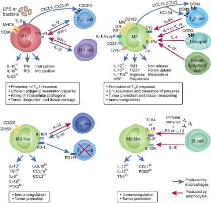
Figure 1-2 Examples of macrophage polarization and common M1 and M2 phenotypic markers.
Macrophages can be polarized to unique phenotypes dependent in response to signals in their immediate microenvironment. Macrophage phenotypes extend across a range of functions, the extremes of which are classified as M1 and M2. These polarization states are considered anti- and pro-tumorigenic, respectively. Tumor-associated macrophages are considered M2-like due to their role in immunosuppression and promotion of angiogenesis and tumor growth. Figure reproduced with permission(Biswas and Mantovani, 2010).
maintenance of iron homeostasis (Recalcati, Locati et al. 2010) and folate metabolism (Puig-Kroger, Sierra-Filardi et al. 2009). The complex nature of macrophage responses to signals in their microenvironment and the functional plasticity they display underscores the need to investigate the contribution of macrophages to tumorigenesis.
1.1.4 Phenotype and Function of Tumor-Associated Macrophages
The M1/M2 macrophage phenotype dichotomy has been useful in characterizing macrophages polarized in vitro as models of disease microenvironments. In reality, TAMs have several unique characteristics that cannot be strictly defined as M1 or M2. Genetic and phenotypic profiling of TAMs have encouraged their classification as M2-like due to their low expression of IL-12, high expression of IL-10 (Mantovani, Sozzani et al. 2002), and, with elevated arginase-1, a reduced capacity to generate ROS/RNS (Movahedi, Laoui et al. 2010). In addition, distinct subsets of TAMs induce angiogenesis through expression of vascular endothelial growth factors (VEGFs), epidermal growth factor (EGF), and IL-8 (Mantovani, Sozzani et al. 2002, Lin, Li et al. 2006, Biswas and Mantovani 2010), and matrix remodeling through release of matrix metalloproteases (MMP) 2, and urokinase plasminogen activator (Hildenbrand, Dilger et al. 1995, Eubank, Galloway et al. 2003, Mantovani, Schioppa et al. 2006). However, in contrast to the “standard” M2 phenotype, TAMs also produce M1 cytokines TNFα and IL-6 (Ikemoto, Yoshida et al. 2003). Additionally, they express several TH1 recruiting chemokines, such as CXCL9 and CXCL10
(Biswas, Gangi et al. 2006), and can express iNOS, which was associated with enhanced T cell suppression (Kusmartsev and Gabrilovich 2005). Importantly, TAMs do not express all markers simultaneously, and as such, represent a heterogeneous population of macrophages polarized by their immediate microenvironment in order to perform specific functions within distinct regions of the tumor (Van Ginderachter, Movahedi et al. 2006), such as promotion of angiogenesis, dampening of immune function, or support of invasion through ECM remodeling.
The observation that TAM phenotype is reprogrammable (Ostrand-Rosenberg 2008, Stout, Watkins et al. 2009, Biswas and Mantovani 2010) has focused attention on TAM-targeted therapies that may, through stimulation with certain cytokines, activation of specific receptors, or blockade of signaling pathways involved in polarization, promote M1 phenotypic dominance over M2 (Kortylewski, Kujawski et al. 2005, Buhtoiarov, Lum et al. 2006, Duluc, Corvaisier et al. 2009), thereby enhancing anti-tumor activity in macrophages.
1.1.5 TAM Regulation of Mammary Tumorigenesis
Considering their tumor-promoting effects, it is unsurprising that TAM density in cancer is correlated with poor prognosis in over 80% of clinical studies (Lin and Pollard 2004) and is associated with higher histological tumor grade, low hormone receptor
expression, and enhanced tumor mitosis in breast cancer (Volodko, Reiner et al. 1998).
Animal studies investigating TAM support of tumorigenesis corroborate the clinical association of TAM density and poor prognosis in cancer. Csfop/Csfop mice, which
bear a natural null recessive mutation in the colony-stimulating factor-1 (CSF-1) gene, depleting the systemic macrophage population, or mice in which macrophages are depleted with liposome-encapsulated clodronate, showed reduced angiogenesis and histological progression to malignancy in spontaneous tumors expressing the Polyoma middle T oncogene (PyMT) under the control of the mouse mammary tumor virus (MMTV), which directs oncogene expression to mammary epithelial cells (MEC) (Lin, Nguyen et al. 2001, Lin, Li et al. 2006, Qian, Deng et al. 2009). Expression levels of CSF-1, a primary macrophage growth factor and chemokine, is also linked to poor prognosis in breast cancer (Kacinski 1997, Beck, Espinosa et al. 2009) and interference with CSF-1 through the use of antisense oligonucleotides or small interfering RNA (siRNA) suppressed growth of mammary tumor xenografts (Aharinejad, Paulus et al. 2004). Studies of CSF-1 depletion or interference, or CSF-1 receptor antagonism, in mouse models of other cancers have yielded similar results (Nowicki, Szenajch et al. 1996, Priceman, Sung et al. 2010). Selective destruction of systemic macrophages, through treatment with a legumain-based DNA vaccine or an attenuated Shignella flexneri, has also shown promising outcomes, resulting in reduced tumor growth, metastasis, and even tumor
regression (Luo, Zhou et al. 2006, Galmbacher, Heisig et al. 2010).
The plasticity and heterogeneity of macrophages open potential treatment options in re-education of TAMs from pro-tumorigenic to anti-tumorigenic (Stout, Watkins et al. 2009). In fact, in certain cancers TAM density is correlated with good prognosis (Lewis and Pollard 2006), indicating that within the appropriate microenvironment, TAM may be inherently anti-tumorigenic (Table 1-1). Multiple in vitro studies of isolated macrophages and TAMs have shown that re-polarization can induce epigenetic changes that sustain the polarization state for subsequent generations (Ivashkiv 2013). Further, attempts to reprogram TAMs in vivo have been successful. Thus, treatment of tumor bearing mice with liposome-encapsulating IL-12 and granulocyte-macrophage CSF (GM-CSF) led to a cytotoxic response and tumor regression that was dependent on type 1 cells such as CTLs and NK cells (Hill, Conway et al. 2002, Tsung, Dolan et al. 2002). In another study, knockout (KO) of p50, a subunit of NF-κB, showed a class switching of TAM towards an M1 phenotype and reduced transplanted tumor growth (Saccani, Schioppa et al. 2006). Adoptive transfer of RBP-/- monocytes, which are deficient in Notch signaling and have reduced production of the M1 cytokines IFNγ, IL-12, and
TNFα, increased tumor volume and weight in Lewis lung carcinoma and B16 melanoma injected mice (Wang, He et al. 2010). Additionally, injection with a CpG oligonucleotide as an activator of TLR9, simultaneously with anti-IL-10 treatment, switched macrophage
Table 1-1 TAMs Correlate with Prognosis in Different Cancers.
High numbers of TAM correlate with poor prognosis in breast and many other cancers. However, in certain cancers, high numbers of TAM correlate with good prognosis. Reproduced with permission and modified (Lewis and Pollard, 2006).
High Numbers of TAMs Correlate with Survival in
Different Cancers
Favorable Prognosis Poor Prognosis
Stomach Breast*,+
Colorectal Prostate*
Melanoma Endometrial*
Bladder*,+ Kidney* Esophageal Superficial+
Squamous cell carcinoma* Malignant uveal melanoma* Follicular lymphoma
*Correlation with increase tumor angiogenesis
+Correlation with increased involvement of local lymph nodes. No correlation with survival was found in colon carcinoma, high-grade astrocytomas, lung carcinoma, or cervical carcinoma
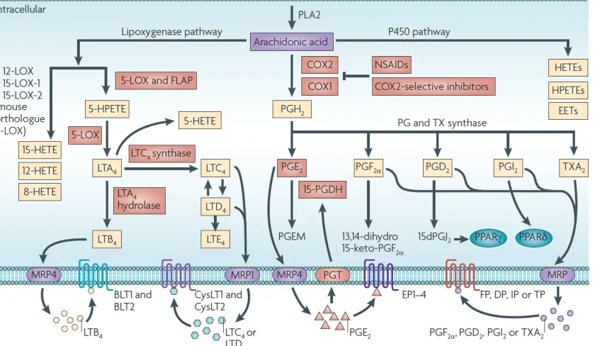
Figure 1-3 Overview of eicosanoid synthesis, degradation, and transport.
COX-1 and -2 are enzymes responsible for the metabolism of arachidonic acid into PGH2, which is further metabolized
by downstream prostaglandin synthases to PGE2, PGF2α, PGD2, PGI2, and TxA2. These prostanoids act on specific
prostanoid receptors, EP1-4, FP, DP1-2, IP, and TP, respectively. 15-PGDH carries out the first step in degradation of PGE2, which has established pro-tumorigenic and immunomodulatory effects while steady state extracellular levels of
PGE2 are maintained by efflux protein MRP4 and influx protein PGT. Arachidonic acid may be metabolized through
other eicosanoid synthesis pathways to, for example, the leukotrienes, HETEs, EETs, HPETEs, and oxo-ETEs, which may be altered through substrate shunting during COX inhibition. Reproduced with permission (Wang and Dubois, 2010).
phenotype from M2 to M1, as evident by enhanced iNOS, TNFα, and IL-12 expression by TAMs (Guiducci, Vicari et al. 2005) and this was coincident with rejection of transplanted mammary tumor carcinoma cell lines TSA and 4T1. Together these studies provide substantial evidence that therapies targeting macrophage phenotype may be of therapeutic benefit in breast or other cancers.
1.2 Targeting Cyclooxygenase-2 in Breast Cancer
1.2.1 Cyclooxygenases, Prostaglandins, and Other Eicosanoids
Cyclooxygenase (COX) is an enzyme responsible for the metabolism of arachidonic acid (AA), released from cell membranes by the action of cytosolic phospholipase A2, to prostaglandin (PG) H2. Two isoforms of COX exists in human and mice: COX-1, which is predominantly responsible for constitutive generation of prostaglandins, and COX-2, which is mainly induced in response to a variety of inflammatory and growth signals (Smyth, Grosser et al. 2009, Wang and Dubois 2010). COX-1/COX-2, also known as prostaglandin-endoperoxide synthase 1/2, catalyzes a two-step process in which AA is first converted into an unstable cyclooxygenated species, PGG2, and then reduced through peroxidation to form stable PGH2. PGH2 is further metabolized by downstream prostaglandin synthases to PGD2, PGE2, PGI2 (also known as prostacyclin). PGF2α, and TxA2 (also known as thromboxane), collectively termed the prostanoids, a family of lipid mediators with diverse and widespread biological functions. The prostanoids act on
specific G-protein coupled receptors (respectively, DP1-2, EP1-4, IP, FP, and TP) modulating physiological and pathological processes. Control of these diverse lipid pathways is directed through cell and context specific regulation of differential COX isoform and PG synthase expression and function, as well as PG receptor expression.
Importantly, AA also can be metabolized through non-COX pathways, including metabolism by lipooxygenases and P450 enzymes (Wang and Dubois 2010), leading to generation of other eicosanoids, such as the leukotrienes, hydroxyeicosatetraenoic acids (HETEs), and eicosatetraenoic acids (EETs) (Figure 1-3). AA metabolism through these pathways can also contribute to human pathologies, including cancer, and may be particularly relevant during COX inhibition when redirection of AA modifies the local lipid profile.
1.2.2 COX-2 and PGE2 in Breast and Other Cancers
Multiple human and animal studies report COX-2 overexpression in breast cancer (Harris 2009, Chen and Smyth 2011). Indeed, targeted mammary epithelial overexpression of COX-2 using the MMTV promoter to control expression, was sufficient to cause mammary tumorigenesis in multiparous mice (Liu, Chang et al. 2001). This was dependent on PGE2 signaling through the EP2 receptor (Chang, Ai et al. 2005) with upregulation of P450 aromatase (Subbaramaiah, Howe et al. 2006), which could be
reversed by COX-2 inhibition. Additionally, selective COX-2 inhibition reduced mammary tumorigenesis in a 7,12-dimethylbenzanthracene (DMBA) carcinogen-induced model of rat tumorigenesis (Harris, Alshafie et al. 2000, Kubatka, Ahlers et al. 2003) while in mice, COX-2 inhibition reduced disease in spontaneous HER2/neu- and LLC xenograft models (Lanza-Jacoby, Miller et al. 2003, Qadri, Wang et al. 2005). Further, global KO of COX-2 reduced size and multiplicity in a HER2/neu model of mammary tumorigenesis with concurrent reduction in tumor vessel density and expression of angiogenic markers (Howe, Chang et al. 2005). Notably, the molecular mechanisms determining reduced tumorigenesis are ill-defined across many studies involving COX-2 pathway disruption, with scant attention generally paid to the tumor stroma and immune microenvironment.
PGE2 is the dominant pro-tumor product of 2 (Wang and Dubois 2010). COX-derived PGH2 is converted to PGE2 through the actions of microsomal prostaglandin E synthase (mPGES) 1 and 2 and cytosolic prostaglandin E synthase (cPGES). Similar to COX-2, mPGES-1 is induced by a variety of stimuli and is the dominant E synthase in tumors (Kamei, Murakami et al. 2003) and functional coupling of COX-2 and mPGES-1 has been reported, while cPGES couples to COX-1 (Murakami, Naraba et al. 2000, Tanioka, Nakatani et al. 2000). Though less well studied, it appears that both COX-1 and COX-2 can couple with mPGES-2 (Wang and Dubois 2010). PGE2 acts through four functionally distinct G protein-coupled receptors, EP1, EP2, EP3, and EP4. EP1 is coupled to Gi, EP3 is coupled to
Gq, while EP2 and 4 are coupled to Gs. PGE2 has been studied thoroughly as a promoter of tumorigenesis (Greenhough, Smartt et al. 2009). PGE2 signaling can suppress glycogen
synthase kinase (GSK) 3β, which is a component of the β-catenin destruction complex (Castellone, Teramoto et al. 2005). The failure of GSK3β to complex with Axin and adenomatosis polyposis coli allows for accumulation of the β-catenin/T cell factor 4 complex, leading to transactivation of the peroxisome proliferator-activated receptor
(PPAR) δ and transcription of pro-tumor genes, such as MMPs, the uPA receptor, and cyclin D1 (Wang, Wang et al. 2004). PGE2 is also known to transactivate the EGF receptor (EGFR), downstream Ras-MAPK pathways, and induce anti-apoptotic protein Bcl-2 (Sheng, Shao et al. 1998, Pai, Soreghan et al. 2002, Wang, Buchanan et al. 2005). Additionally, studies have implicated PGE2 signaling with enhanced angiogenesis, which may occur through EP3-mediated enhanced transcription of VEGF and/or its receptors through ERK/JNK pathways (Pai, Szabo et al. 2001, Amano, Hayashi et al. 2003, Amano, Ito et al. 2009).
Prostaglandin signaling through cell membrane receptors is conditioned by synthesis, transport, and degradation of associated prostaglandins. Solute carrier organic anion transporter 2A1, also known as the prostaglandin transporter (PGT) is responsible for uptake of PGE2, as well as PGD2 and PGF2α, from the extracellular space into the cytosol
PGF2α from the intracellular to the extracellular space, although this process can also
occur through simple diffusion (Reid, Wielinga et al. 2003). 15-hydroxyprostaglandin dehydrogenase (15-PGDH) is the catalyzing enzyme in the first step of PGE2 degradation into its inactive 13,14-dihydro-15-keto-metabolite and thus contributes to lower overall PGE2 levels. Human colorectal cancers are associated with lower expression of 15-PGDH and PGT, which would lead to increased PGE2 available in the extracellular space (Backlund, Mann et al. 2005, Mann, Backlund et al. 2006), as compared to normal colon tissue. Enhanced MRP4 expression was also observed in these tissues. These studies highlight the importance of considering contributors to steady state prostaglandin levels.
Studies have generally focused on PGE2 as the dominant prostaglandin mediator in tumorigenesis, progression, and metastasis. However, other prostanoids may also contribute. TxA2 enhanced angiogenesis in one model of tumorigenesis (Pradono, Tazawa et al. 2002), while PGD2 may be either pro- or anti-tumorigenic dependent on DP1/DP2 receptor expression (Yoshida, Ohki et al. 1998, Murata, Aritake et al. 2011). It is important, therefore, to consider the full complement of COX products, as well as substrate shunting to alternative pathways (e.g. the lipooxygenases), when individual components of the arachidonic acid cascade are modified.
1.2.3 PGE2-dependent Immunomodulation in Cancer
PGE2 is associated with a suppressed M1 response, including reduction of M1 polarization markers and restraint of type 1 cytokine production. TAMs isolated from human ovarian cancers showed decreased NF-κB activation and depressed release of M1 cytokines after treatment with exogenous PGE2, which models the paracrine involvement of COX-2 derived PGE2 (Saccani, Schioppa et al. 2006). Additionally, PGE2 treatment of macrophages suppressed release of several M1 cytokines, such as IL-1β (Knudsen, Dinarello et al. 1986), TNFα, and IL-6 (Bailly, Ferrua et al. 1990) in LPS-stimulated human peripheral blood monocytes, IL-8 in LPS-stimulated human alveolar macrophages (Standiford, Kunkel et al. 1992), and IL-6 and TNFα in LPS-stimulated murine residential peritoneal macrophages (RPMs) (Strassmann, Patil-Koota et al. 1994). Similarly, J774 cells stimulated with LPS showed reduced expression of M1 marker iNOS after PGE2 treatment (D'Acquisto, Sautebin et al. 1998). These effects seemed to be mediated via EP2/EP4-signaling through elevated cyclic AMP (Standiford, Kunkel et al. 1992). PGE2 has also been shown to enhance production of M2 cytokine IL-10 and M2 marker arginase-1 in both murine RPMs (Strassmann, Patil-Koota et al. 1994) and bone marrow-derived macrophages (BMDM) (Wu, Llewellyn et al. 2010). These studies indicate that PGE2 may act in a paracrine manner to favor the pro-tumor M2 macrophage phenotype with co-incident suppression of anti-tumor M1 function.
The distinct autocrine influence of macrophage-derived PGE2 has been studied in vitro and in a transplant setting. Briefly, COX-2 inhibition switched bone marrow cell differentiation towards an F4/80+CD11c+ antigen-presenting cell (APC) phenotype, indicative of an M1 macrophage phenotype (Eruslanov, Daurkin et al. 2010). In the same study, tumor cell-conditioned medium diverted bone marrow cell differentiation away from the APC phenotype with an increase in 15-PGDH/PGT and decrease in MRP4 expression, suggesting that the COX-2-PGE2 pathway was an integral autocrine mediator in macrophage polarization. Interestingly, a dual mPGES-1/5-LO inhibitor (CAY10589) did not recapitulate the phenotype observed with a COX-2 inhibitor, suggesting that degradation enzymes, or other COX-2-derived products, may play a role (Eruslanov, Kaliberov et al. 2009). Additionally, enhanced PGE2 degradation, through induction of the 15-PGDH gene, supported APC phenotype differentiation with a marked reduction in M2 cytokines IL-10 and IL-13 in a xenograft model of colon cancer (Eruslanov, Kaliberov et al. 2009). More recent studies have recapitulated these findings in both humans and murine macrophages (Nakanishi, Nakatsuji et al. 2011, Na, Yoon et al. 2013).
1.2.4 NSAIDs and the Limitations of COX-2 as a Therapeutic Target
The studies discussed above provide strong rationale for the use of COX-2 inhibitors in prevention or treatment of cancer. Non-steroidal anti-inflammatory drugs (NSAIDs) are a class of drugs that inhibit COX function. As the name suggests, these drugs
are alternatives to steroids that provide anti-inflammatory, anti-pyretic, and analgesic effects, reflecting the established functions of prostanoids in inflammation, fever, and pain. NSAIDs currently available for over-the-counter use include ibuprofen and naproxen, and non-selectively inhibit COX-1 and COX-2 enzymes in a reversible manner. Aspirin, which is also a NSAID, irreversibly inhibits COX-1 and COX-2 function through acetylation of Ser 530 or Ser 516, respectively, thereby blocking the active enzymatic sites. A meta-analysis of chemoprevention studies revealed reduced risk of breast, lung, prostate, and colon cancers with non-selective NSAID treatment (Harris 2009). The use of aspirin as a chemopreventative agent is promising but complex – the Nurses’ Health Study found a reduced risk of breast cancer mortality and distant recurrence associated with daily aspirin use (Holmes, Chen et al. 2010), but the larger Women’s Health Study found no effect on breast cancer risk after every other day low dose aspirin (Cook, Lee et al. 2005). Other studies have had varying results (Table 1-2) (Lazzeroni, Petrera et al. 2013). Reductions in breast cancer mortality with aspirin may be due, at least in part, to the anti-platelet effect of aspirin reducing the risk of cancer-associated thrombosis, although the pro-tumor effects of platelets, as well as potential COX-independent effects of aspirin, require further study.
A major adverse effect of NSAID use is gastrointestinal (GI) toxicity, which is attributed to reduced prostaglandin-mediated maintenance of the GI tract. Under
Table 1-2 Epidemiologic studies on breast cancer risk and aspirin use.
Though aspirin use is associated with reduced risk in certain cancers, reports of benefit in breast cancer is inconsistent. Early reports indicate that breast cancer-related mortality and distant recurrence are reduced with aspirin use, although later studies found no association with improved survival and the Women’s Health Study found no effect on breast cancer risk from associated with aspirin use. Reproduced with permission (Lazzeroni, Petrera et al, 2013).
the assumption that the anti-inflammatory effects of NSAIDs resulted from inhibition of the inducible COX-2 enzyme and GI toxicity due to inhibition of COX-1, great effort was dedicated to the development of a new class of COX-2 selective NSAIDs. Case and population control studies showed not only a reduced risk of breast (Harris, Beebe-Donk et al. 2006) and other cancers (Harris 2009), but also a reduced adverse events profile related to GI toxicity (Laine 2002, Rostom, Muir et al. 2007). However, enthusiasm for these drugs has been dampened by the emergence of increased cardiovascular risk (Grosser, Fries et al. 2006, Grosser, Yu et al. 2010). The mechanism that underlies increased cardiovascular toxicity has been elucidated, and is attributed to the loss of COX-2-derived PGI2 and its associated anti-thrombotic and cardioprotective benefits without restriction of thrombogenic COX-1-derived TxA2 in platelets. Furthermore, in a recent meta-analysis, a similar GI risk was reported for COX-2 selective compared to COX-1/2 non-selective NSAIDs (CNT Collaboration, 2013). These studies limit the use of COX-2 inhibitors as a long-term systemic chemopreventative or chemotherapeutic agent and raise important questions about targeting COX-2 inhibitor therapies to tumors, thereby avoiding or limiting systemic side effects.
1.2.5 Deletion of Mammary Epithelial COX-2 Alters TAM Phenotype
To investigate the contribution of tumor cell COX-2 to mammary tumor progression, we engineered mice with selective deletion of COX-2 from the mammary
epithelium (COX-2MECKO) across two models of mammary tumorigenesis (Markosyan, Chen et al. 2011, Markosyan, Chen et al. 2013). Deletion of COX-2 from tumor cells was sufficient to reduce tumorigenesis as indicated by reduced tumor onset in both carcinogen- (medoxyprogesterone implants with oral administration of DMBA) and oncogene-induced (HER2/neu) models of mammary tumorigenesis (Figure 1-4A,B). Additionally, in the HER2/neu model, COX-2MECKO mice had fewer tumors per animal compared to wild type (WT) mice (Figure 1-4C).
Interestingly, reduced tumorigenesis in both tumor models was coincident with a shift in the tumor microenvironment. In the DMBA model, increased expression of CD45, a leukocyte marker, and F4/80, a macrophage marker, was evident in tumors from COX-2MECKO mice (Figure 1-5A). The latter was somewhat surprising, given that increased TAMs are typically associated with enhanced tumor progression (as mentioned above). However, in COX-2MECKO mice, TAMs showed higher expression of several M1 markers,
including CD86, TNFα, and iNOS (Figure 5B) with no change in M2 markers (Figure 1-5C). This suggested that the increase in TAMs was due elevated anti-tumorigenic M1 macrophages, which delayed tumor progression. In concordance with a shift towards type 1 immune function in COX-2MECKO tumors, expression of CD2, a marker of NK cells, was increased, and F4/80 correlated strongly with the anti-tumor TH1 lymphocyte marker TIM3 in COX-2MECKO but not WT tumors (Figure 1-6). In the HER2/neu oncogene model,
Figure 1-4 Deletion of mammary epithelial cell COX-2 reduces tumorigenesis.
Deletion of COX-2 specifically from mammary epithelial cells (COX-2MECKO mice) delayed tumor onset in (A)
DMBA-induced and (B) MMTV-neu-DMBA-induced models of mammary tumorigenesis. Additionally, (C) MEC COX-2 deletion resulted in fewer MMTV-neu induced tumors per animal. Reproduced with permission (Markosyan, Chen et al 2011; Markosyan, Chen et al 2013).
Figure 1-5 COX-2MECKO mice had an enhanced type 1 immune response.
In the carcinogen-induced model, (A) expression of CD2, a marker for natural killer cells, was increased in COX-2MECKO
tumors. (B) Expression of TIM3, a TH1 lymphocyte marker, strongly and positively correlated with F4/80 expression in
COX-2MECKO, but not WT tumors. Reproduced with permission (Markosyan, Chen et al, 2011).
Figure 1-6 Augmented M1 macrophage infiltration in COX-2MECKO tumors.
In the carcinogen-induced model, COX-2MECKO tumors had (A) enhanced expression of the leukocyte marker CD45 and
macrophage marker F4/80. This was coincident with (B) increased expression of several M1 macrophage phenotypic markers with (C) no change in expression of M2 macrophage markers. Reproduced with permission (Markosyan, Chen et al 2011).
reduced angiogenesis (Figure 1-7) was coincident with a similar shift to an enhanced type 1 immune response, with increased CD3+CD4+ helper T cells and CD3+CD8+ CTLs observed in COX-2MECKO tumors (Figure 1-8A). Additionally, CD3-CD8+ cells, which encompass NK cells and dendritic cells, were also increased (Figure 1-8B). Positive CD45 selection of tumor cells revealed a higher Tbet (TH1marker) to Gata3 (TH2 marker) ratio (Figure 1-8C), providing evidence that the increase in helper T cells reflected an increase in TH1 cells promoting a type 1 response. In this model, though no change was observed in M1 macrophage markers, COX-2MECKO tumor expression of M2 macrophage marker RELMα was decreased in leukocytes (CD45-enriched cells) as compared to WT (Figure 1-8D). These studies highlight the individual contribution of COX-2 in one tumor component, the malignant epithelial cell, to mammary tumor progression and the microenvironmental immune response and provide rationale for targeted therapeutics that can activate an anti-tumorigenic TME response.
1.3 Aims of Thesis
1.3.1 The Contribution of Macrophage COX-2 to Mammary Tumorigenesis
COX-2 and macrophages have both been independently associated with enhanced disease in breast cancer (Chen and Smyth 2011). Though a number of studies have suggested that COX-2 expressed by macrophages may support tumor-promoting characteristics (Nakanishi, Nakatsuji et al. 2011, Na, Yoon et al. 2013), the specific
Figure 1-7 COX-2MECKO mice tumors have reduced angiogenesis.
In the oncogene-induced model, COX-2MECKO tumors have (A) reduced expression of several markers of tumor
angiogenesis and have (B) reduced immunostaining for CD31 as a marker of vascular endothelium. Reproduced with permission (Markosyan, Chen et al 2013).
Figure 1-8 CTL, TH1, and NK cells were increased, and M2 tumor-associated macrophages decreased, in COX-2MECKO
mice.
In spontaneous COX-2MECKO tumors, the (A) the proportion of CD4+ and CD8+ T cells, as a proportion of all CD3+ T cells,
was increased in COX-2MECKO tumors, as was the (B) CD8+CD3- population, which encompasses natural killer and
dendritic cells. (C) COX-2MECKO tumors had a higher Tbet:Gata3 ratio, indicating an increase in the amount of TH1 cells
versus TH2 cells. (D) COX-2MECKO tumors had reduced expression of RETNLA (RELMα), an M2 macrophage marker, as
compared to WT tumors. Reproduced with permission (Markosyan, Chen et al 2013).
contribution of macrophage COX-2 to mammary tumorigenesis in vivo is not well defined. In the first aim, COX-2 was specifically deleted in macrophages using Cre-Lox recombination, and the impact on mammary tumorigenesis studied in two mouse models of HER2/neu-driven disease. These studies sought to establish whether and how targeting COX-2 in an important stromal component of mammary tumors, TAMs, can alter tumorigenesis.
1.3.2 Autocrine and Paracrine Influences of COX-2 Products to Macrophage Phenotype Macrophages are a versatile component of innate immunity that play vital roles in both pathological and physiological states (Mantovani, Sozzani et al. 2002, Duluc, Corvaisier et al. 2009, Sica and Mantovani 2012). Depending on signals in the extracellular environment, macrophages display a range of phenotypes, the extremes of which are designated M1 and M2, and typically considered anti-tumorigenic and pro-tumorigenic, respectively. Previous reports have shown a critical role of PGE2 in mediating the inflammatory characteristics of LPS-stimulated macrophages, which resemble the M1 phenotype (Knudsen, Dinarello et al. 1986, Bailly, Ferrua et al. 1990, Chen and Smyth 2011), while pharmacological COX-2 inhibition has been associated with a decreased expression of M2 markers in macrophages (Eruslanov, Daurkin et al. 2010, Na, Yoon et al. 2013). In this aim, the contribution of autocrine and paracrine-derived COX-2 products was investigated invitro in the context of M1 and M2 macrophage polarization though
pharmacological inhibition and genetic deletion of macrophage COX-2, and addition of exogenous prostaglandins. These studies sought to explore the complex contribution of COX-2, from various sources, in modifying macrophage phenotype.
1.3.3 Development of a Potential Macrophage COX-2 Nanotherapeutic
The ultimate goal of these studies is improved options in the prevention and treatment of breast cancer. The clinical use of systemic selective COX-2, particularly for chronic treatment, is limited by unacceptable cardiovascular side effects arising from collateral, and clinically unnecessary, suppression of the anti-thrombotic and cardioprotective PGI2 in the vascular endothelium (Grosser, Fries et al. 2006, Grosser, Yu et al. 2010). Nanoparticle technology can be used to selectively target COX-2 inhibitors to macrophages, an approach that could provide, or even increase, therapeutic benefit, while avoiding unwanted systemic effects. The larger payload associated with nanotherapeutic drug delivery may also allow for reduced dosage, further limiting adverse events, or enhanced efficacy. Reconstituted high-density lipoprotein (rHDL) nanoparticles have been shown to efficiently incorporate with macrophages in complex diseased tissues (Cormode, Skajaa et al. 2008, Duivenvoorden, Tang et al. 2013). In collaboration with Drs David Cormode (U Penn), Willem Mulder (Mount Sinai), and colleagues, this aim presents preliminary data investigating rHDL-conjugated celecoxib and its effectiveness for targeted inhibition of macrophage COX-2.
CHAPTER 2
:
DELETION OF MACROPHAGE COX-2 IN HER2/NEU-MODELS
OF MAMMARY TUMORIGENESIS
2.1 Introduction
COX-2, the primarily inducible form of cyclooxygenase enzyme, converts arachidonic acid into the prostaglandins and is associated with poor prognosis across a wide range of cancers, including breast (Harris 2009). The inhibition of COX-2 in mice, either pharmacologically or through gene deletion, suppresses mammary tumorigenesis (Howe, Subbaramaiah et al. 2002, Lanza-Jacoby, Miller et al. 2003, Howe, Chang et al. 2005). Importantly, COX-2 has been shown to modulate macrophage polarization in vitro and suppress the antigen-presenting phenotype prototypic of anti-tumorigenic M1 macrophages (Eruslanov, Daurkin et al. 2010). TAMs, which have several characteristics similar to pro-tumorigenic M2 macrophages, can influence the function and survival CTLs, a major effector cell in tumor immune destruction, through depletion of arginine, which CTLs utilize to generate cytotoxic RNS (Chang, Liao et al. 2001), and cell surface expression of T cell co-inhibitory molecules (DeNardo, Brennan et al. 2011). TAMs, which respond to tumor-produced CSF-1 for trafficking to the tumor site and growth promotion, can also encourage tumor growth through secretion of epidermal growth factor (EGF), which enhances tumor proliferation (Hernandez, Smirnova et al. 2009). This creates a critical
paracrine loop in which TAMs and tumor cells promote each other’s survival.
HER2 (human epidermal growth factor receptor), also known as ErbB2 or neu, is a receptor tyrosine kinase (RTK) related to the EGF receptor family that is overexpressed in 20-30% of breast cancers (Ursini-Siegel, Schade et al. 2007). HER2 lacks a ligand binding domain but acts as a high affinity co-receptor for other HER family RTKs (Barros, Powe et al. 2010). Its overexpression is correlated with poor prognosis in human breast cancers, and treatment with trastuzumab, a monoclonal antibody against HER2, prolongs disease-free survival in breast cancer patients (Rexer and Arteaga 2012). Mice that are transgenic for an activated form of rat neu, under the control of MMTV to direct mammary epithelial expression, develop tumors within 3 months, suggesting that overexpression of HER2/neu is sufficient, or requires few activating events, for progression to malignancy (Muller, Sinn et al. 1988). Overexpression in human breast cancers are likely due to gene overamplification or alternative splicing that allows for homodimerization (Reese and Slamon 1997). Signaling through HER2 leads to activation of Ras-MAPK signaling, increasing expression of proliferative transcription factors such as c-fos, c-myc, and c-jun (Lewin 1991, Mansour, Matten et al. 1994). HER2 also signals through PI3K-Akt, which increases expression of CyclinD1 and inhibits p27Kip1, disrupting cell cycle control and inhibiting apoptosis (Le, Claret et al. 2003). HER2 overexpression is associated with enhanced angiogenesis and invasion through increasing VEGF and tumor growth factor
(TGF) β, respectively (Yen, You et al. 2000, Ueda, Wang et al. 2004). Given the histological and genetic (Andrechek, Laing et al. 2003) similarities between transgenic HER2/neu murine tumors and human HER2/neu overexpressing breast cancer, genetic models of HER2-induced disease can be useful and relevant tools to study mammary tumorigenesis.
The studies that follow employ Cre/lox recombination technology to achieve targeted deletion of COX-2 in specific cellular subsets. Cre/lox recombination was first described in 1995 (Kuhn, Schwenk et al. 1995), where Cre recombinase, under the control of an IFN-responsive promoter, was used to conditionally excise DNA polymerase β by
flanking the gene target with loxP recognition sites (“flox”). Since then, a number of promoters have been utilized to control Cre recombinase expression to conditional or cell-specific transcription, and a number of floxed gene deletion studies have been reported. Cre recombinase, under the control of the lysozyme M promoter (LysM-Cre) directs Cre expression to a subset of myeloid-derived cells, including monocytes, macrophages, neutrophils, and certain DCs (Clausen, Burkhardt et al. 1999). FitzGerald and colleagues developed a mouse line in which the active site of COX-2 is floxed (COX-2flox, see below), and have characterized COX-2flox/flox mice crossed with LysM-Cre (Hui, Ricciotti et al. 2010). The primary impact of COX-2 deletion in this model was ablation of prostanoid production by macrophages (Figure 2-1), leading to reduced atherogenesis in hyperlipidemic mice. In the study outlined in this Chapter, this model of
Figure 2-1 Prostanoid production after macrophage COX-2 deletion.
(A) LPS-stimulated production of PGE2, TxA2, PGI2, and PGD2 in cultured residential peritoneal macrophage supernatants
was ablated in COX-2MØKO mice. Additionally, (B) Urinary prostanoid metabolite levels were significantly reduced in
LPS-stimulated COX-2MØKO mice. Depression prostanoid formation differed dependent on gender, with significant
depression in all prostanoids apparent in female COX-2MØKO mice. Reproduced with permission (Hui, Ricciotti 2010).
macrophage COX-2 deletion was utilized to study the role of macrophage COX-2 in mammary tumorigenesis.
2.2 Experimental Procedures
2.2.1 Mouse Background and Genotypes
Mouse experiments were conducted in accordance with NIH regulations and were approved by the Institutional Animal Care and Use Committee of the University of Pennsylvania.
COX-2flox/flox mice on the C57/BL6 background have introns 5 and 8 of the COX-2 gene flanked by loxP sites (“flox”) and have been previously described (Ishikawa and Herschman 2006). COX-2flox/flox mice were fully backcrossed to the FVB/N background (>9 generations) and are denoted as WT mice. Subsequently, WT mice were crossed with mice expressing activated rat c-neu oncogene (Val664-Glu) under the control of the mouse mammary tumor virus promoter (MMTV-neu), which directs expression of neu oncogene to mammary epithelial cells (Muller, Sinn et al. 1988) (Jackson Laboratories, Strain #005038). COX-2flox/flox mice positive for MMTV-neu are denoted WTneu. Further, C57/BL6 mice expressing Cre recombinase under the control of the LysM promoter, which directs expression of Cre to cells of myeloid lineage (Clausen, Burkhardt et al. 1999), were backcrossed on to the FVB/N background through 7 generations, utilizing the JAX Speed
Congenics Service (Jackson Laboratories) to ensure >99.9% FVB/N. LysM-Cre mice, which express the unfloxed wild type COX-2 gene, were retained as a second set of control mice and are denoted LysM-WT and LysM-WTneu. Crossing COX-2flox/flox (WT) mice with LysM-Cre (LysM-WT) mice results in specific deletion of COX-2 in subsets of myeloid-derived cells, with the primary effect in macrophages and monocytes (Hui, Ricciotti et al. 2010), and are denoted COX-2MØKO or COX-2MØKOneu, as appropriate. For all experiments, LysM-Cre and MMTV-neu were maintained heterozygous and genotypes verified by PCR of lysed tail DNA (Hui, Ricciotti et al. 2010, Markosyan, Chen et al. 2011).
2.2.2 Cell Lines and Culture
NAF and SMF, two cell lines derived from mammary carcinomas harvested from MMTV-neu transgenic mice (Elson and Leder 1995), were kindly provided by Dr. Lewis Chodosh (University of Pennsylvania). SMF cells were cultured in high-glucose DMEM (Invitrogen) with 10% calf serum, 0.5% L-glutamine, 1% Pen/Strep, and 4 µg/mL insulin (”SMF medium”). NAF cells were maintained in high-glucose DMEM with 10% fetal bovine serum, 0.5% L-glutamine, and 1% Pen/Strep (“10% FBS/DMEM”). Cells were split by incubating in 0.25% trypsin for 10 minutes. To make conditioned medium, SMF (6 x 10⁷ cells in a T175 flask in 20 mL SMF medium) were grown for 24 hours, washed twice with serum-free SMF medium and then incubated in fresh serum-free SMF medium for 24 hours. The resultant conditioned medium (SMF-CM) was filtered and aliquoted for use in
migration experiments (see below).
To generate stable transfects expressing luciferase, luciferase-pcDNA3 (Addgene; Plasmid #26612) plasmid was inserted into pLKO.1-puro lentiviral plasmid vector and packaged into MISSION TRC Lentiviral Particles (Sigma-Aldrich, #CSTVRS). NAF cells were transduced using MISSION TRC Lentiviral Particles, according to manufacturer’s instructions. In brief, cells were treated with 8 µg/mL protamine sulfate (Sigma-Aldrich, #P4020) before transduction with 4.6 x 10⁵ TU of luciferase lentiviral particles for 18 hours. Selection was carried out under 2 µg/mL puromycin (Sigma-Adrich, P7130) for at least 2 passages. The resultant cells were termed NAFLuc and luciferase expression was confirmed by treatment with 150 μg/mL D-Luciferin (Sigma-Aldrich, #L6882) with subsequent fluorescence detection at 550 nm (VICTOR3 1420 Counter, Perkin Elmer).
L929 cells (American Type Culture Collection, #CCL-1) were maintained in 10% FBS/DMEM as a biological source of CSF-1 for bone marrow-derived macrophage (BMDM) culture (Davies and Gordon 2005). L929 cells cultured to 100% confluency in a T75 flask were split 1:5 and cell supernatants collected and stored after another 4 days of culture.
2.2.3 Bone Marrow-Derived Macrophage Isolation, Culture, and Treatments
BMDM were isolated and cultured as described (Davies and Gordon 2005, Zhang, Goncalves et al. 2008). Briefly, bone marrow cells were flushed from female mouse