ANIMAL SCIENCE
Monitoring of monthly SCC in she-camel in relation to milking practice,
udder status and microbiological contamination of milk
Saeed K. Saleh1,2*, Ghamri Al-Ramadhan1and Bernard Faye1,3
1
Camel and Range Research Center, P.O. Box 322, Al-Jouf, Sakaka, Saudi Arabia 2
Animal Health Research Institute (AHRI), Dokki, Giza, Egypt 3
CIRAD-ES, Campus international de Baillarguet, TA C/dir B 34398 Montpellier, France
Abstract
Somatic cell counts and bacteriological examinations were measured in 28 camel milk samples from 2 farms, one with milking machine (farm A) and the second with hand milking (farm B). The milk was analyzed for 6 months after the parturition, every month, the first one occurring one week approximately after delivery. The somatic cell count was higher at the first sampling in the two farms but significantly more in farm B. The microbiological contamination was also higher in farm B (37% samples were contaminated) than in farm A (12%). The somatic cell count decreased all along the lactation stage and increased with the parity but the trends were not significant due to the high variability of the values. On average, the normal level of somatic cell counts is low compared to cow.
Key words: Camel, Subclinical mastitis, Bacterial contamination, Somatic cell count
Introduction
The consumption of camel milk is a long-standing tradition in Saudi Arabian and the demand for camel milk and other products in the Kingdom and elsewhere is rising (Faye and Bonnet, 2012). Even if many factors affect milk production and quality, such as breed, age, management, nutrition, parity, and lactation stage, mastitis is the single most important factor affecting production in dairy animals, including camels. While clinical mastitis can be easily recognized, subclinical mastitis almost always passes unnoticed, which accounts for its high prevalence among lactating camel herds in many countries (Guliye et al., 2002; Mohammed et al., 2005; Abdel Gadir et al., 2006; Hawari and Hassawi, 2008; Abera et al., 2009).
Traditional hand milking, use of anti-suckling devices, presence of teat lesions, and failure to apply basic hygienic measures are important predisposing factors for mastitis in camels (Mohammed et al., 2005; Abdel Gadire et al., 2005).
Somatic Cell Count (SCC) is widely used in cow for monitoring the milk quality in dairy industry. In camel, the use of SCC is not very common, but was applied as indirect diagnostic tool for detecting uninfected and infected quarters (Abdurahman, 1995; Abdurahman et al., 1995; Saleh and Faye, 2011). However, because the basal levels of cells and their physiological variations are not yet established in this species (Abdurahman et al., 1992), the interpretation could be problematic. Notably, the variation along the lactation was rarely documented. The aim of the present work was to monitor monthly SCC in camel milk for six month after calving and udder contamination in two farms with similar practices except the milking practice, hand milking vsmachine milking.
Materials and Methods Animals
Fourteen lactating camels (Camelus dromedarius) were kept at the farm of Camel and Range Research Center (farm A) at Al-Jouf region (Saudi Arabia) where animal was milked by machine and fourteen lactating camels from one surrounding herd milked manually (farm B). The lactating camels were of various parties and suckling their calves and they were housed together Received 24 February 2012; Revised 12 April 2012; Accepted
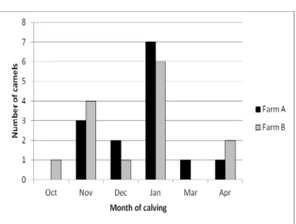
distribution was different (Figure 1). The calving season was concentrated between October and April in the both farms and the distribution of parturitions was similar also (Figure 2).
Figure 1. Distribution of the camel parities between farms A and B.
Figure 2. Seasonal distribution of the camel calving in farms A and B.
Sampling procedure
A total of 168 milk samples from the 28 lactating camels were collected at the morning milking through six months after calving to monitor the monthly somatic cell count (SCC) and bacterial contamination in milk. The camel calves were allowed to suckle in order to stimulate milking. The udder and teats were washed and cleaned with 70% alcohol. The first few strips of milk were discarded. About 15ml of milk (four quarters mixed) was then collected into sterile glass vials. The samples were kept on ice during transportation. The milk samples
were subjected to bacteriological isolation and also tested for SCC.
Bacteriological examination
An aliquot of the milk samples (0.01 ml) from each animal were streaked on blood agar and Baird-Parker agar plates. Plates were incubated for 24-48 h at 37°C. The plates were then examined for growth colony morphology. Individual colonies will be picked for identification according to the Scandinavian recommendations on examination of bovine milk samples (Klastrup, 1975).
Somatic Cell Count (SCC)
The somatic cell counts (cell/ml) for the milk samples were determined using NucleoCounter SCC-100 (coulter electronic-Chemometec A/s, Denmark). As the SCC values are log-normal (Shook, 1982), means and S.D were calculated after logarithm transformation of the raw data (Neperian logarithm base e=2.718). However, the values were expressed in cells/ml to be more explicit. To get values in cells/ml, the inverse transformation according to the function exwas applied.
Statistical analysis
To compare the monthly results between farms A and B, a variance analysis on repeated measures (ANOVA) was applied on the log-transformed data. When the number of cells/ml was below the detection limit of the SCC counter (10,000 cells/ml), the value of 10,000 was retained for calculation.
For all statistical analysis, the XLstat software (Addinsoft©) was used (repeated anova procedure)
Results
Bacteriological examination
Table 1. Bacteriological finding of camel udder milk samples in two farms with different milking process.
Farm B Farm A
Isolated bacteria
Milking process by hand Milking process by machine
% No.
% No.
26.2 22
17.8 15
Coagulase-negative Staphylococci
3.6 3
-Staphylococcus aureus
7.1 6
4.8 4
Micrococcus
63.1 53
77.4 65
No growth (non infected)
100 84
100 84
Total
Somatic cell count
SCC varied from 11,000 to 298,000 cells/ml. in farm A and from 14, 000 cell/ml to 643,000 in farm B after parturition. The values decreased highly after parturition in both farms (Table 2), but this decrease was more marked in farm B. The difference between the farm A and farm B was significant at month 1 (P <0.05) and 3 (P <0.001) (Figure 3). After 6 months, the values were similar in the 2 farms (approximately 15,000 cells/ml).
Figure 3. Somatic cell count (in log) in camel farms A (milking machine) and B (hand milking) according to the
physiological stage.
At the month 1, the SCC increased with parity, passing on average from 91.000 cells/ml. in primiparous camel to 115.000 for parity more than 4, but the difference was not significant (Figure 4). By including all the data along the 6 first month of lactation, no significant change was observed.
Regarding the seasonal variations, there was a tendency of SCC decreasing all along the year (Figure 5) since October but it was no significant. The SCC appeared higher in camel with early calving (October and November) and in late calving (April), but this trends were not significant also (Figure 6).
Table 2. Compared values of mean SCC in camel milk samples in two farms with different milking process.
Monthly mean SCC/ml.
M1 M2 M3 M4 M5 M6
Farm A 53,068 34,628 14,762 15,090 14,721 14,372
Farm B 164,110 30,929 50,843 20,461 23,292 15,571
Discussion
Udder health status of camel
The present results showed a clear difference between the udder health status in the two sampled farms, the farm with hand milking being more affected than the farm with milking machine, at least at the beginning of the lactation. However, the type of milking was not probably only in cause. Indeed, the general hygiene would be more probably responsible of the results revealing that herd B with poor hygiene of milking process had a higher prevalence of subclinical mastitis (Aljumaah et al., 2011) and intramammary infections. Indeed, poor hygiene during milking is identified as a risk factor for occurrence of mastitis as well in bovine (Abdurhman, 2006) as in camel (Tourette et al., 2002; Saber et al., 2010).
This might be due in farm B to absence of udder washing, milking of she camels with milkers which have cuts and chaps on their hands and using of common udder cloths, which could be vectors of spread especially for contagious mastitis. The bacteriological findings were comparable with the results obtained on SCC. CNS and S. aureuswere the main bacteria recovered in farm B using milking by hand.
Bacterial contamination of camel milk
Kospakov (1976) isolated staphylococcal strain from udder tissue, bulk milk and udder skin of Bactrian camels. The microorganism found in the present study was regarded as important pathogens causing mastitis in dromedary camels (Bakhiet et al., 1992; Abdurahmann, 1996; Abera et al., 2010).
As described by Younan et al. (2001), the prevalence of Staphylococci varies according to different studies, but there is nearly no publication on bacteriological hygiene of milk where Staphylococci are not mentioned (Eberlein, 2007).
Also, Radostits et al. (2000) asserted that S. aureuswas well adapted to survive in the udder and usually establishes a mild subclinical infection of long duration, from which it shed in milk, facilitating transmission to healthy animal mainly during milking process. This agrees with the data presented in this study.
Somatic cell count variation
SCC in milk is widely used as an indicator of the degree of inflammation of the udder and to predict udder infection since long time in cattle (Poutrel and Rainard, 1982). It is also the basis of most indirect tests for subclinical mastitis. The references in camel are more recent (Merin et al., 2004) and the variation factors, except in case of intramammary infections were not widely studied.
In our study, the first sampling occurring one week after parturition had on average the highest values. The first stage of lactation could be associated with decreased resistance of mammary gland to infection as result of immune depression following the stresses and hormonal changes that occur around the time of parturition and onset of lactation may leads to high prevalence of subclinical mastitis (Sordillo, 2005; Burvenich et al., 2007). At reverse, the change within lactation characterized by a significant decrease after the first month was not reported in cow where, on average, the pattern was reverse to lactation curve (Serieys, 1985).
An increase in the number of SCC in camel milk with infected quarter has been reported by Mostafa et al. (1987). The increase of SCC or mastitis with age of dairy animals or parity was widely observed in cow (Serieys, 1985; Faye et al., 1986) like in our study on camel in spite it was not significant due to the high variability within parity.
Conclusion
It appeared that camel SCC can be in less quantity than in normal cow milk as many of our samples are below the detection threshold of 10,000 cells/ml. Anyway, it is recommended that in order to reduce the prevalence of mastitis, improved milking hygiene, prevention of skin lesions, culling of chronic mastitis carriers and treating of clinically infected she camel should be practiced. Post calving milk SCC could be useful surveillance tool for monitoring mastitis, although the values require appropriate interpretation due to the lactation stage dependent physiological variation after calving.
Acknowledgements
authors thank Mr. Sallal Issa Al-Mutairi, Head of the CRRC for his support, and Dr. M. Bengoumi, regional supervisor of the FAO project for his encouragements. We express our grateful to the Prince Mansour who give us the opportunity to collect milk in his camel farm.
References
Abdurahmann, O. A. S., R. Cooray and S. Bornstein. 1992. The ultrastructure of cells fragments in mammary secretions of Camelus bactrianus. J. Vet. Med. A 39:648-655.
Abdurahmann, O. A. S. 1995.
N-acetyl-B-D-glucosaminidase and serum albumin as
indicatiors of subclinical mastitis in the Camel. J. Vet. Med. A 42:643-647
Abdurahmann, O. A. S. 1996. The detection of subclinical mastitis in the Bactrian camel (Camelus bactrianus) by somatic cell count and California mastitis test. Vet. Res. Comm. 20:9-14.
Abdurahmann, O. A. S., H. Ageb, B. Abbas and G. Astron. 1995. Relations between udder infection and somatic cells in camel (Camelus dromedarius) milk. Acta Vet. Scand. 36:648-655.
Abdurahman, O. A. S. 2006. Udder health and milk quality among camels in the Errer vally of eastern Ethiopia, Liv. Res. Ru. Develop. 18:1-9.
Abera, M., O. Abdi, F. Abunna and B. Megersa. 2010. Udder health problems and major bacterial causes of camel mastitis in Jijiga, Eastern Ethiopia: implication for impacting food security, Trop. Anim. Hlth Prod. 42:341-347.
Abdel Gadir, A. E, G. Hildebrandt, J. N. Kleer, B. Molla, M. K. Yule and M. Baumann. 2005. Prevalence and risk factors of camel (Camelus dromedaries) mastitis based on bacteriological examination s in selected regions of Ethiopia. J. Camel Pract. Res.12:203-207.
Abdel Gadir, A. E., G. Hildebrandt, J. N. Kleer, B. Molla, M. K. Yule and M. Baumann. 2006. Comparison of California Mastitis Test (CMT), Somatic Cell Count (SCC) and bacteriological examination for detection of camel (Camelus dromedaries) mastitis in
Aljumaah, R. S., F. F. Almutairi, M. Ayadi, M. A. Alshaikh, A. M. Aljumaah and M. F. Hussein. 2011. Factors influencing the prevalence of subclinical mastitis in lactating dromedary camels in Riyadh Region, Saudi Arabia. Trop. Anim. Health Prod. 43(8):1605-1610.
Bakheit, M. R. H. Aqab and I. E. Mamoun. 1992. Camel mastitis in western Sudan. Sudanese J. Vet. Sc. Anim. Husb. 31:58-59.
Burvenich, C., D. D. Bannerma, J. D., Lippolis, L. Peelman, B. J. Nonnecke, J. Kehrli and M. J. Paape. 2007. Cumulative physiological events influence the inflammatory response of the bovine udder to Escherichia coli infections during the transition period. J. Dairy Sc. 90:39-54.
Eberlein,V. 2007. Hygienic status of camel milk in Dubai (United Arab Emirates) under tow different milking management systems. Doctoral thesis, veterinary faculty, Ludwig-Maxmillians-Universitat Munchen, pp.120.
Faye, B., J. C. Fayet, M. Genest and M. Chassagne. 1986. Enquête écopathologique continue: 10. Variations des fréquences pathologiques en élevage bovin laitier en fonction de la saison, de l'année et du numéro de lactation. Ann. Rech. Vét. 17:233-246.
Faye, B. and P. Bonnet. 2012. Camel sciences and economy in the world: current situation and perspectives. Proc. 3rd ISOCARD conference. Keynote presentations. 29th January -1st February, 2012, Mascate (Sultanate of Oman), 2-15.
Guliye, A. Y., C. Van Creveld and R. Yagil. 2002. Detection of subclinical mastitis in dromedary camels (Camelus dromedaries) using somatic cell counts and the N-acetyl-beta-D-glucosaminidase test. Trop. Anim. Health Prod. 34(2):95-104.
Hawari, A. D. and D. S. Hassawi. 2008. Mastitis in one humped she-camel (Camelus dromedaries) in Jordan. J. Biol. Sc. 8:958-961.
Klastrup, O. 1975. Nordic recommendations of quarter milk samples. Annu. Bull. Int. Dairy Fed. 85:41-52.
Merin U., S. Sela, B. Rosen, R. Pinto and G. Leitner. 2004. Standards for camel milk. In: B. Faye and P. Esenov (Eds), pp.152-158. Proc. Intern. Workshop, Desertification combat and food safety: the added value of camel producers”. Ashkabad (Turkmenistan), 19-22 April 2004. “Vol. 362 NATO Sciences Series, Life and Behavioural Sciences”. IOS press Publ., Amsterdam (The Netherlands).
Mohammed, A., M. Ruiz-Bascaran, and B. Abera. 2005. Cross-sectional study of mastitis in camels (Camelus dromedaries) in Somali Region, Southeastern Ethiopia, Bull. Anim. Hlth. Prod. Africa 53:195-201.
Mostafa, A. S., A. M. Ragab, E. E. Safwat, Z. El-Sayed, M. Abd-el-Rahman, N. A. El-Danafand and M. T. Shouman. 1987. Examination of raw she-camel milk for detection of subclinical mastitis. J. Egypt. Vet. Med. Assoc. 47:117-128.
Poutrel, B. and P. Rainard. 1982. Predicting the probability of quarter infection (by major pathogens) from somatic cell concentration. Am. J. Vet. Res. 43:1296.
Radostits, O. M., D. C. Blood, C. Gay and K. W. Hinchcliff. 2000. Veterinary medicine: a text book of the disease of cattle, sheep, pigs, goats, andhorses. 9theds., (Saunders, London), 603-700.
Saber, K., S. Mohammed and A. Ahmed. 2010. Sanitary conditions of lactating dromedary she-camel environment with special reference to milk quality and subclinical mastitis monitoring. Emir. J. Food Agric. 22(3):207-215.
Saleh, S. K. and B. Faye. 2011. Detection of subclinical mastitis in dromedary camels (Camelus dromedaries) using somatic cell counts, California mastitis test and udder pathogen. Emir. J. Food Agric. 23(1):48-58.
Serieys F. 1985. Concentration cellulaire du lait individual de vache: influence de l’état d’infection mammaire, du numéro, du stade de lactation et de la production laitière. Ann. Rech. Vet. 16(3):255-261.
Sordillo, L. M. 2005. Factors affecting mammary gland immunity and mastitis susceptibility, Livestock Prod. Sci. 98:89-99.
Tourette, I., S. Messad and B. Faye. 2002. Impact des pratiques de traite des éleveurs sur la qualité sanitaire du lait de chamelle en Mauritanie. Rev. Elev. Med. Vét. Pays Trop. 55:229-233.