Published by Oriental Scientific Publishing Company © 2019
This is an Open Access article licensed under a Creative Commons license: Attribution 4.0 International (CC-BY).
A Comparative Evaluation of Reliability of an Android-based
App and Computerized Cephalometric Tracing Program for
Orthodontic Cephalometric Analysis
Pavithra Shettigar1, Shravan Shetty2*, Roopak D. Naik1, Shrinivas M. Basavaraddi1 and Anand K. Patil1
1Department Of Orthodontics and Dentofacial Orthopaedics, S.D.M. College of
Dental Sciences and Hospital, Dharwad, Karnataka, India-580009.
2Department Of Orthodontics and Dentofacial Orthopaedics, Manipal College of Dental Sciences,
Mangalore, Manipal Academy of Higher Education, Manipal, Karnataka, India-576104. *Corresponding E-mail: shravan.shetty@manipal .edu
http://dx.doi.org/10.13005/bpj/1645
(Received: 29 December 2018; accepted: 02 February 2019)
This study aimed to assess the reliability of the android smartphone-based app OneCeph by comparing it with computer cephalometric tracing program Dolphin Imaging software. 50 cephalometric radiographs were randomly selected. On each cephalogram 20 landmarks were marked. 15 parameters indicating skeletal, dental and soft tissue parameters were selected and measured. The values obtained from Dolphin imaging software and the OneCeph app were compared with respect to the assessment of measurements of various parameters by paired t-test. It was observed thatfour parameters out of the fifteen showed significant differences between Dolphin imaging software and OneCeph app (p<0.05). For all the other parameters selected, no differences were observed between Dolphin and OneCeph digital methods and also there is a significant and positive correlation between the measures obtained from the Dolphin and OneCeph app for each landmark parameter. The results obtained by the OneCeph app showed most parameters are comparable with the Dolphin Imaging software. Therefore, it can be concluded that this app is reliable, user-friendly which facilitates its use by the clinician on a regular basis. This user-friendly OneCeph app can be utilized with sufficient accuracy for the cephalometric analysis of most of the measurements required in day-to-day clinical orthodontic practice.
Keywords: Cephalometry, Computers, Digital Radiography, Smartphone.
Cephalometric radiography is an essential tool in orthodontics which has been extensively used for orthodontic diagnosis and treatment planning.1
The conventional cephalometric analysis is carried out on acetate sheets in which the landmarks are marked and linear and angular measurements are determined. In spite of its extensive application in the field of orthodontics,it can be prone to systematic and random error and is also time
consuming. Landmark identification, technical
measurements and radiographic acquisition
are the main sources of errors. Identification of
landmarks, being the major source of errors, is dependent on the experience of the operator,
definition of the landmark, density of the image and image sharpness. To add to this difficulty, it is a
compression of a three-dimensional (3D) structure to a two-dimensional (2D) image.2
by the digital method. Incidence of individual error can be minimized by using computers in treatment planning. It provides fast, precise and standardizedevaluation with a high rate of reproducibility.2
Earlier in computerized radiography, the transfer of the analogue data to digital format was done using digitizer pads, digital cameras and scanners. Recent advancements havepermitted us to use direct digital images, which provides instant image acquisition, facilitated image enhancement,reduction in radiation doses, image sharing and archiving and removal of technique-sensitive developing procedures. It also reduces potential errors due to operator fatigue.3
Recently there has been a rise in the usage of newer technologyin all aspects of our lives. This is true for particularly for smartphones, which are not only meant for phone calls.4An app is
typically a small specialized program downloaded on to a smartphone device. It is accessed using a smartphone that connects to a library of apps via the
internet and enables the users to find specific apps
for their needs that serve their needs. When there is a need for quick reference or desktop computer access is not feasible, these smartphone apps are idea tools due to their speed, ability to update and portability.5
Not only there has been a rise in the use of smartphone apps but also there are apps which have been designed for medical and dental
field. These apps have been one of the fastest
developing categories of programs and they include various programs which are planned and designed
specifically for orthodontics.6
Given the rise of computer-assisted cephalometric tracing programs usage in day-to-day orthodontic practice, there is a need to assess the accuracy of these commercially available cephalometric tracing software to allow the clinician to decide the suitable software and methods of analysis.3
Objective
The study was aimed to assess the reliability of the android mobile based app OneCeph by comparing it with thecomputer cephalometric tracing program Dolphin Imaging software.
Materials and MethOds
Fifty cephalometric radiographs were taken randomly from patients who had consulted the Department of Orthodontics and Dentofacial Orthopaedics. Patients with unerupted or missing incisors, poor quality of radiographs, craniofacial
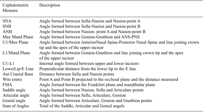
table 1. Landmark selection
Cephalometric Description Measure
SNA Angle formed between Sella-Nasion and Nasion-point A SNB Angle formed between Sella-Nasion and Nasion-point B ANB Angle formed between Nasion- point A and Nasion-point B Max Mand Plane Angle formed between Gonion-Gnathion and ANS-PNS
U1/Max Plane Angle formed between AnteriorNasal Spine-Posterior Nasal Spine and line joining crown tip and the apex of the upper incisor
L1/Mand Plane Angle formed between Gonion-Gnathion and line joining crown tip and the apex of the upper incisor
U1-L1 Internal angle formed between upper and lower incisors LowerLip/E Line Perpendicular distance from the lower lip to the E line Ant Cranial Base Distance between Sella and Nasion points
Wits (mm) Point A and Point B projected to the occlusal plane and the distance measured FMA Angle formed between the Frankfort plane and mandibular plane
Saddle angle Angle formed between Nasion, Sella and Articulare points Articular angle Angle formed between Sella, Articulare, Gonion
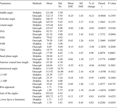
Fig. 2. Digital tracing with OneCeph app
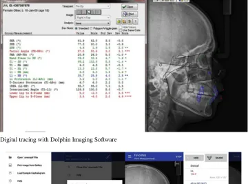
Fig. 1. Digital tracing with Dolphin Imaging Software
deformity, and non-permanent dentition with impacted teeth were excluded to ensure the accurate and consistent measurement by minimizing the margin of error.
Minimizing random error
Each participant was positioned in the cephalostat with the sagittal plane at a right angle to the path of the X-rays with the Frankfort plane
parallel to the floor and teeth in centric occlusion
and the lips gently sealed. The radiographs were
obtained with a magnification of 102.16%. Calibration for accuracy
The actual size of each image was calibrated in millimeters based on theknown
distance of 10 mm between the two fixed points
on the cephalostat rod in the radiograph. This calibration was standardized for all the images.
Landmark identification was performed manually
on digital images and then stored in the Dolphin Imaging program.
All measurements taken on both devices were carried out by the same investigator. The
brightness, magnification, contrast and zoom in/
out could be enhanced by the observer in both the programs.
Procedure
The digital images (50 cephalograms)
were imported into Dolphin Imaging software and the digital tracing was done using Dolphin Imaging Software Version 11.5 (Dolphin Imaging) (Figure 1).
table 2. Comparison of Dolphin and OneCeph methods with respect to assessment
of measurements of various parameters by paired t test (*p<0.05)
Parameters Methods Mean Std. Mean SD % of Paired P-value
Dv. Diff. Diff. change t
Saddle angle Dolphin 121.88 5.50
Oneceph 122.13 5.93 -0.25 1.83 -0.21 -0.9646 0.3395
Articular angle Dolphin 146.47 9.18
Oneceph 145.93 9.45 0.53 2.37 0.36 1.5863 0.1191
Gonial angle Dolphin 125.44 8.61
Oneceph 125.47 8.08 -0.03 2.40 -0.02 -0.0825 0.9346
SNA Dolphin 83.53 3.95
Oneceph 83.18 4.08 0.35 1.43 0.42 1.7272 0.0904
SNB Dolphin 79.62 4.82
Oneceph 79.20 5.05 0.43 1.26 0.54 2.3860 0.0209*
ANB Dolphin 3.90 3.96
Oneceph 4.05 4.04 -0.15 0.83 -3.84 -1.2850 0.2048
FMA Dolphin 19.75 6.26
Oneceph 17.99 6.02 1.76 2.07 8.90 6.0078 0.0001*
Basal plane angle Dolphin 28.84 6.13
Oneceph 28.19 6.48 0.66 1.30 2.27 3.5774 0.0008*
Anterior cranial base length Dolphin 63.50 6.70
Oneceph 64.04 4.23 -0.55 4.51 -0.86 -0.8562 0.3961
Interincisal angle Dolphin 113.49 16.53
Oneceph 113.92 16.80 -0.43 2.16 -0.38 -1.4190 0.1622
U1-NP Dolphin 25.59 3.37
Oneceph 25.35 3.26 0.24 1.05 0.95 1.6450 0.1064
L1-MP Dolphin 36.10 4.00
Oneceph 35.61 4.05 0.49 1.40 1.35 2.4577 0.0176*
Wits appraisal Dolphin 1.71 5.56
Oneceph 1.99 5.75 -0.28 1.18 -16.49 -1.6834 0.0987
Sum of the angles Dolphin 393.85 6.42
Oneceph 393.72 6.72 0.13 1.97 0.03 0.4530 0.6525
Lower lip to e-line(mm) Dolphin 1.71 3.53
Oneceph 1.70 3.41 0.01 0.43 0.82 0.2284 0.8203
as described above and digital tracing was done (Figure 2).
20 landmarks were marked on each
cephalogram and 15 parameters indicating skeletal, dental and soft tissue parameters were selected and measured (Table 1).
statistical analysis
The values for each analysis done by both Dolphin and OneCeph app was tabulated. All the values were then analyzed. Before the statistical analysis, the normality assumption was tested using Kolmogorov Smirnov test. It showed that the normality assumption had been met, so a parametric test (paired t test) was carried out. The values obtained from Dolphin imaging software and OneCeph app was compared with respect to
assessment of measurements of various parameters by paired t-test.
results
It is observed that the basal plane
angle, SNB, L1 to MP, FMA showed significant
differences between manual and digital methods
(p<0.05). For all the other parameters selected, no
differences were observed between Dolphin and OneCeph digital methods. (Table 2)
table 3. Correlation between the measurements obtained using Dolphin and OneCeph by applying
Karl Pearson's method. (*p<0.05)
Parameters r-value t-value p-value
Saddle angle 0.9513 21.3892 0.0001* Articular angle 0.9680 26.7331 0.0001* Gonial angle 0.9606 23.9471 0.0001*
SNA 0.9369 18.5596 0.0001*
SNB 0.9684 26.8867 0.0001*
ANB 0.9789 33.1723 0.0001*
FMA 0.9439 19.8062 0.0001*
Basal plane angle 0.9804 34.4487 0.0001* Anterior cranial 0.7493 7.8401 0.0001*
base length
Interincisal angle 0.9917 53.4987 0.0001*
U1-NP 0.9504 21.1633 0.0001*
L1-MP 0.9393 18.9623 0.0001*
Wits appraisal 0.9787 33.0054 0.0001* Sum of the angles 0.9562 22.6261 0.0001* Lower lip to 0.9928 57.3487 0.0001*
e-line(mm)
disCussiOn
Lateral cephalometry is a vital toolto assess the relationship between skeletal, dental and soft tissue structures and also to identify the sagittal and vertical discrepancies. Progress in technology has resulted in increased usage of digital cephalometric analysis softwares, which
have numerous benefits: reduction in radiation
doses, improvement in thedata storage and easy manipulation of images.7 Whether a digital or a
smartphone app is selected, it should be precise, safe, reliable, and highly reproduceable.8In the
literature of orthodontics, there are manyresearches which test thereproducibility and reliability of the Dolphin Imaging software.They have proven to be reliable and more frequently usedthan other digital cephalometric imaging software8,9 The present
study compares the reliability of the cephalometric app OneCeph with the Dolphin Imaging software.
The cephalometric software programs can be either completely automated or semiautomated.10This study used semiautomated
software. Initially manual location of landmarks is done and then the cephalometric analysis was performed by the computer system. This leads to lesser measurement errors than the conventional (manual) cephalometric analysis. Overall, by using
computer programs the errors which result from drawing and measuring with a ruler and a protractor may be eliminated .11
The uncertainty in landmark identification
causes significant tracing errors, which needs skills relying on operator’s experience,12quality
of original radiographs,resolution of the digital images,nature of cephalometric landmark.7 A
study by Erkan and his associates had stated the importance of standardization in comparative studies like this study. The intra-examiner error is lesser than the inter-examiner error, thus, to reduce the possibility of errors, this study was standardized by having only one examiner for both Dolphin cephalometric method and Oneceph app cephalometric method.13
In this study SNB, FMA, Basal Plane Angle, L1 to MP showed a difference between dolphin and OneCeph. Sekiguchi and Savara showed that nasion (N) might be challenging to locate when the nasofrontal suture is not clearly seen and it has been reported thatmenton, nasion and posterior nasal spine were also sources of errors this might have contributed for the difference in SNB.14
The difference in measurement of Basal Plane angle may be attributed to palatal plane angle, ANS point identification, which shown poor consistency and is often affected by the superimposition of other anatomical structures.2
Also the position of gnathion which is used to form a line with gonion to measure the mandibular plane angle. The gonion shows variation in its’ position in the vertical and horizontal axes. This may be due
to the difficulty in identifying the landmark on the
curved anatomical region.7
It has been found that the gonion,orbitale, porion, menton and lower incisor apex were the most inconsistent and unreliable points7,15 this
might have contributed to the difference in FMA and L1 to MP.
In general, the study showed statistically
significant values on the correlation of Dolphin
An android based Oneceph app provides a convenient interface with most commonly used analyses, Burstones cephalometrics for orthognathic surgery, cephalomorphic, Downs, Holdway, Jarabak, McNamara, Ricketts, Steiners, Schwarz, Tweed, Wits Appraisal, Beta angle and Yen angle, etc. This app also demonstrates the potential of a smartphone to simplify a complex, time-consuming diagnostic task such as cephalometric analysis while simultaneously providing structured reference and e-learning capabilities.
COnClusiOn
The results obtained by the OneCeph app showed most parameters are comparable with the Dolphin Imaging software. Therefore, it can be concluded that this app is reliable, user-friendly which facilitates its use by the clinician on a regular basis.
Clinical Significance
This user-friendly OneCeph app can be utilized with sufficient accuracy for the cephalometric analysis of most of the measurements required in day-to-day clinical orthodontic practice.
reFerenCes
1. Bruks A, Enberg K, Nordqvist I, Hansson AS, Jansson L, Svenson B. Radiographic examinations as an aid to orthodontic diagnosis and treatment planning. Swed Dent J.; 23
(2-3):77-85 (1999).
2. Albarakati SF, Kula KS, Ghoneima AA. The reliability and reproducibility of cephalometric
measurements: a comparison of conventional
and digital methods. Dentomaxillofac Radiol.;
41(1):11-7 (2012).
3. Polat-Ozsoy O, Gokcelik A, ToygarMemikoglu
TU. Differences in cephalometric measurements:
a comparison of digital versus hand-tracing methods. Eur J Orthod.; 31(3):254-9 (2009).
4. Singh P. Orthodontic apps for smartphones. J
Orthod; 40(3):249-55 (2013).
5. Baheti MJ, Toshniwal N. Orthodontic apps at
fingertips. Prog Orthod;15(1):36 (2014).
6. Ozdalga E, Ozdalga A, Ahuja N. The smartphone
in medicine: a review of current and potential use
among physicians and students. J Med Internet Res.;14(5):e128 (2012).
7. Chen YJ, Chen SK, Chang HF, Chen KC. Comparison of landmark identification in traditional versus computer-aided digital cephalometry. Angle Orthod; 70(5):387-92 (2000).
8. Celik E, Polat-Ozsoy O, ToygarMemikoglu TU. Comparison of cephalometric measurements with digital versus conventional cephalometric analysis. Eur J Orthod;31(3):241-6 (2009).
9. Nouri M, Hamidiaval S, Baghban AA, Basafa M, Fahim M. Efficacy of a newly designed cephalometric analysis software for McNamara analysis in comparison with dolphin software. J Dent (Tehran); 12(1):60 (2015 ).
10. Sommer T, Ciesielski R, Erbersdobler J,
Orthuber W, Fischer-Brandies H. Precision of cephalometric analysis via fully and semiautomatic evaluation of digital lateral cephalographs. Dentomaxillofac Radiol.;
38(6):401-6 (2009).
11. Liu JK, Chen YT, Cheng KS. Accuracy of computerized automatic identification of cephalometric landmarks. Am J Orthod Dentofacial Orthop.;118(5):535-40 (2000).
12. Arponen H, Elf H, Evalahti M, Waltimo-Siren J. Reliability of cranial base measurements on lateral skull radiographs. Orthod Craniofac Res;
11(4):201-10.
13. Erkan M, Gurel HG, Nur M, Demirel B. Reliability of four different computerized cephalometric analysis programs. Eur J Orthod.;
34(3):318-21 (2012).
14. Sekiguchi T, Savara BS. Variability of cephalometric landmarks used for face growth studies. Am J Orthod.;61(6):603-18 (1972).
15. Chen YJ, Chen SK, Yao JC, Chang HF. The
effects of differences in landmark identification
on the cephalometric measurements in traditional versus digitized cephalometry. Angle Orthod.;