RESEARCH ARTICLE
EVALUATION OF DIFFERENT EXTRACTION METHODS FROM
PUNICA GRANATUMPEELS
AND ITS
ANTI PROLIFERATIVE ACTIVITY AGAINST COLORECTAL CANCER CELL LINE HCT-116
1, 2, *
Salam NASREDDINE and
1Amale MCHEIK
1Doctoral School of Science and Technology, Research Platform for Environmental Science (PRASE), Faculty of Sciences,
Lebanese University, Lebanon
2Anti-cancer Therapeutic Approaches Group (ATAC), PEACE Laboratory, Biology Department, Faculty of Sciences,
Lebanese University, Lebanon
ARTICLE INFO
ABSTRACT
Objective: to investigate the effect of the different types of solvents (water, ethanoland methanol) and extraction methods (ultrasound and maceration) on P. granatum peels and their antiproliferative activity against colorectal cancer cell line HCT116.
Methods: The antiproliferative (determined by Neutral red assay) effect of different extraction methods from Punicagranatum peels was tested on the epithelial ovarian cancer (SKOV-3) cell line.
Results: The results obtained revealed that the solvents and the extraction methods had a significant effect against HCT116. For the extraction methods applied, the maceration extraction method showed that it has a better anti-proliferative effect compared to the ultrasound method. In addition, the methanol peels extracted by maceration technique showed the highest antiproliferative activity on the HCT-116 cell lines with an IC50 of 31.94 ± 4.98 μg/ml.
Conclusion: This study shows than P.granatum peels extracts have great potential as future natural antitumor and antioxidant agents.
Copyright © 2018, Salam NASREDDINE and Amale MCHEIK. This is an open access article distributed under the Creative Commons Attribution License, which permits unrestricted use, distribution, and reproduction in any medium, provided the original work is properly cited.
INTRODUCTION
The pomegranate, Punicagranatum, belongs to the Punicaceae
family. This plant is mainly found in Assia, India, China, United State, Mexico and throughout the Mediterranean region. The pomegranate possesses different size, color and taste (Lansky, 2007; Mena, 2013). Pomegranate fruits can be also divided into three parts: the seeds (about 3% of the fruit
weight), the juice (about 30% of the fruit weight) and the peel,
which also include the interior network of membranes.
Pomegranates were always considered as a sacred fruit that grow in the gardens of Paradise, inHinduism, Persian, Greek, Jewish, Christian, Chineseand in Muslim culture (Seeram, 2006). In many countries and particularly in the Mediterranean region, the pomegranate has been used extensively in the traditional medicine to treat many diseases such as fertility. In the ancient Indian Ayurvedic tradition, the pomegranate was used as an antiparasitic agent and to treat ulcers and diarrhea (Naqvi, 1991; Caceres, 1987).
*Corresponding author:1, 2Salam NASREDDINE
1
Doctoral School of Science and Technology, Research Platform for Environmental Science (PRASE), Faculty of Sciences, Lebanese University, Lebanon
2Anti-cancer Therapeutic Approaches Group (ATAC), PEACE Laboratory, Biology
Department, Faculty of Sciences, Lebanese University, Lebanon
In Greco-Arab Medicine, the pomegranate was used to treat diabetes (Zaid, 2013). The pomegranate fruit has been widely used as a traditional remedy against diarrhea, acidosis, microbial infections, helminth infection, snakebite, fever, leprosy, burns, hemorrhage, dysentery and respiratory pathologies (Kim, 2009; Jain, 1984; Siang, 1983; Singh, 1980; Arseculeratne, 1985). Furthermore, the dried pomegranate peels are considered beneficial for the treatment of diarrhea, ulcers, colitis, headache and dysentery (Ismail, 2012). In the Egyptian culture, the dried pomegranate peel was used to treat several disorders such as inflammation, cough, intestinal
worms and infertility. The traditional importance
of pomegranate as a medicinal plant is now being reinforced by data obtained by modern science. Several studies have demonstrated that the different parts of pomegranate and especially the seeds are rich inpolyphenols (including flavonoids and tannins) which have been proved to be responsible for its antioxidant properties (El-Nemr, 2006).The antioxidant activity of pomegranate juice is at least 20% higher than the other beverages like black apple juice, orange juice, cherry juice, cranberry juice, blueberry juice, grape juice, red wines and iced tea (Seeram, 2008). Pomegranate peel and juice
contain high amounts of bioactive compounds
especiallyphenolic compounds. The most common phenolic
compounds presentincludeflavonoids (anthocyanins and
ISSN: 0976-3376
Vol. 09, Issue, 10, pp.8902-8909, October,Asian Journal of Science and Technology 2018ASIAN JOURNAL OF
SCIENCE AND TECHNOLOGY
Article History:
Received 20th July, 2018
Received in revised form
18th August, 2018
Accepted 14th September, 2018
Published online 30th October, 2018
Key words:
Punicagranatum peels, Antiproliferative activity, Epithelial ovarian cancer.
Citation: Salam NASREDDINE and Amale MCHEIK, 2018. “Evaluation of different extraction methods from Punica granatumpeels and its anti
catechins) and hydrolyzable tannins (punicalin, pedunculagin, punicalagin, gallic and ellagic acid) (Seeram, 2004). These compounds are responsible for more than 90% of the
antioxidant potential of the fruit (Afaq, 2005; Negi, 2003;
Zahin, 2010). The ellagitannins including gallic acid and
ellagic acid, are responsible for the anticancer, antimutagenic, antidiabetic, anti-inflammatory, antifungal and antimicrobial activity of this fruit (Kasimsetty, 2010; Gil, 2000; Adams, 2010; Yuan, 2012; Adams, 2006; Glazer, 2012; Machado, 2002; Bialonska, 2009). The wide spectrum of health benefits of pomegranate peels have been attributed to its composition of a wide range of phytochemicals, which is why we focused our studyonoptimizing the extraction procedure onthe bioactive compounds in order to obtain the extracts with the best antiproliferative activity. Different types of solvents (water, ethanol and methanol) and extraction methods (maceration and ultrasound) were employed and the obtained extracts were evaluated in terms of in vitro antiproliferative activity againstthe human colorectal cancer HCT-116 cells.
MATERIALS AND METHODS
Fruit collection and powder preparation: Fresh fruits were collected from south Lebanon from an altitude of 300meters. After their collection, fruits were cleaned, washed with water, peeled and the peels were dried in the shade at room temperature and away from sunlight. Dried peels were then grinded to powders and were preserved in clean plastic containers and kept away from light, heat and moisture till use.
Preparation of crude extract by maceration: 15 g of the
powdered peels of P. granatum were placed in different
beakers, each containing150 mL of one of the three different solvents, water, ethanol and methanol. The solution was macerated at room temperature for 24 hrs with continuous agitation. After the preparation of the macerate, the solution was filtered using 0.45 μm filter paper and then concentrated by a rotary evaporator at 40°C with reduced pressure. The obtained extracts were then stored at -20°C to be used in
different tests (Zeidan et al., 2014).
Preparation of crude extracts by ultrasound: 1 g of the
powdered peels of P. granatum dissolved in 50 ml of the
different solvents (water, ethanol and methanol) was placed for an hour at 60 ̊C in the ultrasound apparatus. The obtained extracts were then filtered and evaporated to be stored at - 20
°C (Bandar et al., 2013).
Cells and Cell Culture: Human colorectal cancer cell line
HCT-116 was maintained in RPMI-1640 medium (Sigma
Chemical Company) supplemented with 0.1mg/ml
streptomycin, 100 U/ml penicillin and 10% fetal bovine serum
(FBS). HCTT-116 was cultured at 37°C with 5% CO2.
Neutral Red Analysis: Cell viability was performed using
Neutral Red assay based on the initial protocol described
earlier (Maleket al., 2011; Nasreddine et al., 2015). Neutral
Red, a chromogenic dye, is an indicator of a lysosomal activity. Lived cells demonstrated a chromogenic change with Neutral Red which were detected spectrophotometrically. Briefly, cells were detached from the tissue culture flask with 2 ml of trypsin solution. The cell pellet was obtained by centrifugation at 1.000 rpm for 5 minutes. The density of the viable cells was counted by the trypan blue exclusion in a
haemocytometer. Cells were then plated in a 96-well microtiter
plate, at a concentration of 8 × 10³cells/well and incubated ata
humidified 37°Cwith 5% CO2which allows the cells to adhere.
After 24 hrs, the cells were treated with five different
concentrations of P. granatum peel extracts: 50, 100, 150, 200
and 250μg/ml each being tested in triplicates. The plates were
incubated for 24, 48 and 72hrs at 37°C with 5% CO2. The
untreated cells were used as a negative control, whilst cells incubated only with buffer (water, ethanol and methanol) were used as a vehicle control. No effect was observed in the buffer. At 24, 48 and 72hrs, the old medium was replaced by100 μl of fresh medium which contains40 μg/ml neutral red and was incubated for 3 hrs. This was done to allow the uptake of the vital dye into the lysosomes of viable and undamaged cells. The media was then discarded and the cells were washed twice with 100 μl of 1X PBS. The intracellular accumulation of neutral red dye was extracted with200 μllysing solution (50% ethanol-1% acetic acid). The optical density (OD) of the eluted dye was measured at 490 nm using a microplate reader. The experiments were conducted in triplicates. The percentage of inhibition of each of the test samples was calculated according to the following formula using the OD values obtained:
Percentage of inhibition (%) = (OD control − OD sample)/OD control × 100.
Statistical analysis: All results were presented as mean ± standard error of the mean (SEM). Statistical analyses were performed with Graph Pad Prism 7 (Graph Pad Software Inc., CA, USA). With Two-way ANOVA test to determine time and treatment effects, respectively. Groups that are significantly different from control are indicated in the figures as * p < 0.05, ** p < 0.01, *** p < 0.001 and **** P < 0.0001.
RESULTS
Impact of the different types of solvents and extraction
methods of P.granatum plantpeelson theviability of
HCT-116 cancer cells: An evaluation of the antiproliferative
activity of the different types of solvents and extraction
methods of P.granatum plant peels was done by measuring the
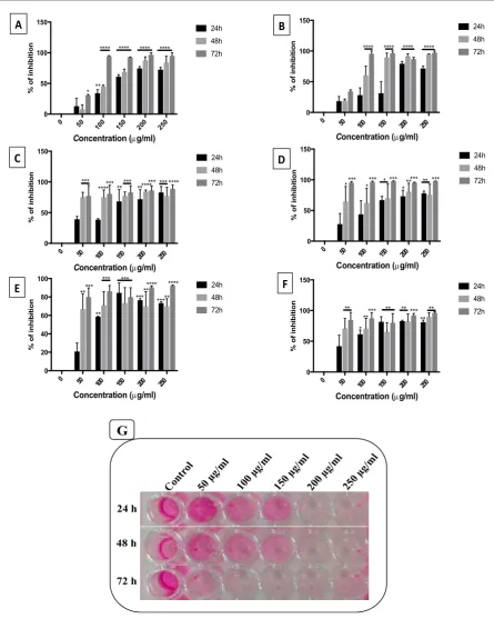
viability of the HCT-116 cell line. This evaluation was done using the Neutral Red Cytotoxicity/ Viability Assay after the treatment of the cancerous cell line for 24, 48 and 72 hrs with increasing concentrations (50, 100, 150, 200, and 250 μg/ml) of these extracts. The effect of inhibition by these extracts was dose and time-dependent (Figures 1 and Figure 2). The percentage of inhibition was calculated according to the formula mentioned above. The results of the neutral red assay showedthat the majority of the different types of solvents (water, ethanol, methanol) and extraction methods (ultrasound and maceration) have a significant antiproliferative effect at different doses and different times (Figure 1) while these extracts have no significant antiproliferative effect after 24 hrs of treatment at a concentration of 50 μg/ml (Figure 1 A-F). As shown in Figures 1A & 1B, the concentration of 50 μg/ml of water extract, produced by ultrasound technique during48 hrs and maceration extraction technique during48 and 72 hrs, has
no antiproliferative effect. Also, a non significant
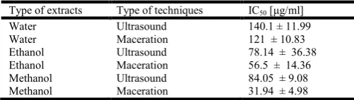
The different types of solvents and extraction methods used at a concentration of 250 μg/ml resulted within 72 hrsin ≥ 90 % inhibition ofHCT-116 cells (Figure 1). In addition, the maceration extraction technique, using the different types of solvents, showed an antiproliferative activity greater than that of the ultrasound extraction technique. In order to determine the concentration required to achieve a 50% inhibition of cells induced by each type of solvents and extraction methods, the dose response curve was plotted. The results of the cytotoxic
assay are presented as IC50 (μg/ml) in Table 1. It was clear
from the results obtained that the methanol extract produced by the maceration extraction technique (within24 hrs) has the highest antiproliferative activity on the HCT-116 cell lines
with an IC50 of 31.94 ± 4.98 μg/ml.
HCT-116 cells were incubated for 24, 48 and 72 hrs with different concentrations (0-250 μg/ml) of different solvents
prepared with P. granatum plant peels and using different
extraction methods. Cell viabilitywasestimated by
theneutralred (NR). Percent inhibition of viable cells is calculated through the formula: % Inhibition = OD (opticaldensity) of non-treatedcells - OD of treated cells / OD of non-treated cells × 100. Results represent % inhibtionof cell survivalofHCT-116 cellscompared to control.Each column
represents different concentrations of P.granatum extract and
each line represents different incubation times. (A) Represents the water extract produced by ultrasound extraction technique; (B) represents the water extract produced by maceration extraction technique; (C) represents the ethanol extract
Figure 1.Effects of the different types of solvents and extraction methodson the viability of HCT-116 cancer cellsusingP.granatumplant peels. A, F.
0 50
100 150 200 250
0 50 100 150
Concentration (mg/ml)
% o f in h ib it io n 24h 48h 72h **** **** **** **** 0 50
100 150 200 250
0 50 100 150
Concentration (mg/ml)
% o f in h ib it io n 24h 48h 72h ** * **** **** **** ***** *** 0 50
100 150 200 250
0 20 40 60 80 100
Concentration (mg/ml)
% o f in h ib it io n 24h 48h 72h *** ***** ** *** *** ****** ***** **** 0 50
100 150 200 250
0 50 100 150
Concentration (mg/ml)
% o f in h ib it io n 24h 48h 72h ** **** **** **** **** * 0 50
100 150 200 250
0 50 100 150
Concentration (mg/ml)
% o f in h ib it io n 24h 48h 72h ** ** *** *** *** *** *** **** **** **** 0 50
100 150 200 250
0 50 100 150
Concentration (mg/ml)
% o f in h ib it io n 24h 48h 72h ** ** * ***** ** ** *** **
A
B
C
D
E
F
50 µ
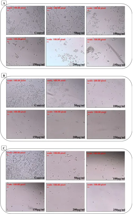
Figure 2. Microscopicview of HCT-116 cellsafter 24 (A), 48 (B) and 72 hours (C) of incubation with increasing concentrations (50, 100, 150, 200 and 250 μg/ml) ofmethanol extract of P.granatum produced by maceration extraction technique. The results presented
are from one experiment out of three that were carriedout and photographed microscopically (× 40)
Control 50µg/ml 100µg/ml
150µg/ml 200µg/ml 250µg/ml
A
Control 50µg/ml 100µg/ml
150µg/ml 200µg/ml 250µg/ml
B
Control 50µg/ml 100µg/ml
150µg/ml 200µg/ml 250µg/ml
Table 1. Concentrations of P.granatum extracts produced by different types of solvents and different extraction methods, which induced 50% decrease in HCT-116 cancer cell survival, determined by Neutral red cytotoxicity assay
Type of extracts Type of techniques IC50 [μg/ml]
Water Ultrasound 140.1 ± 11.99
Water Maceration 121 ± 10.83
Ethanol Ultrasound 78.14 ± 36.38
Ethanol Maceration 56.5 ± 14.36
Methanol Ultrasound 84.05 ± 9.08
Methanol Maceration 31.94 ± 4.98
produced by ultrasound extraction technique; (D) represents the ethanol extract produced by maceration extraction technique; (E) represents the methanol extract produced by ultrasound extraction technique; (F) represents the methanol extract produced by maceration extraction technique. (G) Represents the NR assay plate of stained cells. Each column represents the different concentrations of methanol extract of
P.granatum produced by maceration extraction technique and
each line represents the different incubation times. Experiments were conducted in triplicates and results represent the mean ± SEM (standard error of the mean) of n = 3 independent experiments. The resultant P-value was expressed as * P <0.05; ** P < 0.01; P*** <0.001 was considered to be statistically highly significant and **** P < 0.0001 extremely significant (Two-way ANOVA).
DISCUSSION
In 2015, the World Health Organization (WHO) has identified colorectal cancer as the third leading cause of death globally (774 000 deaths) after lung (1.69 million deaths) and liver (788 000 deaths) cancers. While, in Lebanon, a study conducted by
Shamseddine et al. (2014), showed that the colorectal cancer
ranked as the fourth most prevalent kind of cancer among males and the second among females. Moreover, according to the national cancer registry of the Lebanese Ministry of Public Health (MOPH), the frequency of reported incidents of colorectal cancer increase after the age of 60 in both males and females (Lebanese Ministry of Public Health, 2012). Over the last few years, many studies support the argument that regular consumption of fruits, vegetables, and grains that are rich in
polyphenols may reduce the risk of colon cancer (Gossé, 2015;
Ramos, 2007; Bobe, 2010). The Pomegranate, which is very
rich in polyphenol have also been studied for its protective
effects against colon cancer. Seeram et al. (Seeram, 2005)
reported the effect of pomegranate juice (PJ) and its purified polyphenols on human colon cancer cell lines (HT-29, HCT116, SW480, SW620), and found that PJ displayed the highest antiproliferative and proapoptotic effects compared to its purified polyphenols. So what this study showed that separation of individual polyphenols from PJ may decrease the overall anti-proliferative activity, owing to the requirement of multifactorial effects and chemical synergy of the action of multiple compounds compared to that with the most active single agent alone. Treatment of HT-29 cancer cells with PJ indicates that this juice plays an anti-inflammatory activity in colon cancer cells (Adams, 2006). The anti-inflammatory effect of PJ is also demonstrated in a mouse model, by the down regulation of inflammatory mediators such as COX-2 and iNOS, in colon tissue (Marín, 2013). Several studies suggested that the anticarcinogenic effect of PJ could largely be due to their hydrolysis product ellagic acid, which induced apoptosis in colon cancer cells (Larrosa, 2006). These effects
were mediated through the main mechanism of apoptosis, such as introducing cytochrome c in cell cytosol and by up-regulation of pro-apoptotic Bax protein and down-up-regulation of anti-apoptotic Bcl-2 protein (Larrosa, 2006; Malik, 2005; Seeram, 2004, 2005). In vivo studies have shown the chemo preventive effects of pomegranate and its potential role in colon cancer prevention. The consumption of PJ and pomegranate seed oil (PSO) suppressed the number of aberrant
cryptfoci in Fisher 344 male rats with colon
carcinogenesis induced by azoxymethane (Banerjee, 2013;
Kohno, 2004). Additional studies based in chemo preventive effects of twenty-six patients with colorectal cancer (CRC) after consumption of pomegranate extract (PE). The study found high level of urolithin in malignant colon tissues after
intake of the PE, which exhibit cancer chemopreventive
activity (Nuñez-Sánchez, 2014). The anti-proliferative effect of pomegranate peel (PP) was studied in different types of cancer, such as prostate (Albrecht, 2004), bladder (Masci, 2016) lung (Zahin, 2014) and breast (Masci, 2016) while its effect in colon cancer has not yet been studied. The aim of this work was to develop the best extraction method for PP in terms to obtain the highest antiproliferative capacity. For the extraction methods applied, the maceration extraction method showed that it has a better antiproliferative effect compared to the ultrasound method. In addition, the methanol extract showed the highest antiproliferative activity, followed by ethanol and water extract, confirming previous results in literature (Shuhau, 2010; Masci, 2016). This study demonstrated that the use of good extraction method is important to give a best antiproliferative activity.
REFERENCES
Adams, L. S., Seeram, N. P., Aggarwal, B. B., Takada, Y., Sand, D. and Heber, D. 2006. Pomegranate Juice, Total Pomegranate Ellagitannins, and Punicalagin Suppress Inflammatory Cell Signaling in Colon Cancer Cells.
Journal of Agricultural and Food Chemistry, 54(3),
980-985. doi:10.1021/jf052005r
Adams, L. S., Zhang, Y., Seeram, N. P., Heber, D. and Chen, S. 2010. Pomegranate Ellagitannin-Derived Compounds Exhibit Antiproliferative and Antiaromatase Activity in
Breast Cancer Cells In vitro. Cancer Prevention Research,
3(1), 108-113. doi:10.1158/1940-6207.capr-08-0225
Afaq, F., Saleem, M., Krueger, C. G., Reed, J. D. and Mukhtar, H. 2004. Anthocyanin- and hydrolyzable tannin-rich pomegranate fruit extract modulates MAPK and NF-?B pathways and inhibits skin tumorigenesis in CD-1
mice. International Journal of Cancer, 113(3), 423-433.
doi:10.1002/ijc.20587
Albrecht, M., Jiang, W., Kumi-Diaka, J., Lansky, E. P., Gommersall, L. M., Patel, A., Campbell, M. J. 2004. Pomegranate Extracts Potently Suppress Proliferation, Xenograft Growth, and Invasion of Human Prostate Cancer
Cells. Journal of Medicinal Food, 7(3), 274-283.
doi:10.1089/jmf.2004.7.274
Al-Zoreky, N. 2009. Antimicrobial activity of pomegranate
(Punicagranatum L.) fruit peels. International Journal of
Food Microbiology, 134(3), 244-248. doi:10.1016/j.ijfood
micro.2009.07.002
Arseculeratne, S. N., Gunatilaka, A. and Panabokke, R. G. 1985. Studies on medicinal plants of srilanka. part 14:
Toxicity of some traditional medicinal herbs. Journal of
Ethno pharmacology, 13(3), 323-335. doi:
Bakkiyaraj, D., Nandhini, J. R., Malathy, B. and Pandian, S. K. (2013). The anti-biofilm potential of pomegranate
(Punicagranatum L.) extractagainsthumanbacterial and
fungalpathogens. Biofouling, 29(8), 929-937. doi:10.1080/
08927014.2013.820825
Bandar, H., Hijazi, A., Rammal, H., Hachem, A., Saad, Z. and Badran, B. 2013. Techniques for the Extraction of Bioactive Compounds from Lebanese Urticadioica. American Journal of Phytomedicine and Clinical
Therapeutics,1(6), 507-513.
Banerjee, N., Kim, H., Talcott, S. and Mertens-Talcott, S.
2013. Pomegranate polyphenolics suppressed
azoxymethane-induced colorectal aberrant crypt foci and inflammation: Possible role of miR-126/VCAM-1 and
miR-126/PI3K/AKT/mTOR. Carcinogenesis, 34
(12),2814-2822. doi:10.1093/carcin/bgt295
Bate-Smith, E. 1973. Haemanalysis of tannins: The concept of
relative astringency. Phytochemistry, 12(4), 907-912.
doi:10.1016/0031-9422(73)80701-0
Bialonska, D., Kasimsetty, S. G., Khan, S. I. and Ferreira, D. 2009. Urolithins, Intestinal Microbial Metabolites of Pomegranate Ellagitannins, Exhibit Potent Antioxidant
Activity in a Cell-Based Assay. Journal of Agricultural
and Food Chemistry, 57(21), 10181-10186. doi:10.1021/
jf9025794
Bobe, G., Albert, P. S., Sansbury, L. B., Lanza, E., Schatzkin, A., Colburn, N. H. and Cross, A. J. 2010. Interleukin-6 as a Potential Indicator for Prevention of High-Risk Adenoma Recurrence by Dietary Flavonols in the Polyp Prevention
Trial. Cancer Prevention Research, 3(6), 764-775.
doi:10.1158/1940-6207.capr-09-0161
Braga, L., Shupp, J., Cummings, C., Jett, M., Takahashi, J., Carmo, L. and Nascimento, A. 2005. Pomegranate extract inhibits Staphylococcus aureus growth and subsequent
enterotoxin production. Journal of Ethnopharmacology,
96(1-2), 335-339. doi:10.1016/j.jep.2004.08.034
Burapadaja, S. and Bunchoo, A. 1995. AntimicrobialActivity
of Tannins fromTerminaliacitrina. Planta Medica,61(04),
365-366. doi:10.1055/s-2006-958103
Cáceres, A., Girón, L. M., Alvarado, S. R. and Torres, M. F. 1987. Screening of antimicrobial activity of plants popularly used in guatemala for the treatment of
dermatomucosal diseases. Journal of Ethnopharmacology,
20(3), 223-237. doi:10.1016/0378-8741(87)90050-x
Chokr, A., Watier, D., Eleaume, H., Pangon, B., Ghnassia, J., Mack, D. and Jabbouri, S. 2006. Correlation between biofilm formation and production of polysaccharide intercellular adhesin in clinical isolates of
coagulase-negative staphylococci. International Journal of Medical
Microbiology, 296(6), 381-388. doi:10.1016/j.ijmm.
2006.02.018
Christensen, G. D., Simpson, W. A., Younger, J. J., Baddour,
L. M., Barrett, F. F., Melton, D. M., et al. 1985. Adherence
of coagulase-negative staphylococci to plastic tissue culture plates: A quantitative model for the adherence of
staphylococci to medical devices. Journal of Clinical
Microbiology,22(6), 996-1006.
Cockerill, F. R. 2012. Methods for dilution
antimicrobialsusceptibility tests for bacteria that
growaerobically: Approved standard - ninthedition.
Wayne, PA: CLSI.
Dahham, S. S., Ali, M. N., Tabassum, H. and Khan, M. 2010. Studies on antibacterial and antifungal activity of
pomegranate (Punicagranatum L.). American-Eurasian
Journal of Agricultural & Environmental Sciences,9(3),
273-281.
El-Nemr, S. E., Ismail, I. A. and Ragab, M. 1990. Chemical
composition of juice and seeds of pomegranate fruit. Food
/ Nahrung, 34(7), 601-606. doi:10.1002/food.19900340706
El-Sherbini, G. M., Ibrahim, K. M., El-Sherbiny, E. T., Abdel-Hady, N. M. and Morsy, T. A. 2010. Efficacy of Punicagranatum extract on in-vitro and in-vivo control of
Trichomonasvaginalis. Journal of the Egyptian Society of
Parasitology, 40(1), 229-244. doi: DOI:
10.1055/s-0031-1282309
Endo, E. H., Ueda-Nakamura, T., Nakamura, C. V. and Filho, B. P. 2012. Activity of Spray-dried Microparticles Containing Pomegranate Peel Extract against Candida
albicans. Molecules, 17(9), 10094-10107. doi:10.3390/
molecules170910094
Gil, M. I., Tomás-Barberán, F. A., Hess-Pierce, B., Holcroft, D. M. and Kader, A. A. 2000. Antioxidant Activity of Pomegranate Juice and Its Relationship with Phenolic
Composition and Processing. Journal of Agricultural and
Food Chemistry, 48(10), 4581-4589. doi:10.1021/
jf000404a
Glazer, I., Masaphy, S., Marciano, P., Bar-Ilan, I., Holland, D., Kerem, Z. and Amir, R. 2012. Partial Identification of Antifungal Compounds from Punicagranatum Peel
Extracts. Journal of Agricultural and Food Chemistry,
60(19), 4841-4848. doi:10.1021/jf300330y
Gossé, F., Guyot, S., Roussi, S., Lobstein, A., Fischer, B., Seiler, N. and Raul, F. 2005. Chemopreventive properties of apple procyanidins on human colon cancer-derived metastatic SW620 cells and in a rat model of colon
carcinogenesis. Carcinogenesis, 26(7), 1291-1295. doi:10.
1093/carcin/bgi074
Haslam, E. 1996. Natural Polyphenols (Vegetable Tannins) as
Drugs: Possible Modes of Action. Journal of Natural
Products,59(2), 205-215. doi:10.1021/np960040
Ismail, T., Sestili, P. and Akhtar, S. 2012. Pomegranate peel and fruit extracts: A review of potential anti-inflammatory
and anti-infective effects. Journal of Ethnopharmacology,
143(2), 397-405. doi:10.1016/j.jep.2012.07.004
Jain, S. and Puri, H. 1984. Ethnomedicinal plants of
Jaunsar-Bawar Hills, Uttar Pradesh, India. Journal of
Ethnopharmacology,12(2), 213-222.
doi:10.1016/0378-8741(84)90049-7
Kasimsetty, S. G., Bialonska, D., Reddy, M. K., Ma, G., Khan, S. I. and Ferreira, D. 2010. Colon Cancer Chemopreventive Activities of Pomegranate Ellagitannins and Urolithins.
Journal of Agricultural and Food Chemistry,58(4),
2180-2187. doi:10.1021/jf903762h
Kim, Y. H. and Choi, E. M. 2009. Stimulation of osteoblastic differentiation and inhibition of interleukin-6 and nitric oxide in MC3T3-E1 cells by pomegranate ethanol
extract. Phytotherapy Research, 23(5), 737-739. doi:10.
1002/ptr.2587
Kohno, H., Suzuki, R., Yasui, Y., Hosokawa, M., Miyashita, K. and Tanaka, T. 2004. Pomegranate seed oil rich in conjugated linolenic acid suppresses chemically induced
colon carcinogenesis in rats. Cancer Science,95(6),
481-486. doi:10.1111/j.1349-7006.2004.tb03236.x
Lansky, E. P. and Newman, R. A. 2007. Punicagranatum (pomegranate) and its potential for prevention and
treatment of inflammation and cancer. Journal of
Ethnopharmacology, 109(2), 177-206. doi:10.1016/j.jep.
Larrosa, M., González-Sarrías, A., García-Conesa, M. T., Tomás-Barberán, F. A. and Espín, J. C. 2006. Urolithins, Ellagic Acid-Derived Metabolites Produced by Human Colonic Microflora, Exhibit Estrogenic and Antiestrogenic
Activities. Journal of Agricultural and Food Chemistry,
54(5), 1611-1620. doi:10.1021/jf0527403
Larrosa, M., Tomás-Barberán, F. A. and Espín, J. C. 2006. The dietary hydrolysable tannin punicalagin releases ellagic
acid that induces apoptosis in human colon
adenocarcinoma Caco-2 cells by using the mitochondrial
pathway. The Journal of Nutritional Biochemistry, 17(9),
611-625. doi:10.1016/j.jnutbio.2005.09.004
Lebanese Ministry of Public Health: National cancer registry -
cancer surveillance; 2012. http://www.moph.gov.lb/
Prevention/ Surveillance/Pages/Cancer.aspx.
Liu, Y., Gallardo-Moreno, A. M., Pinzon-Arango, P. A., Reynolds, Y., Rodriguez, G. and Camesano, T. A. 2008. Cranberry changes the physicochemical surface properties
of E. coli and adhesionwithuroepithelialcells. Colloids and
Surfaces B: Biointerfaces, 65(1), 35-42. doi:10.1016/
j.colsurfb.2008.02.012
Machado, T. D., Leal, I. C., Amaral, A. C., Santos, K. R., Silva, M. G. and Kuster, R. M. 2002. Antimicrobial
Ellagitannin of Punicagranatum Fruits. Journal of the
Brazilian Chemical Society, 13(5), 606-610. doi:10.1590/
s0103-50532002000500010
Machado, T., Pinto, A., Pinto, M., Leal, I., Silva, M., Amaral,
A., et al. 2003. In vitro activity of Brazilian medicinal
plants, naturally occurring naphthoquinones and their analogues, against methicillin-resistant Staphylococcus
aureus. International Journal of Antimicrobial Agents,
21(3), 279-284. doi:10.1016/s0924-8579(02)00349-7
Malek, S. N., Lee, G. S., Hong, S. L., Yaacob, H., Wahab, N. A., Weber, J. F. and Shah, S. A. 2011. Phytochemical and
Cytotoxic Investigations of Curcuma mangga
Rhizomes. Molecules, 16(6), 4539-4548. doi:10.3390/
molecules16064539
Malik, A., Afaq, F., Sarfaraz, S., Adhami, V. M., Syed, D. N. and Mukhtar, H. 2005. Pomegranate fruit juice for
chemoprevention and chemotherapy of prostate
cancer. Proceedings of the National Academy of Sciences,
102(41), 14813-14818. doi:10.1073/pnas.0505870102
Marín, M., Giner, R. M., Ríos, J. and Recio, M. C. 2013. Intestinal anti-inflammatory activity of ellagic acid in the acute and chronic dextranesulfate sodium models of mice
colitis. Journal of Ethnopharmacology, 150(3), 925-934.
doi:10.1016/j.jep.2013.09.030
Masci, A., Coccia, A., Lendaro, E., Mosca, L., Paolicelli, P. and Cesa, S. 2016. Evaluation of different extraction methods from pomegranate whole fruit or peels and the
antioxidant and antiproliferative activity of the
polyphenolic fraction. Food Chemistry, 202, 59-69.
doi:10.1016/j.foodchem.2016.01.106
Mena, P., Vegara, S., Martí, N., García-Viguera, C., Saura, D. and Valero, M. 2013. Changes on indigenous microbiota, colour, bioactive compounds and antioxidant activity of
pasteurised pomegranate juice. Food Chemistry, 141(3),
2122-2129. doi:10.1016/j.foodchem.2013.04.118
Naqvi, S. A., Khan, M. S. and Vohora, S. B. 1991. Antibacterial, antifungal and anthelmintic investigations on
Indian medicinal plants. Fitoterapia, 62(3), 221-228.
Nasreddine, S., Salameh, F., Hassan, K. H., Daher, A., Nasser, M., Rammal, H. and Hijazi, A. 2015. Valproic acid induces apoptosis and increases CXCR7 expression in epithelial
ovarian cancer cell line SKOV-3. European Scientific
Journal, 323-327.
Naz, S., Siddiqi, R., Ahmad, S., Rasool, S. and Sayeed, S. 2007. Antibacterial Activity Directed Isolation of
Compounds from Punicagranatum. Journal of Food
Science, 72(9). doi:10.1111/j.1750-3841.2007.00533.x
Negi, P., Jayaprakasha, G. and Jena, B. 2003. Antioxidant and antimutagenic activities of pomegranate peel extracts.
Food Chemistry, 80(3), 393-397.
doi:10.1016/s0308-8146(02)00279-0
Nitin, K., Pooja, U. and Gaurav, S. 2018. Phytochemical Screening of Pomegranate Peel using crude
Hydro-alcoholic Extract and pharmacological activities.
International Journal of Scientific and Research
Publications, 8(1), 193-199.
Nuñez-Sánchez, M. A., García-Villalba, R., Monedero-Saiz, T., García-Talavera, N. V., Gómez-Sánchez, M. B., Sánchez-Álvarez, C. and Espín, J. C. 2014. Targeted metabolic profiling of pomegranate polyphenols and urolithins in plasma, urine and colon tissues from
colorectal cancer patients. Molecular Nutrition & Food
Research, 58(6), 1199-1211. doi:10.1002/mnfr.201300931
Pai, V., Chanu, T. R., Chakraborty, R., Raju, B., Lobo, R. and Ballal, M. 2011. Evaluation of the antimicrobialactivity of Punicagranatumpeelagainst the entericpathogens: An in
vitro study. Asian Journal of Plant Science and Research,
57(62), 1-2.
Ramos, S. 2007. Effects of dietary flavonoids on apoptotic
pathways related to cancer chemoprevention. The Journal
of Nutritional Biochemistry, 18(7), 427-442. doi:10.1016/
j.jnutbio.2006.11.004
Seeram, N. P., Aviram, M., Zhang, Y., Henning, S. M., Feng, L., Dreher, M. and Heber, D. 2008. Comparison of Antioxidant Potency of Commonly Consumed
Polyphenol-Rich Beverages in the United States. Journal of
Agricultural and Food Chemistry, 56(4), 1415-1422.
doi:10.1021/jf073035s
Seeram, N. P., Lee, R. and Heber, D. 2004. Bioavailability of ellagic acid in human plasma after consumption of ellagitannins from pomegranate (Punicagranatum L.)
juice. ClinicaChimica Acta,348(1-2), 63-68.
doi:10.1016/j.cccn.2004.04.029
Seeram, N. P., Schulmann, N. R. and Heber, D.
2006. Pomegranates: AncientRoots to Modern Medicine.
Boca Raton, USA: CRC Press.
Seeram, N., Adams, L., Henning, S., Niu, Y., Zhang, Y., Nair, M. and Heber, D. 2005. In vitro antiproliferative, apoptotic and antioxidant activities of punicalagin, ellagic acid and a total pomegranate tannin extract are enhanced in combination with other polyphenols as found in
pomegranate juice. The Journal of Nutritional
Biochemistry, 16(6), 360-367. doi:10.1016/j.jnutbio.2005.
01.006
Shamseddine, A., Saleh, A., Charafeddine, M., Seoud, M., Mukherji, D., Temraz, S. and Sibai, A. M. 2014. Cancer trends in Lebanon: A review of incidence rates for the period of 2003–2008 and projections until 2018.
Population Health Metrics, 12(1), 1-8. doi:10.1186/
1478-7954-12-4
Shuhua, Q., Hongyum, J., Yanning, Z. and Weizhi, H. 2010. Inhibitory effects of Punicagranatum peel extracts on
Botrytis cinerea. Plant Diseases and Protection, 36(1),
Siang, S. T. 1983. Use of combined traditional Chinese and
western medicine in the management of burns. Panminerva
Medica, 25(3), 197-202.
Singh, V. P., Sharma, S. K. and Khare, V. S. 1980. Medicinal
plants from Ujjain District Madhya Prades (Vol. 5). Part
II. Indian Drugs and Pharmaceuticals India.
Taguri, T., Tanaka, T. and Kouno, I. 2004. Antimicrobial Activity of 10 Different Plant Polyphenols against Bacteria
Causing Food-Borne Disease. Biological &
Pharmaceutical Bulletin, 27(12), 1965-1969. doi:10.1248/
bpb.27.1965
Vasconcelos, L. C., Sampaio, M. C., Sampaio, F. C. and Higino, J. S. 2003. Use of Punicagranatum as an antifungal agent againstcandidosisassociatedwith denture stomatitis. VerwendungvonPunicagranatumalsAntimykotikumgegen
Candidose in Verbindung mit
Zahnprothesen-Stomatitis. Mycoses, 46(5-6), 192-196.
doi:10.1046/j.1439-0507.2003.00884.x
Voravuthikunchai, S. P. and Limsuwan, S. 2006. Medicinal Plant Extracts as Anti–Escherichia coli O157:H7 Agents
and Their Effects on Bacterial Cell Aggregation. Journal of
Food Protection, 69(10), 2336-2341.
doi:10.4315/0362-028x-69.10.2336
Voravuthikunchai, S. P., Sririrak, T., Limsuwan, S., Supawita, T., Iida, T. and Honda, T. 2005. Inhibitory Effects of Active Compounds from Punicagranatum Pericarp on
Verocytotoxin Production by Enterohemorrhagic
Escherichia coli O157: H7. Journal Of Health Science,
51(5), 590-596. doi:10.1248/jhs.51.590
Yuan, T., Ding, Y., Wan, C., Li, L., Xu, J., Liu, K., et al. 2012.
Antidiabetic Ellagitannins from Pomegranate Flowers:
Inhibition of α-Glucosidase and Lipogenic Gene
Expression. Organic Letters, 14(20), 5358-5361.
doi:10.1021/ol302548c
Zahin, M., Ahmad, I., Gupta, R. C. and Aqil, F. 2014. Punicalagin and Ellagic Acid Demonstrate Antimutagenic Activity and Inhibition of Benzo[a]pyrene Induced DNA
Adducts. BioMed Research International,2014, 1-10.
doi:10.1155/2014/467465
Zahin, M., Aqil, F. and Ahmad, I. 2010. Broad spectrumantimutagenic activity of antioxidant active
fraction of Punicagranatum L. peel extracts. Mutation
Research/Genetic Toxicology and Environmental
Mutagenesis, 703(2), 99-107. doi:10.1016/j.mrgentox.
2010.08.001
Zaid, H. and Saad, B. 2013. State of the Art of Diabetes
Treatment in Greco-Arab and Islamic Medicine. Bioactive
Food as Dietary Interventions for Diabetes, 327-337.
doi:10.1016/b978-0-12-397153-1.00036-6
Zeidan, S., Hijazi, A., Rammal, H., Kobaissi, A. and Badran, B. 2014. Extraction of Phenolic Compounds and Flavonoids From Eryngiumcreticum L. by Conventional
and Non-conventional Extraction Techniques. World
Journal of Pharmacy and Pharmaceutical Sciences,3(7),
1889-1898.