http://www.sciencepublishinggroup.com/j/wjfst doi: 10.11648/j.wjfst.20180202.14
ISSN: 2637-6016 (Print); ISSN: 2637-6024 (Online)
Possible Mimics of Duffy Binding Protein-II for
Plasmodium
vivax
Binding Endothelial Cells or Binding
Plasmodium
falciparum
by Mimicking Epitope on Erythrocyte Binding
Antigen-175 A
Jesus’ Christus, Michael Arden Madson
*Research and Development Department, BioLogistics LLC, Ames Iowa, USA
Email address:
*
Corresponding author
To cite this article:
Jesus’ Christus, Michael Arden Madson. Possible Mimics of Duffy Binding Protein-II for Plasmodium vivax Binding Endothelial Cells or Binding Plasmodium falciparum by Mimicking Epitope on Erythrocyte Binding Antigen-175 A. World Journal of Food Science and Technology. Vol. 2, No. 2, 2018, pp. 44-55. doi: 10.11648/j.wjfst.20180202.14
Received: July 19, 2018; Accepted: August 15, 2018; Published: September 19, 2018
Abstract:
Two molecules from the same source, Κ casein, are suggested as treatments to prevent infection by Plasmodium vivax, malaria virus, through the prevention of Duffy Binding Protein II (DBPII) monomers 1 and 2 from binding. By preventing DBPII monomer 1 and 2 binding there would be half the Duffy Antigen Receptor for Chemokines (DARC) binding to the DBPII trimer. This may prevent infection by this virus. Here the essential, structural, characterization of two potential binding mimics of the DBPII monomer 2 to prevent its binding to DBPII monomer 1, are provided. Κ casein is treated with NaBH4 in 1 N NH4OH (pH 11.4) to produce the two molecules; N-acetamido deoxy neuraminyl (α 2->3’) N-acetamido deoxyneuraminyl (α 2->6’) galactosyl 4’ (hydrido) sulfo (β 1->3) N-acetamido deoxy galactosaminyl 6 (hydrido) sulfo di-(hydrido) di-phospho serinyl (di-hydrido) sulfo tyrosine dipeptide and N-acetamido deoxy neuraminyl (α 2->3’) N-acetamido deoxy neuraminyl (α 2->6’) 4’ (di-hydrido) sulfo galactosyl (β 1-> 3) 6 (d-hydrido) sulfo 1,5 anhydro N-acetamido deoxy galactosaminitol. Only the first of the two molecules presents a (di-hydrido) sulfo tyrosine, yet both have two (di-hydrido) sulfo groups. Still the dipeptide may mimic another sulfo tyrosine on the DBPII monomer 2, thought to be away from T266 (threonine 266) a significant distance, possibly Y363 of DBPII monomer 2. These molecules may also mimic the appropriate epitope on Erythrocyte Binding Antigen (EBA)-175 of Plasmodium falciparum to prevent infection by this virus.
Keywords:
Carbohydrate Mimics, Malaria Infection, Κ Casein, Hydride Chemistry, Mass Spectrometry, HPAEC-PAD1. Introduction
New treatments for malaria are necessary because of the expense of the treatment regimen required over the life-time of the disease etiology. Because of the cost of these treatments those who are poverty stricken cannot afford complete treatment and the disease gains strength and develops into drug resistant malaria. Clearly, lower cost medicines to fight against malaria are needed.
Here the target is the probable requirement of a sulfo-tyrosine of DBPII monomer 2, Y278 (sulfo-tyrosine 278), and T266 (threonine 266) of DBPII monomer I, to be in close proximity to each other through hydrogen bonding. The
DBPII monomers 1 and 2 are locked and ready for correct quaternary structure, of the hetero-trimer, in order to bind the second Duffy Antigen/Receptor of Chemokines (DARC) [1], [2] for infection by malaria virus.
Another target to prevent malaria infection is the Neu (α 2-> 3) Gal (β 1-2-> 4) epitope, sequences found in glycophorin A, which has been found on the malaria erythrocyte binding antigen on Plasmodium falciparum. [3] As a mimic of this epitope Κ casein oligosaccharide di-phospho serinyl sulfo tyrosine after treatment with NaBH4 in 1N NH4OH of the
The authors [1, 2] do their calculations without sulfate ester substitution of tyrosine but this does not preclude the need for sulfate because there is a known antibody to sulfo-tyrosine substituted DBP, [4] available commercially (Millipore-Sigma), which prevents DBP-II:DARC binding.
Another tyrosine is not far away in the monomer 2 DBPII binding cleft, Y (tyrosine) 271. [1, 2] These authors, also found that exchanging Y363 (tyrosine 363) for D (aspartic acid) in monomer 1 results in complete loss of DARC binding. This tyrosine, Y363, may be an effective candidate for mimicry by the molecules from Κ casein isolate, the Κ casein having been treated with NaBH4 in pH 11.4 NH4OH.
Also, others [3, 5] have discovered that the Neu (2->3) Gal (1->4) Glc epitope, from glycophorin A, is important in erythrocyte binding and infection by malaria virus. We hope to use the molecules originating from Κ casein to substitute. They could be similar in structure to the N-glycan of DARC, and, be important in DARC binding to DBPII monomer 1. [6] The molecules could replace DBPII monomer 2’s sulfo-tyrosine, Y278, to prevent the hetero-trimer from complete binding.
Κ casein, treated with NaBH4 in pH 11.4 NH4OH, yields
possible DBPII monomer 2 mimics and these mimics could prevent attachment of Plasmodium vivax to red blood cells and may prevent infection by this virus. Essential to this work is the structural characterization of these two molecules from Κ casein. [7]Workers have proposed partial structural characterization of Κ casein O-linked oligosaccharide using traditional Carlson β-elimination, NaBH4 in 1N NaOH,
presumably pH 14, for overnight or longer. Evidence for a more complete view of the structures of the two title molecules from novel chemistry using NaBH4 in pH 11.4
NH4OH (1N) is shown here.
Because of the source of the title molecules, simply isolated from cow’s milk, prevalent, even in the third world, the two components could be simply prepared from the milk isolate and used as anti-malaria medicines.
2. Materials and Methods
2.1. Preparation of Samples
A sample of Κ casein (< 0.300 g, Sigma-Aldrich, St. Louis, MO, USA) is treated with NaBH4 (3µL, 4N solution)
in NH4OH (1.oo mL, 2N, pH 11.4-11.6) for 8 hours at
ambient temperature. The reaction mixture was pushed through an NH4+ cation exchange cartridge (Thermo Fisher,
Sunnyvale, CA USA). The NH4+ cartridge is prepared from
an H+ cation exchange cartridge (ThermoFisher, Sunnyvale, CA, USA) by pushing 2N NH4OH (20mL). [8, 9] The
effluent was immediately frozen and only partially thawed for analysis. The O-linked oligosaccharide di-peptide and 1, 5 anhydro N-acetamido deoxy galactosaminitol derived tetrasaccharide doubly substituted with (di-hydrido) sulfo groups are in the same sample and are isolated as noted
below.
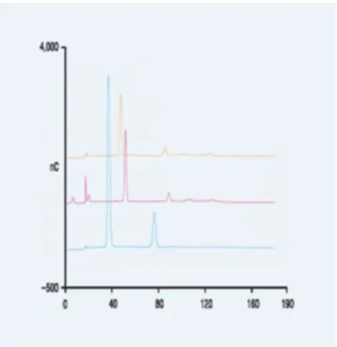
Figure 1. HPAEC-PAD traces of O-linked oligosaccharide dipeptides. Blue trace is Κ casein oligosaccharide dipeptide and 1, 5 anhydro oligosaccharide.
2.2. Isolation of HPAEC-PAD of Components
A sample of the treated Κ casein components was taken and chromatographed and each component collected by retention time on an HPAEC-PAD using a CarboPac MA 1 column (ThermoFisher, Co., Sunnyvale, CA, USA) with a flow rate of 150 µL/ min. with a 310 mM NaOH isocratic elution for at least 190 min. A chromatogram, figure 1, and mass spectra, figures 2 and 3, were obtained and appear after the Results and Discussion section.
Figure 3. This is the mass spectrum generated from isolating the smaller of two peaks from the MA1 chromatogram; K Casein oligosaccharide 1, 5 anhydro N acetamido deoxy galactosaminitol.
2.3. API Mass Spectrometry and MALDI-TOF Mass Spectrometry
The effluent from the HPAEC-PAD was pushed through
an NH4+ cartridge, prepared as noted above and the
effluent was evaporated to no less than 200 µL, H2O (1.00
mL, 18 megaΩ resistivity) added to the vial and the solution pushed through a Na+ cartridge (ThermoFisher, Sunnyvale, CA, USA) prepared by pushing NaOH (freshly prepared from 50% NaOH, 20 ml, 1N) through the H+ cation exchange cartridge, ThermoFIsher, Sunnyvale, CA, USA, and then water was pushed through the Na+ cartridge (using 18 megaΩ resistivity H2O, Milipore Co,
MA, USA)). The effluents from the Κ casein isolates are immediately frozen. The samples should not in any way be concentrated to dryness. A sample of the partially frozen sample was also taken, both peaks (shorter and longer retention times) isolated, singly, for the mass spectrometer, from the HPAEC-PAD system and analyzed by API 2000 triple quadrapole ms as noted. [9, 10] The samples for the API 2000 mass spectrometer were infused with 1% methanolic H2O. A MALDI mass spectrum,
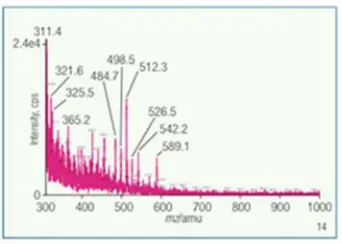
figure 4, was also obtained for the mixture of the oligosaccharide dipeptide and 1, 5 anhydro oligosaccharides.
Figure 4. MALDI mixture of K-casein O-linked oligosaccharide sulfo tyrosine dipeptide and trisaccharide 1, 5 anhydro N acetamido deoxy galactosaminitol.
3. Results and Discussion
There are two components from the treatment of Κ casein with NaBH4 in 1N NH4OH, figure 1 and figure 4. The first of
these two components is N-acetamido deoxy neuraminyl (α 2->3’) N-acetamido deoxy neuraminyl (α 2->6’) galactosyl 4’ (di-hydrido) sulfo (β 1->3) 6 (di-hydrido) sulfo 2 acetamido 2 deoxy galactosaminyl di (hydrido) di phospho serinyl (di-hydrido) sulfo tyrosine dipeptide. The second is the oligosaccharide portion of the above noted molecule with a 1, 5 anhydro N acetamido deoxy galactosaminitol at the ‘reducing end’ of the tetrasaccharide. Both of these molecules are the sulfo and, for the former molecule, sulfo and phospho derivative substituted molecule. [11]
Hydrido derivatives of oligosaccharides are used. They may stabilize the sulfate and phosphate esters toward nucleophilic attack by hydroxide anion. We describe the hydride insertion reaction chemistry. [11, 12]
One group of workers [13] isolated the N-glycan by PNGase F or Endoglycosidase H treatment of K562 expressing wild type recombinant Duffy protein after their purification. They found strong reactions to appropriate lectins indicating Fuc (α 1->6), Gal (β 1->4)-GlcNAc and NeuAc (α 2->6) and no lectin reaction for Man or branched Man and none also for NeuAc (α->3).
dipeptide and 1, 5 anhydro alditol are supported. The dipeptide contains not only this derivatized, sulfated carbohydrate, but also, the isolated dipeptide includes a tyrosine substituted with derivatized sulfate.
An interpretation of our mass spectral data for Κ casein has been published. [10] It suggested a sulfo tyrosine substituent of a dipeptide linked to serine tetrasaccharide. The linkage between the tetrasaccharide and serine must not be a direct linkage because the Κ casein was treated with pH 11.4 NH4OH which is two to three orders of magnitude less
base than the 1 N NaOH at a much lower time than with the standard Carlson β-elimination. The di-phospho linkage between Κ casein tetrasaccharide and serine sulfo tyrosine dipeptide are proposed here
The 1, 5 anhydro molecule is the result of novel nucleophilic attack of hydride anion on the anomeric carbon, C-1, linked to (hydrido) phosphate group. These molecules may be helpful in preventing the binding of DBPII monomer 2 to DBPII monomer 1 or EBA-175 binding and prevent malaria virus progression.
The larger of the two molecules are linked to oligosaccharide to serine, and subsequently sulfo tyrosine, by di-(hydrido) di-phospho group. This linkage is suggested because the original Carlson β-elimination of O-linked oligosaccharides from peptides, using 1 N NaOH containing NaBH4 with a presumed pH of approximately 14, requires,
also, an overnight treatment. In our case we use pH 11.4 to pH 11.6. The amount of hydroxide used is nearly three orders of magnitude less than traditional Carlson β-elimination. This higher pH and more time is required because of a very high pKa, possibly higher than 13, for the abstraction of the serine α hydrogen which is required to initiate β-elimination.
In this protocol the molecule would remain intact, to include the di-phospho groups converted to the di-(hydrido) di-phospho groups and the sulfo groups converted to the (di-hydrido) sulfo substituents, when using NaBH4 in NH4OH.
There would be minimal Carlson β-elimination at pH 11.6 or using pH 11.4 NaOH, without NaBH4, and shorter times, [14]
except if the carbohydrate protein linkage is through a di-phospho linkage, as in non-reductive β-elimination method. [14]
Since the reaction conditions require an uncapped reaction vial, the pH will drop permitting hydrolysis of peptide linkages driven by the release of NH3 to the atmosphere. This
phenomenon is known to occur in hydrazine hydrate, pKa ~ 8, hydrolysis of glycan N acetamido groups from glycoproteins.
The second molecule found has two (di-hydrido) sulfo substitutions on the oligosaccharide with a 1, 5 anhydro N-acetamido deoxy galactosaminitol on the ‘reducing end’ of the oligosaccharide. We demonstrate this structure as shown by the cumulative support for ion structures for ten ions from
the ms, figure 3, of the smaller peak, and longer retention time, from the HPAEC-PAD chromatography, a trace (blue) is shown in figure 1.
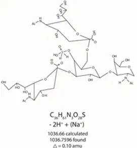
A MALDI spectrum in figure 4 which is from the mixture of substituted oligosaccharides from Κ casein shows two peaks one at m/z 1036.7596 and m/z 698.8596. Figures 5 and 13 describe pictorially the identities of two components of standard Κ casein after reaction under the new conditions.
The smaller of the two peaks, smaller peak in HPAEC-PAD as well (figure 1, blue trace), is shown to have two (di-hydrido) sulfo substituents at O-6 of N-acetamido deoxy 1, 5 anhydro galactosaminitol and O-4’ of the galactose residue. It is also substituted at the O-3’ and O-6’ positions of the galactosyl residue with N-acetamido deoxy neuraminyl groups.
The 1, 5 anhydro galactosaminitol originates from the N-acetamido deoxy galactosaminyl trisaccharide sulfo di-phospho serinyl sulfo tyrosine dipeptide from Κ casein and its reaction with NaBH4 in pH 11.4 NH4OH.
The chemistry for H- nucleophilic substitution of the hydrido phospho substituted anomeric carbon of N-acetamido galactose, C-1, is novel. We suggest a possible mechanism to involve steroelectronics of the O-5, C-1, O-1 and P-1 atoms of the whole di (di-hydrido) sulfo oligosaccharide di-(hydrido) di-phospho serinyl (di-hydrido) tyrosine dipeptide molecule. [15, 16] An O-1 non-bonding electron orbital can be anti-periplanar to the P-1 to O double bond. Along with the H atom on the P atom, O-1 donates electron density preventing development of partial positive charge on the P-1 atom, δ+. This weakens the O-1 to C-1 bond. An O-5 non-bonding electron orbital is in an anti-periplanar alignment with the C-1 atom giving the O-5-C-1 bond, double bond character which may lead to an SN1
nucleophilic H- substitution at 1 and while breaking the C-1 to O-C-1 bond resulting in the C-1, 5 anhydro alditol.
This would be also important as an inhibitor of DBPII monomer 1 and monomer 2 binding to prevent Plasmodium vivax infection. This molecule could help determine the importance of, remote to the Y 278 to T266 interaction, another sulfo group possible requirement for DBPII monomer 1 and monomer 2 binding and also the contribution of the 1, 5 anhydro glycan to the possible inhibition of malaria infection.
Figure 5. MALDI ms peak of Κ casein oligosaccharide dipeptide mixture.
Figure 6. Four ions from API 2000 ms of 1, 5 anhydro alditol isolated from Κ casein.
Two of the ions, m/z 465.3 and m/z 526.5, figures 7 and 8, are substituted with one (di-hydrido) sulfo group rather than two (di-hydrido) sulfo group substitutions on the 1, 5 anhydro glycan. This reflects fragmentation not another compound since we isolated this molecule in pure form by HPAEC-PAD preparative chromatography. For one ion, m/z 589.1, figure 9, one of 5-acetamido deoxy neuraminyl groups has been fragmented away while the other remains. Yet it still retains the (di-hydrido) sulfo, though it is drawn hydrated.
Figure 8. m/z 526.5 from HPAEC-PAD small peak isolate from Κ casein treated with NaBH4 in NH4OH.
An exception to the tetrasaccharide remaining intact in all ten ions, the ion, m/z 589.1, is fragmented at the C-3 to O-1’ bond, noted in figure 9. The 4’ hydrated (di-hydrido) sulfo, 6’ N-acetamido deoxy neuraminyl substituted galactose is drawn to represent the ion. Three ions, m/z 498.5, m/z 426.3 and m/z 484.7, figures 10, 11 and 12, are drawn comprising 4’ (di-hydrido) sulfo galactosyl (β 1-> 3) 2-acetamido 1, 5 anhydro galactosaminitol structure. The ion, figure 12, m/z 484.7 has these attributes; its (di-hydrido) sulfo group hydrated and ionized as a diammonium ion salt.
Figure 9. m/z 589.1 from HPAEC-PAD small peak isolate from Κ casein treated with NaBH4 in NH4OH.
Figure 10. m/z 498.5 from HPAEC-PAD small peak isolate from Κ casein treated with NaBH4 in NH4OH.
Figure 11. m/z 426.3 from HPAEC-PAD small peak isolate from Κ casein treated with NaBH4 in NH4OH.
The larger of the two MALDI, figure 4, peaks, m/z 698.8596, drawn in figure 13 shows the whole molecule less one (di-hydrido) sulfo group. The larger of the two peaks, isolated by chromatography via HPAEC-PAD, figure 1, was subjected to mass spectrometry on an API 2000 triple quadrapole mass
spectrometer. The mass spectrum, API 2000 ms, of the isolated component, is shown in figure 2. The ion, m/z 719.1, figure 14, drawn, represents the total isolated molecule less three ketene molecules. The loss of ketene, CH2=C=O, is common for
N-acetamido substituted carbohydrates.
Figure 13. m/z 698.8596 from MALDI ms of mixture of title components.
The following figures, figures 14-20, depict the ions drawn to support the structural characterization of the larger of the two components from treatment of Κ casein with NaBH4 in pH 11.4 NH4OH. The two ions; m/z 401.1 and 613.2, figures 15 and
16, are drawn. The whole tetrasaccharide, less two ketene, m/z 613.2, three ketenes, m/z 401.1, and two water molecules, from both ions and without the sulfo tyrosine component of the molecule are drawn with the di (hydrido) di-phospho linking serine to the tetrasaccharide with two (di-hydrido) sulfo constituents.
Figure 15. m/z 401.1 ion from ms of HPAEC-PAD isolate (the larger of two peaks).
Figure 16. m/z 613.2 ion from ms of HPAEC-PAD isolate (the larger of two peaks).
In figure 17 the structure of an N-acetamido deoxy galactosaminyl di (hydrido) di-phospho (hydrated) serinyl (di-hydrido) sulfo tyrosine ion, m/z 335.0 is drawn. This ion helps to corroborate the dipeptide portion of the molecule isolated from NaBH4
Figure 17. m/z 335.0 ion from ms of HPAEC-PAD isolate (the larger of two peaks).
In the structure drawn to represent ion m/z 931.0, figure 18, the tetrasaccharide is shown less three ketenes yet intact as a fragment without the di (hydrido) di-phospho serinyl (di-hydrido) sulfo tyrosine. The last two ions, m/z 506.9 and m/z 825.0, are drawn, figures 19 and 20, showing a trisaccharide, one 3’ linked and one 6’ linked, of an N-acetamido deoxy neuraminyl group with linkage to galactosyl N-acetamido deoxy galactosaminyl di (hydrido) di-phospho group. One di (hydrido) phospho group is hydrated, m/z 506.9, and one is dehydrated, m/z 825.0.
Figure 18. m/z 931.0 ion from ms of HPAEC-PAD isolate (the larger of two peaks).
Figure 19. m/z 506.9 ion from ms of HPAEC-PAD isolate (the larger of two peaks).
Figure 20. m/z 825.0 ion from ms of HPAEC-PAD isolate (the larger of two peaks).
Methods are described [9-11] to isolate and analyze these derivatives from Κ casein.
A sulfo-tyrosine is asserted as a mediary for binding DARC to Duffy Binding Protein (DBPII). [17] attest to This hypothesis is attested to and this fact is proposed to be the title compounds, from Κ casein, as inhibitors of Plasmodium vivax binding of DARC and subsequent progression by this virus, malaria.
so that the second DARC molecule can bind to the DBPII trimer and then chemokines.
The di-hydrido derivative of the sulfated title compound may be stable to hydrolysis, due to the difference between the electro-negativities of a hydroxyl versus two hydrogens, making the sulfuryl group, OH substituted and not two H substituted, sulfur develop partial positive charge, δ+, compared to the substituents with two hydrogen substitutions, making the sulfuryl group of sulfate a better nucleofuge, in the case of hydroxyl substitution.
4. Conclusion
The use of the two molecules, described and structurally established above, may mimic the DBP-RII monomer 2 to provide the sulfo-tyrosine dipeptide oligosaccharide and 1, 5 anhydro tetrasaccharide, both from Κ casein, in order to prevent DBP-RII monomer 2 from binding the DBP-RII monomer 1, in Plasmodium vivax. If this quaternary structure is blocked by the these compounds, Plasmodium vivax
cannot effectively bind its DBP-RII monomer 2, preventing Y 278 or Y271 or Y363 sulfate interaction with DBPII monomer 1, and preventing progression toward infection by the malaria virus. The 1, 5 anhydro tetrasaccharide, without the (di-hydrido) sulfo tyrosine epitope, could also bind to
Plasmodium vivax because of a report of the presence of 5-N-acetamido neuraminyl-Gal epitope [6] on the N-glycans of recombinant DBPII.
It is suggested that these two molecules may be mimics of erythrocyte binding antigen, EBA-175, with the epitope of
Plasmodium falciparum, Neu (α 2->3’) Gal (β 1->4), present in both isolated molecules from Κ casein.
References
[1] Batchelor, J.; Zahm, J.; Tolia, N.; Dimerization of
Plasmodium vivax DBP is induced upon receptor binding and drives recognition of DARC Nature Structural and Molecular Biology (2011) 18:908-914.
[2] Batchelor, J.; Malpede, B.; Omatagge, N.; DeKover, G.; Henzler-Wildman, K.; Tolia, N. Red blood cell invasion of
Plasmodium vivax structural basis for DBP engagement of DARC, PLoS Pathogens 10, 1.e1003869 (2014).
[3] Orllandi, P.; Klotz, F.; Haynes, J.; A Malaria Invasion Receptor, the 175 kilodalton Erythrocyte Binding Antigen of
Plasmodium falciparum Recognizes the Terminal Neu5Ac (α2-3) Gal- sequences of glycophorin A J. Cell Biology 116 (4) 901-909 (1992).
[4] Ntumngia, F.; Thomson-Lugue, R.; Tomes, L.; Gunalan, K.; Carvalho, L.; Adams, J.; A novel erythrocyte binding protein of Plasmodium vivax suggests an alternative pathway into
Duffy positive reticulocytes mbio.asm.org 1-5 American Society for Microbiology 7: (4) e01261-16 (2016 (b)).
[5] Wanaguru, M.; Crosnier, C.; Johnson, S.; Rayner, J.; Wright, G. Biochemical Analysis of the Plasmodium falciparum
Erythrocyte-binding Antigen-175 (EBA175)-Glycophorin-A Interaction IMPLICATIONS FOR VACCINE DESIGN J. Biol. Chem. 288 32016-32117 (2013).
[6] Grodecka, M.; Czerwiński, M,; Duk, M.; Lisowska, E.; Waśniowska, K; Analysis of recombinant Duffy protein-linked N-glycans using lectins and glycosidases Acta Biochimica Polonica 57 (1) 49–53 (2010).
[7] Saito, T.; Itoh, T.; Variations and Distributions of O-Glycosidically Linked Sugar Chains in Bovine K-Casein J. Dairy Sci. 75:1768-1774 (1992).
[8] Madson, M.; Rao S.; Avdalovich, N.; Pohl, C.; A simple method for the isolation of N- and O-linked oligosaccharides from glycoproteins poster Glycobiology San Diego CA, USA (2005).
[9] Madson, M. Method of isolating and analyzing oligosaccharides from glycoproteins patent US 9, 625, 468 B1 (April 18th 2017).
[10] Madson, M. Mass Spectrometry; Techniques for Structural Characterization of Glycans, Elsevier, May 30th New York USA (2016).
[11] Madson, M. Method of discerning substitution of carbohydrate esters patent US 9, 726, 761 B2 (August 8th 2017).
[12] Madson M.; Manufacturing of MeOH, formaldehyde, formic acid and ammonium pentaborate tetrahydrate from CO2 patent
US 8, 685, 355 B2 (2014).
[13] Ntumngia, F.; Thomson-Luque, R.; Pires, C.; Adams, J.; The role of the human Duffy antigen receptor for chemokines in malaria susceptibility: current opinion and future treatment prospects J. of Receptor, Ligand and Channel Research 9: 1-11 (2016 (a)).
[14] Madson, M.; System and method for non-reductive β-elimination US 9, 488, 653 B2 patent (November 8th 2016).
[15] Deslongchamps, P.; Stereoelectronic control in the cleavage of tetrahedral intermediates in the hydrolysis of esters and amides Tetrahedron 31 (20) 2463-2490 (1975).
[16] Deslongchamps, P.; Rowan, D.; Pothier, N.; G.; Saunders, J.; 1, 7-Dioxaspiro [5.5] undecanes. An excellent system for the study of stereoelectronic effects (anomeric and exo-anomeric effects) in acetals Canadian Journal of Chemistry 59 (7) 1105-1121 (1981).


