R E S E A R C H
Open Access
Identification of novel
GNAS
mutations in
intramuscular myxoma using
next-generation sequencing with single-molecule
tagged molecular inversion probes
Elise M. Bekers
1,2*, Astrid Eijkelenboom
1, Paul Rombout
1, Peter van Zwam
3, Suzanne Mol
4, Emiel Ruijter
5,
Blanca Scheijen
1and Uta Flucke
1Abstract
Background:Intramuscular myxoma (IM) is a hypocellular benign soft tissue neoplasm characterized by abundant myxoid stroma and occasional hypercellular areas. These tumors can, especially on biopsy material, be difficult to distinguish from low-grade fibromyxoid sarcoma or low-grade myxofibrosarcoma.GNASmutations are frequently involved in IM, in contrast to these other malignant tumors. Therefore, sensitive molecular techniques for detection ofGNASaberrations in IM, which frequently yield low amounts of DNA due to poor cellularity, will be beneficial for differential diagnosis.
Methods:In our study, a total of 34 IM samples from 33 patients were analyzed for the presence ofGNAS
mutations, of which 29 samples were analyzed using a gene-specific TaqMan genotyping assay for the detection of GNAShotspot mutations c.601C > T and c602G > A in IM, and 32 samples using a novel next generation sequencing (NGS)-based approach employing single-molecule tagged molecular inversion probes (smMIP) to identify mutations in exon 8 and 9 ofGNAS. Results between the two assays were compared for their ability to detectGNASmutations with high confidence.
Results:In total, 23 of 34 samples were successfully analyzed with both techniques showingGNASmutations in 12 out of 23 (52%) samples. The remaining 11 samples were analyzed with either TaqMan assay or smMIP assay only. The TaqMan assay revealedGNASmutations in 16 out of 29 samples (55%), with six samples c.601C > T (p.R201C; 38%) and ten samples c.602G > A (p.R201H; 62%) missense mutations. The smMIP assay identified mutations in 16 out of 28 samples (57%), with five samples c.601C > T (p.R201C; 31%) and seven samples c.602G > A (p.R201H; 44%) missense mutations. In addition, four samples (25%) revealed novel IM-associated mutations, including c.601C > A (p.R201S), c.602G > T (p.R201L), c.602G > C (p.R201P) and c.680A > G (p.Q227R). Combining the results of both tests, 23 out of 34 sporadic IM samples (68%) showed aGNASmutation.
Conclusions:Both the TaqMan and the smMIP assay a show a high degree of concordance in detectingGNAS hotspot mutations in IM with comparable sensitivity. However, since the NGS-based smMIP assay permits mutation detection in whole exons ofGNAS, a broader range ofGNASmutations can be identified by the smMIP approach.
Keywords:Next generation sequencing, TaqMan genotyping, smMIP assay,GNASmutation, Intramuscular myxoma
* Correspondence:Elise.Bekers@radboudumc.nl;e.bekers@nki.nl 1
Department of Pathology, Radboud University Medical Center, Geert Grooteplein Zuid 10, 6525, GA, Nijmegen, The Netherlands
2Department of Pathology, The Netherlands Cancer Institute, Amsterdam, The Netherlands
Full list of author information is available at the end of the article
Background
Intramuscular myxoma (IM) is a benign soft tissue neoplasm that belongs to the group of myxoid tumors characterized by a marked abundance of extracellular myxoid matrix. These tumors share several histological features, and depending on their clinical presentation and place of origin, can be subdi-vided into intramuscular, superficial-cutaneous, odontogenic and juxta-articular myxoma [1,2]. These myxomas all repre-sent distinct entities with different characteristic gene lesions involved in their pathogenesis. Therefore, gene mutation analysis can be very helpful in differential diagnosis to sup-port the histopathology of these tumors [3,4].
IM is characterized by bland spindle- and/or stellate-shaped cells embedded in a hypovascular, abun-dant myxoid stroma. The nuclei are small showing no or minimal nuclear atypia. Often areas with increased cellu-larity can be observed and when hypercellular areas pre-dominate it is designated as cellular myxoma [2, 5, 6], which can easily be confused with low-grade fibromyx-oid sarcoma or low-grade myxofibrosarcoma, especially in very small biopsies. IM is a somatic mosaic disorder generally occurring as a sporadic solitary neoplasm, al-though it can be part of Mazabraud’s syndrome charac-terized by a combination of polyostotic fibrous dysplasia with multiple IM’s [7,8]. Mazabraud’s syndrome and the closely related McCune-Albright syndrome, which is as-sociated with fibrous dysplasia, café au lait macules and endocrine disorders, are caused by activating missense mutations in codon 201 of theGNASgene [8–12].
GNAS encodes the stimulatory G-alpha subunit of the heterotrimeric G-protein complex, which regulates activa-tion of adenylyl cyclase that converts adenosine triphos-phate (ATP) into cyclic adenosine monophostriphos-phate (cAMP). Overproduction of second messenger cAMP and activation of downstream signaling pathways has been ob-served in cells harboring GNAS mutations [13, 14]. In 2000, Okamoto et al. first described somatic post-zygotic
GNASmutations in IM with and without fibrous dysplasia [8]. Thereafter, three more studies showed thatGNAS le-sions occur frequently in sporadic IM, which were de-tected in 36–61% of the cases, and exclusively involved c.601C > T (p.R201C) and c.602G > A (p.R201H) muta-tions [8,15,16]. On the other hand,GNASmutations are absent in low-grade myxofibrosarcoma, which can be use-ful in the differential diagnosis with (cellular) IM [17,18]. Notably, juxta-articular myxoma and cardiac myxoma also lackGNASdriver mutations [4,16].
A complicating factor for mutation detection in IM is the mosaicism of GNASmutations combined with hypocellu-larity of the tumor, where low concentrations of genomic DNA are isolated from these tissue specimens, especially in the case of biopsy material. In the past decades, several techniques forGNASmutation detection have been devel-oped and used [8,19–21]. In 2009, Delaney et al. tested 28
IM’s for GNAS mutations by using conventional PCR followed by mutation-specific restriction enzyme digestion (PCR-MSRED) and COLD-PCR/MSRED and showed that COLD-PCR/MSRED was more sensitive than the conven-tional PCR (61% vs. 29% mutations) [15]. Thus, this tumor type may benefit from the development of more robust and sensitive techniques for mutation detection, such as next generation sequencing (NGS). Recently, our molecular diagnostic laboratory has developed a novel NGS-based ap-proach employing single-molecule molecular inversion probes (smMIP) that combines multiplex analysis with single-molecule tagging, also named Unique Molecule Identifiers (UMI) [22,23]. By using this method, duplicate reads can be identified and merged into a single consensus, reducing falpositive calls originated during PCR and se-quencing and allowing a technical sensitivity of 1% mutant allele. In addition, the actual number of sequenced genomic DNA (gDNA) molecules can be determined, which is espe-cially relevant when analyzing limited amounts of gDNA. Furthermore, the strand-specific amplifications allows the distinction between genuine C > T and G > A mutations from deamination artifacts frequently observed when se-quencing gDNA in older formalin-fixed paraffin-embedded (FFPE) tissue specimens. [22,23].
In this study, we applied both TaqMan-based assays and the smMIP technique forGNASmutation detection in IM, and compared both methods for reliable mutation detection in a diagnostic setting.
Methods Patient samples
This study included 34 samples of sporadic intramuscular myxoma from 33 patients that were collected retrospect-ively (from 1998 till 2018) from archives of the Pathology Departments in the Netherlands of the Radboud University Medical Centre, Jeroen Bosch Hospital (Den Bosch), PAMM institute (Eindhoven) and Rijnstate Hospital (Arnhem). None of the patients were prior diagnosed with fibrous dysplasia or developed this during follow-up. From one patient, two samples (sample 28 and 29) were analyzed, which yielded identical data for mutation analyses. For each case, a 4μm thick section of FFPE material was stained with haematotoxylin and eosin (H&E). The histological diagnoses were revised (UF, EB) and classified according to the 2013 World Health Organization criteria [2]. The sam-ples included in this study complied with the standards of the Committee for Human Research Ethics (CMO).
DNA isolation
15μg/mL GlycoBlue (AM9516; Thermo Fisher, Waltham, MA), and 400μg proteinase K (19,133; Qiagen, Valencia, CA), followed by inactivation at 95 °C for 10 min. DNA concentration for TaqMan assay was assessed with the NanoDrop Microvolume Spectrophotometer (Peqlab Bio-technologies, Erlangen, Germany) and for smMIP assay with the Qubit Broad Range Kit (Q32853; Thermo Fisher). To concentrate the DNA samples for the robotized proto-col of the smMIP procedure, supernatant was cooled on ice and precipitated in the presence of 70% EtOH and 1/ 10 volume 3 M NaAc (pH 5.2). Pellets were washed with cold 70% EtOH and dissolved in 80μL Tris-EDTA. DNA quality of the samples was tested using a size ladder con-trol PCR, in which gene segments of house-keeping genes were amplified, yielding different fragment sizes (100, 200, 300 and 400 bp), depending on the extent of fragmenta-tion of the DNA.
TaqMan genotyping assay
Pre-designed and validated gene-specific TaqMan Genotyp-ing Assays from Thermo Fisher Scientific was used for quantitative real-time RT-PCR. Every set contained gene specific forward 5’-CTTTGGTGAGATCCATTGACCTCA A-3′ and reverse primers 3’-CACCTGGAACTTGG TCTCAAAGATT-5′ and fluorescence labeled probes (Table1). Probes are spanning an exon junction to detect genomic DNA. The PCR reaction volume was 20μl and contained 1μl DNA (10 ng/μl), 10μl TaqMan Universal PCR Mastermix NoAmpErase UNG (Applied Biosystems, Foster City, CA), 0,5μl predesigned and validated gene-specific TaqMan Gene Expression Assay mix (Applied Biosystems), 0,5μl TE buffer (Promega) and 8μl water. ABI Prism 7500 Real-Time PCR system (Applied Biosystem) was used to amplify codon 201 of exon 8 of theGNASgene from each sample on a 96-well reaction plate with the following protocol: 10 min denaturation at 95 °C, 40 cycles of 15 s denaturation at 95 °C, 1 min annealing and exten-sion at 60 °C. Dilution studies were performed using fibrous dysplasia samples harboring the two previously described
GNASmutations. The limit of detection was reliable at a variant allele frequency (VAF) of 5%.
Next generation sequencing with single-molecule molecular inversion probes
The single-molecule molecular inversion probe (smMIP) procedure was performed as described elsewhere [22]. In short, a pool of smMIPs covering 41 mutational hotspot re-gions of 23 distinct genes, including GNAS, was
phopshorylated with T4 polynucleotide kinase. A total of 100 ng genomic DNA was used as input in the capture re-action with the diluted phosphorylated smMIP pool. After extension, ligation and exonuclease treatment, PCR reac-tions were performed with barcoded reverse primers and iProof high-fidelity master-mix (Biorad). PCR reactions of the different samples were pooled, and purified with 0.8 x volume of Agencourt Ampure XP Beads (Beckman Coulter, Brea, CA). The purified libraries were prepared for sequen-cing on a NextSeq 500 instrument (Illumina, San Diego, CA) according to the manufacturer’s protocol (300 cycles Mid Output sequencing kit, v2), resulting in 2 × 150 bp paired-end reads. Data analyses were performed as previ-ously described [22]. Variants were called at a VAF of > 1% and≥3 mutant gDNA molecules and a minimum of 20 gDNA molecules analyzed at that position. Samples that did not fulfil the standard settings with respect to gene coverage in combination with tumor load were marked as inconclusive [22].
Results
Histopathology and clinical information of intramuscular myxoma cases
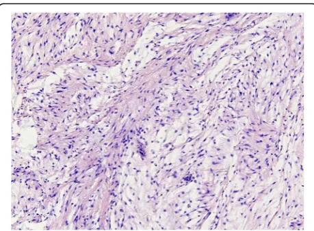
Histopathology of H&E-stained slides confirmed that a selected set of 34 samples from 33 patients showed the classical features of IM, which were composed of uniform, sparsely distributed cytological bland spindle- or stellate-shaped cells with tapering eosinophilic cytoplasm and small nuclei embedded in an abundant myxoid stroma. One case showed prominent hypercellular areas with more collagenous stroma and was diagnosed as cellu-lar myxoma according to the criteria defined by Nielsen et al (Fig. 1) [5, 6]. Of the 34 myxoid tumors, 31 samples were obtained by local excision and 3 samples by needle biopsy (Table2). From one patient (sample 28 and 29), a biopsy and the following excision were analyzed forGNAS mutational status. No recurrences were reported in any of the cases and no additional treatment was given. Follow up time ranged between 2 months and 21 years.
GNAS mutation detection in intramuscular myxoma
All samples (n = 34) were tested for the presence of
GNAS mutations, 32 samples using the smMIP assay and 29 samples with the TaqMan genotyping assay. First,
GNAS mutation detection was performed for 29 DNA samples with the TaqMan assay, where specific fluores-cently labeled probes were used for the detection of c.601C > T (p.R201C) and c.602G > A (p.R201H) hotspot
Table 1Fluorescent reporter probes for TaqMan assay
TaqMan assay Reporter probe wild-type Reporter probe mutant
GNASc.601C > T 5’-CAGGACACGGCAGCGA-3’ 5’-CAGGACACAGCAGCGA-3’
GNASc.602G > A 5’-TTCGCTGCCGTGTCCT-3’ 5’-CGCTGCCATGTCCT-3’
mutations (Fig. 2). Each sample was analyzed in two in-dependent assays together with both negative and posi-tive control samples. GNAS genetic alterations were identified in 16 out of 29 samples (55%), with six sam-ples c.601C > T (38%) and ten samsam-ples c.602G > A (62%) mutations (Table3). From one patient both samples (bi-opsy and excision) were positive for c.601C > T mutation (Table3; sample 28 and 29).
Next, we determined the presence ofGNAS mutations in exon 8 and exon 9 by the smMIP assay. Within the smMIP Cancer Hotspot Panel, two smMIPs covered
GNAS exon 8 (providing sequencing analysis of both DNA strands for a total 74 bp) and two smMIPs GNAS exon 9 (sequencing analysis of 59 bp), respectively (Fig.3). Other mutational hotspot regions that were covered by smMIPs included BRAF, CTNNB1, EGFR, HRAS, KRAS,
NRAS, IDH1, IDH2 and KIT (for the complete list see [22]). The smMIP assay was performed on 32 samples in total, including the 27 samples that also showed a success-ful TaqMan assay and an additional set of 5 IM samples. From these 32 samples, the NGS data of 5 samples (for which a successful TaqMan assay was available) was based on a very limited number of gDNA molecules, and there-fore could not reliably be interpreted, most likely because of very low cellularity of the IM sample and/or inferior DNA quality (Table3; inconclusive, Inc). In total, 16 out of 28 samples (57%) showed aGNAS mutation, with five samples c.601C > T (31%) and seven samples c.602G > A (44%) mutations. In addition, four samples (25%) revealed novel IM-associated mutations, including c.601C > A (p.R201S), c.602G > T (p.R201L), c.602G > C (p.R201P) and c.680A > G (p.Q227R).
Combining the above, 23 samples were successfully analyzed with both techniques showingGNASmutations in 12 out of 23 (52%) samples. Collectively, our data
demonstrate that in 23 out of 34 IM samples (68%) a
GNASmutation was detected using either TaqMan and/ or smMIP assay. All samples that were successfully ana-lyzed with both approaches and harbored the classical c.601C > T or c.602G > T mutations were identified with both methods.
In total, eight samples showed the classical c.601C > T mutation (35%) and eleven samples harbored the c.602G > T mutation (48%). All hotspot mutations de-tected by smMIP were also identified by the TaqMan assay, including samples with a VAF of 5% (sample 2). On the other hand, due to the more stringent settings of the smMIP assay, five cases with hotspot mutations identified by TaqMan assay did not yield sufficient data by the smMIP approach for reliable interpretation. In contrast, the smMIP assay allowed the detection of four novel (po-tential) pathogenicGNAS mutations (17% of the 23 mu-tated samples) beyond the c.601C > T and c.602G > A mutations, not previously described for IM. Thus, both as-says provide merits in the molecular diagnostics of IM.
Discussion
Intramuscular myxoma (IM) mostly occurs sporadically in the skeletal muscle of the thigh. These lesions affect mainly middle-aged adults, women more often than man [1,24]. The prevailing view is that driver mutations of this neoplasm are exclusively located in codon 201 of the
GNAS gene, encoding the stimulatory G-protein alpha subunit that activates the enzyme adenylate cyclase. Due to the low cellularity and somatic mosaicism in most of these lesions, mutation detection can be quite challenging and the presence of a mutation can be easily missed.
In our study we used two different techniques (TaqMan and smMIP assay) to compare the detection sensitivity of
GNASmutations in these lesions. In our series, 23 out of 34 sporadic IM cases (68%) showed aGNASmutation, 16 out of 29 samples (55%) in the TaqMan assay and 16 out of 28 samples (57%) in the smMIP assay of which 23 sam-ples were successfully analyzed with both techniques showingGNASmutations in 12 out of 23 (52%) samples. The test-specific detection rate was 55% with the TaqMan assay and 57% for the smMIP approach. The VAF for the TaqMan assay was determined at > 5% in this study and the required input was only 10 ng gDNA. The VAF for smMIP was set at > 1% and a minimum of 3 mutant gDNA molecules, and a coverage of 20 gDNA molecules. This demonstrates that both tests are sensitive methods and useful for molecular diagnostics of tumor samples harboring mutations with a low mutant allele frequency.
In comparison, Walther et al found GNASmutations in 37% (23/63) of IMs with direct Sanger sequencing and Delaney et al detected mutations in 61% (17/28) using COLD-PCR/MSRED [15, 16]. However, the smMIP technique, because of the whole exon
Fig. 1Representative photomicrograph of heamatoxylin and eosine
sequencing nature of this test, allowed detection of four additional mutations that previously have not been de-scribed in IM. By using smMIP we identified one c.680A > G mutation in exon 9, and three novel muta-tions in exon 8, one mutation at position c.601, namely c.601C > A, and two mutations at position c.602, which included c.602G > C and c.602G > T. The c.601C > A mutation has previously been reported in fibrous dyspla-sia, while the c.602G > C and c.602G > T mutations were only reported in sporadic endocrine tumors so far [9,10, 19,25]. These mutations were not detected by TaqMan,
since this assay was designed to report only the two clas-sical hotspot mutations c.601C > T and c.602G > T.
The smMIP approach allows the distinction between genuine C > T and G > A mutations from deamination arti-facts frequently observed when sequencing gDNA from FFPE tissue specimens [22]. All cases harboring a C > T or G > A mutation in GNAS, mutant reads originating from both DNA strands were observed, showing that these rep-resent genuine mutations. Since the TaqMan approach does not allow this distinction, deamination artifact could poten-tially cause false positive results. In our study, all hotspot
Table 2Patient characteristics
Patient Age at presentation (years) Gender Tumor localization Tumor size (cm) excision Follow-up (months)
1 74 M Lower leg 14 207
2 42 F Thigh 5 201
3 51 F Upper arm 0.8 128
4 45 M Upper leg 5.5 126
5 40 F Thigh Bx 99
6 47 F Lower leg 10 93
7 51 M Shoulder 3 85
8 64 M Thigh 5.2 83
9a 53 M Upper arm 2 83
10 45 M Thigh 5.8 82
11 57 F Thigh 9 67
12 72 M Thigh 3.5 63
13 71 M Back 3 210
14 55 M Thigh 4 182
15 55 M Thigh 4 157
16 53 F Upper arm 3 156
17 47 F Upper arm 5.5 100
18 38 F Upper arm 2 108
19 57 M Thigh 3.2 126
20 39 F Upper arm 7 191
21 69 F Chest 2 63
22 46 F Shoulder 3.5 200
23 64 F Thigh 3 194
24 46 M Thigh 5 181
25 67 F Lower arm 1.7 127
26 49 F Thigh 3 73
27 58 F Thigh 3 244
28b
33 F Thigh 7.5 224
29 65 M Thigh 5.5 31
30 59 F Thigh 7 12
31 40 F Upper arm Bx 260
32 71 F Thigh 1 6
33 54 M Thigh Bx 2
Bx: biopsy,a
cellular myxoma,b
mutations detected with the Taqman assay were confirmed with the smMIP technique, indicating no false-positive re-sults with Taqman. Even samples with a VAF of around 5% could be detected by both TaqMan assay and smMIP. The four samples with a hotspot mutation that were detected by TaqMan, but did not yield a reliable smMIP assay result, all had a mutated VAF of approximately 10–30% as judged by Taqman assay, and were therefore interpreted as true mutations.
Significant benefits of the TaqMan assay include low cost and short turn-around time (≤2 working days). A limitation of Taqman is that within one assay only one or two hotspot
mutations can be detected. For smMIP analysis the turn-around time in our laboratory is≤7 working days. A large initial investment was needed and high numbers of samples are required for parallel analyses to have a cost-efficient test. Because multiple genes can be tested at once with the smMIP assay, large amounts of samples are relatively easy obtained in routine clinical setting with the current demand of molecular diagnostics [22]. Because of the sensitive characteristics of the smMIP technique and its accuracy of mutation detection on FFPE material as well as the broader coverage of theGNASgene, this technique to our opinion is preferable.
Fig. 2Schematic overview of the Taqman assay. In addition to the genomic DNA template, four additional oligonucleotide components are
The most important differential diagnoses of IM, espe-cially the cellular variant, are low-grade fibromyxoid sar-coma and low-grade myxofibrosarcoma. In biopsy material the distinction can be challenging and in those cases molecular diagnostics can be beneficial. A specific
immunohistochemical and molecular signature is well known for low-grade fibromyxoid sarcomas with expres-sion of MUC4 and the presence ofFUS/EWSR1-CREB 3
L2/1fusions making a distinction from IM easily possible [17, 18]. In contrast, for low-grade myxofibrosarcoma,
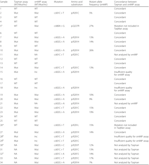
Table 3Mutation analysis intramuscular myxoma
Sample Taqman assay (WT/Mut/Inc)
smMIP assay (WT/Mut/Inc)
Mutation Amino acid substitution
Mutant allele frequency (smMIP)
Concordance between Taqman and smMIP assay
1 WT WT Concordant
2 Mut Mut c.601C > T p.R201C 5% Concordant
3 WT WT Concordant
4 WT WT Concordant
5a WT Mut c.680A > G p.Q227R 27% Mutation not included in
TaqMan assay
6 WT WT Concordant
7 Mut Mut c.602G > A p.R201H 13% Concordant
8 Mut Mut c.602G > A p.R201H 14% Concordant
9 WT WT Concordant
10 Mut Mut c.602G > A p.R201H 26% Concordant
11 Mut NA c.601C > T p.R201C Not analyzed by smMIP
12 WT WT Concordant
13 WT WT Concordant
14 Mut Mut c.601C > T p.R201C 13% Concordant
15 Mut Inc c.602G > A p.R201H Insufficient quality
for smMIP assay
16 WT WT Concordant
17 WT WT Concordant
18 Mut Inc c.602G > A p.R201H Insufficient quality
for smMIP assay
19 Mut Mut c.602G > A p.R201H 10% Concordant
20 Mut Mut c.602G > A p.R201H 9% Concordant
21 Mut NA c.602G > A p.R201H Not analyzed by smMIP
22 Mut Mut c.601C > T p.R201C 15% Concordant
23 Mut Mut c.602G > A p.R201H 19% Concordant
24 WT WT Concordant
25 WT WT Concordant
26* WT Mut c.602G > T p.R201L 15% Mutation not included
in TaqMan assay
27 Mut Mut c.602G > A p.R201H 14% Concordant
28b Mut Inc c.601C > T p.R201C Insufficient quality for smMIP assay
29b Mut Inc c.601C > T p.R201C Insufficient quality for smMIP assay
30a NA Mut c.602G > C p.R201P 12% Not analyzed by Taqman
31 NA Mut c.601C > T p.R201C 13% Not analyzed by Taqman
32a NA Mut c.601C > A p.R201S 14% Not analyzed by Taqman
33 NA Mut c.601C > T p.R201C 17% Not analyzed by Taqman
34 NA Mut c.602G > A p.R201H 7% Not analyzed by Taqman
WT: wild-type; Mut: mutation identified; Inc.: inconclusive; NA: not analyzed
a
Samples with novel mutations in smMIP assay which are not included in the TaqMan assay
b
specific immunohistochemical or molecular characteris-tics are lacking. Sensitive molecular tests, like smMIP and TaqMan assays forGNASmutation analysis, might be very helpful in assessing the diagnosis, which has therapeutic consequences when considering malignancy [21]. Never-theless there are also cases in which no GNAS mutation could be detected, suggesting that there are still other ab-errations to be identified in IM.
Panagopoulos et al recently found abnormal karyotypes in 21 out of 68 cases, with nine cases showing nonrandom involvement of chromosome 8 (which harbors theGNAS gene) with seven cases showing trisomy 8, one with a dele-tion and one with a translocadele-tion. Only one case in their
series showed a c.601C > T GNAS mutation [26]. Thus, chromosomal aberrations could be an alternative explan-ation for at least a subset of the non-mutated cases.
The smMIP-NGS cancer hotspot panel that was employed to check for GNASmutations, also contained smMIPs that covered mutational hotspots in the genes
AKT1, BRAF, CTNNB1, EGFR, ERBB2, GNA11, GNAQ, H3F3A, H3F3B, HRAS, IDH1, IDH2, JAK2, KRAS, MPL,
MYD88, NRAS, PDGFRA and PIK3CA. In none of the
32 samples that could be reliably analyzed by smMIPs, additional mutations were detected in these regions. Thus,GNASmutations represent a unique driver muta-tion for this benign tumor type.
a
b
Fig. 3Schematic overview of the smMIP assay. (a) First, the single molecule molecular inversion probe (smMIP) capture procedure is performed.
smMIPs are long oligonucleotides consisting of two targeting arms (extension probe and ligation probe), joined by a backbone. The probe sequences are complementary to genomic DNA sequences surrounding the target region that covers a hotspot location (indicated by the yellow asterisk). During the capture reaction, smMIPs are hybridized to genomic DNA (gDNA), followed by an extension and ligation reaction, which results in circular smMIPs. Subsequent exonuclease treatment will remove all linear gDNA and unused smMIPs. Between the backbone and probe sequences are primer sequences (green bars) that are used to amplify the target region, followed by library preparation and next-generation sequencing (NGS). (b) By including a single-molecule tag of 8 random nucleotides (N8) at the end of the ligation probe, duplicate reads can be
Conclusion
In conclusion, both TaqMan and smMIP assay are com-parably sensitive molecular methods with valuable ap-plicability in diagnostic pathology for IM. Furthermore, due to a broader coverage of the GNAS gene by the smMIP approach, four novel IM-associated missense mutations of GNAScould be identified (17% of all mu-tated samples), which previously have only been re-ported in McCune-Albright syndrome and sporadic endocrine tumors.
Abbreviations
ATP:adenosine triphosphate; cAMP: cyclic adenosine monophosphate; CMO: Committee for human research ethics; FFPE: formalin-fixed paraffin-embedded; FRET: fluorescence resonance energy transfer;
H&E: haematotoxylin and eosin; IM: intramuscular myxoma; MGB: minor groove binder; NGS: next generation sequencing; smMIP: single-molecule molecular inversion probes; UMI: Unique molecule identifiers; VAF: variant allele frequency
Acknowledgements
Not applicable.
Funding
Not applicable.
Availability of data and materials
The datasets used and/or analyzed during the current study are available from the corresponding author on reasonable request.
Authors’contributions
EB contributed to study design, laboratory work, revised pathology and is the main contributor to writing the manuscript. UF contributed to study design, writing the manuscript, and revised pathology. BS contributed to study design, laboratory work and was a major contributor in writing the manuscript. AE contributed to laboratory work and provided data and for and helped writing methods section in manuscript. PR contributed to laboratory work and writing methods section in manuscript. SM, PZ and ER provided tissue samples. All authors have read and approved the final version to be published.
Ethics approval and consent to participate
Ethical approval was obtained by the Committee for Human Research Ethics (CMO). No consent to participate was needed from the participants.
Consent for publication
Not applicable.
Competing interests
The authors declare that they have no competing interests.
Publisher’s Note
Springer Nature remains neutral with regard to jurisdictional claims in published maps and institutional affiliations.
Author details
1
Department of Pathology, Radboud University Medical Center, Geert Grooteplein Zuid 10, 6525, GA, Nijmegen, The Netherlands.2Department of Pathology, The Netherlands Cancer Institute, Amsterdam, The Netherlands. 3Department of Pathology, PAMM Michelangelolaan 2, 5623 EJ Eindhoven, The Netherlands.4Department of Pathology, Jeroen Bosch Hospital, Henri Dunantstraat 1, 5223, GZ, Den Bosch, The Netherlands.5Department of Pathology, Rijnstate Hospital, Wagnerlaan 55, 6815, AD, Arnhem, The Netherlands.
Received: 5 October 2018 Accepted: 24 January 2019
References
1. Goldblum JR, Folpe AL, Weiss SW, Enzinger FM, Weiss SW. Enzinger and Weiss's soft tissue tumors. 6th ed. Philadelphia, PA: Saunders/Elsevier; 2014. xiv, 1155 p. 2. Fletcher CDM, World Health Organization. International Agency for Research
on Cancer. WHO classification of tumours of soft tissue and bone. 4th ed. Lyon: IARC Press; 2013. 468 p
3. Boson WL, Gomez RS, Araujo L, Kalapothakis E, Friedman E, De Marco L. Odontogenic myxomas are not associated with activating mutations of the Gs alpha gene. Anticancer Res. 1998;18(6A):4415–7.
4. Okamoto S, Hisaoka M, Meis-Kindblom JM, Kindblom LG, Hashimoto H. Juxta-articular myxoma and intramuscular myxoma are two distinct entities. Activating Gs alpha mutation at Arg 201 codon does not occur in juxta-articular myxoma. Virchows Arch. 2002;440(1):12–5.
5. Nielsen GP, O'Connell JX, Rosenberg AE. Intramuscular myxoma: a clinicopathologic study of 51 cases with emphasis on hypercellular and hypervascular variants. Am J Surg Pathol. 1998;22(10):1222–7. 6. van Roggen JF, McMenamin ME, Fletcher CD. Cellular myxoma of soft
tissue: a clinicopathological study of 38 cases confirming indolent clinical behaviour. Histopathology. 2001;39(3):287–97.
7. Cabral CE, Guedes P, Fonseca T, Rezende JF, Cruz Junior LC, Smith J. Polyostotic fibrous dysplasia associated with intramuscular myxomas: Mazabraud's syndrome. Skelet Radiol. 1998;27(5):278–82.
8. Okamoto S, Hisaoka M, Ushijima M, Nakahara S, Toyoshima S, Hashimoto H. Activating Gs(alpha) mutation in intramuscular myxomas with and without fibrous dysplasia of bone. Virchows Arch. 2000;437(2):133–7.
9. Schwindinger WF, Francomano CA, Levine MA. Identification of a mutation in the gene encoding the alpha subunit of the stimulatory G protein of adenylyl cyclase in McCune-Albright syndrome. Proc Natl Acad Sci U S A. 1992;89(11):5152–6.
10. Shenker A, Weinstein LS, Moran A, Pescovitz OH, Charest NJ, Boney CM, et al. Severe endocrine and nonendocrine manifestations of the McCune-Albright syndrome associated with activating mutations of stimulatory G protein GS. J Pediatr. 1993;123(4):509–18.
11. Shenker A, Weinstein LS, Sweet DE, Spiegel AM. An activating Gs alpha mutation is present in fibrous dysplasia of bone in the McCune-Albright syndrome. J Clin Endocrinol Metab. 1994;79(3):750–5.
12. Weinstein LS, Shenker A, Gejman PV, Merino MJ, Friedman E, Spiegel AM. Activating mutations of the stimulatory G protein in the McCune-Albright syndrome. N Engl J Med. 1991;325(24):1688–95.
13. O'Hayre M, Vazquez-Prado J, Kufareva I, Stawiski EW, Handel TM, Seshagiri S, et al. The emerging mutational landscape of G proteins and G-protein-coupled receptors in cancer. Nat Rev Cancer. 2013;13(6):412–24. 14. Weinstein LS, Chen M, Xie T, Liu J. Genetic diseases associated with
heterotrimeric G proteins. Trends Pharmacol Sci. 2006;27(5):260–6. 15. Delaney D, Diss TC, Presneau N, Hing S, Berisha F, Idowu BD, et al. GNAS1
mutations occur more commonly than previously thought in intramuscular myxoma. Mod Pathol. 2009;22(5):718–24.
16. Walther I, Walther BM, Chen Y, Petersen I. Analysis of GNAS1 mutations in myxoid soft tissue and bone tumors. Pathol Res Pract. 2014;210(1):1–4. 17. Bartuma H, Moller E, Collin A, Domanski HA, Von Steyern FV, Mandahl N, et al.
Fusion of the FUS and CREB3L2 genes in a supernumerary ring chromosome in low-grade fibromyxoid sarcoma. Cancer Genet Cytogenet. 2010;199(2):143–6. 18. Mertens F, Fletcher CD, Antonescu CR, Coindre JM, Colecchia M, Domanski
HA, et al. Clinicopathologic and molecular genetic characterization of low-grade fibromyxoid sarcoma, and cloning of a novel FUS/CREB3L1 fusion gene. Lab Investig. 2005;85(3):408–15.
19. Candeliere GA, Roughley PJ, Glorieux FH. Polymerase chain reaction-based technique for the selective enrichment and analysis of mosaic arg201 mutations in G alpha s from patients with fibrous dysplasia of bone. Bone. 1997;21(2):201–6. 20. Idowu BD, Al-Adnani M, O'Donnell P, Yu L, Odell E, Diss T, et al. A sensitive
mutation-specific screening technique for GNAS1 mutations in cases of fibrous dysplasia: the first report of a codon 227 mutation in bone. Histopathology. 2007;50(6):691–704.
22. Eijkelenboom A, Kamping EJ, Kastner-van Raaij AW, Hendriks-Cornelissen SJ, Neveling K, Kuiper RP, et al. Reliable next-generation sequencing of formalin-fixed, paraffin-embedded tissue using single molecule tags. J Mol Diagn. 2016;18(6):851–63.
23. Hiatt JB, Pritchard CC, Salipante SJ, O'Roak BJ, Shendure J. Single molecule molecular inversion probes for targeted, high-accuracy detection of low-frequency variation. Genome Res. 2013;23(5):843–54.
24. Enzinger FM. Intramuscular Myxoma; a review and follow-up study of 34 cases. Am J Clin Pathol. 1965;43:104–13.
25. Gorelov VN, Dumon K, Barteneva NS, Palm D, Roher HD, Goretzki PE. Overexpression of Gs alpha subunit in thyroid tumors bearing a mutated Gs alpha gene. J Cancer Res Clin Oncol. 1995;121(4):219–24.