RESEARCH
Myocardial injury in diabetic patients
with multivessel coronary artery disease
after revascularization interventions
Paulo Cury Rezende, Whady Hueb
*, Rosa Maria Rahmi, Thiago Luis Scudeler,
Diogo Freitas Cardoso de Azevedo, Cibele Larrosa Garzillo, Carlos Alexandre Wainrober Segre,
Jose Antonio Franchini Ramires and Roberto Kalil Filho
Abstract
Background: Diabetic patients may be more susceptible to myocardial injury after coronary interventions. Thus, the aim of this study was to assess the release of cardiac biomarkers, CK-MB and troponin, and the findings of new late gadolinium enhancement (LGE) on cardiac magnetic resonance (CMR) in patients with type 2 diabetes mellitus after elective revascularization procedures for multivessel coronary artery disease (CAD).
Methods: Patients with multivessel CAD and preserved systolic ventricular function underwent either elective percu-taneous coronary intervention (PCI), off-pump or on-pump bypass surgery (CABG). Troponin and CK-MB were system-atically collected at baseline, 6, 12, 24, 36, 48 and 72 h after the procedures. CMR with LGE was performed before and after the interventions. Patients were stratified according to diabetes status at study entry. Biomarkers and CMR results were compared between diabetic and nondiabetics patients. Analyses of correlation were also performed among glycemic and glycated hemoglobin (A1c) levels and troponin and CK-MB peak levels. Patients were also stratified into tertiles of fasting glycemia and A1c levels and were compared in terms of periprocedural myocardial infarction (PMI) on CMR.
Results: Ninety (44.5%) of the 202 patients had diabetes mellitus at study entry. After interventions, median peak tro-ponin was 2.18 (0.47, 5.14) and 2.24 (0.69, 5.42) ng/mL (P= 0.81), and median peak CK-MB was 14.1 (6.8, 31.7) and 14.0 (4.2, 29.8) ng/mL (P= 0.43), in diabetic and nondiabetic patients, respectively. The release of troponin and CK-MB over time was statistically similar in both groups and in the three treatments, besides PCI. New LGE on CMR indicated that new myocardial fibrosis was present in 18.9 and 17.3% (P= 0.91), and myocardial edema in 15.5 and 22.9% (P= 0.39) in diabetic and nondiabetic patients, respectively. The incidence of PMI in the glycemia tertiles was 17.9% versus 19.3% versus 18.7% (P= 0.98), and in the A1c tertiles was 19.1% versus 13.3% versus 22.2% (P= 0.88).
Conclusions: In this study, diabetes mellitus did not add risk of myocardial injury after revascularization interventions in patients with multivessel coronary artery disease.
Trial Registration Name of Registry: Evaluation of cardiac biomarker elevation after percutaneous coronary intervention or coronary artery bypass graft; URL: http://www.controlled-trials.com.ISRCTN09454308
Keywords: Type 2 diabetes mellitus, Coronary artery disease, Percutaneous coronary intervention, Coronary artery bypass, Myocardial infarction
© The Author(s) 2017. This article is distributed under the terms of the Creative Commons Attribution 4.0 International License (http://creativecommons.org/licenses/by/4.0/), which permits unrestricted use, distribution, and reproduction in any medium, provided you give appropriate credit to the original author(s) and the source, provide a link to the Creative Commons license, and indicate if changes were made. The Creative Commons Public Domain Dedication waiver (http://creativecommons.org/ publicdomain/zero/1.0/) applies to the data made available in this article, unless otherwise stated.
Open Access
Background
Myocardial revascularization interventions are routinely indicated during the course of the treatment of patients with chronic coronary artery disease (CAD). The most important benefit observed with these interventions is the improvement in angina and exercise capacity in patients with limiting symptoms [1, 2].
However, during these interventions, myocardial dam-age may occur, and this may also influence outcomes during follow-up [3]. Periprocedural myocardial injury (PMI) has been extensively studied but is still not com-pletely understood. The diagnosis is still challenging, and the appropriate treatment is yet to be defined. Especially with the emergence of highly sensitive markers of myo-cardial necrosis, the diagnosis of myomyo-cardial injury and infarction after coronary interventions has become even more debatable [4, 5].
Many known and unknown factors may influence myo-cardial injury following interventions. Although this is still controversial, diabetes mellitus may change myo-cardial functions and potentially make the myocardium more susceptible to ischemic injuries [6].
Experimental studies that addressed this question have shown contradictory results about the ischemic myocar-dial sensitivity of diabetic animal models. While some studies have demonstrated higher cardiac damage after ischemic insults [7, 8], others have shown no differences [9] or even a protective role of diabetes [10–12].
Additionally, clinical studies in the scenario of acute coronary syndromes have shown indirect evidence of a higher susceptibility of diabetic patients to such things as greater risk for the development of heart failure [13] and higher morbidity and mortality rates [14]. How-ever, uncertainties still remain about whether these are caused by a higher sensitivity to myocardial ischemia due to diabetes mellitus, or if these are related to other reasons such as more diffuse and aggressive coronary disease or due to the microvascular complications poten-tially caused by diabetes. Moreover, direct evidence from human studies about myocardial responses to ischemic insults in diabetic populations is still inconclusive and scarce.
In this context, the medicine, angioplasty or surgery study V is a prospective trial that assessed myocardial injury after myocardial revascularization interventions in patients with chronic multivessel CAD who underwent percutaneous coronary intervention (PCI), on-pump, or off-pump coronary artery bypass surgery (CABG) [15]. Cardiac biomarkers, troponin, and CK-MB were system-atically collected before and after procedures to assess myocardial injury by the perspective of high-sensitive biomarkers released after interventions. Patients also underwent cardiac magnetic resonances (CMR) with late
gadolinium enhancement (LGE) before and after proce-dures to assess new areas of myocardial infarction by the perspective of a high-sensitive imaging study. In this post hoc analysis, myocardial injury was analyzed in patients with type 2 diabetes mellitus and compared with patients without diabetes.
Methods
The medicine, angioplasty, or surgery study V (MASS-V) is a prospective nonrandomized trial that aimed at study-ing PMI after PCI, on-pump, or off-pump CABG [15]. Patients with angiographically documented multives-sel coronary stenosis of more than 70% by visual assess-ment and myocardial ischemia due to angina symptoms or positive stress tests were included. Only patients with stable symptoms and preserved systolic ventricular func-tion were included. They were excluded if they have experienced a recent acute coronary syndrome or other thromboembolic phenomena in the last 3 months, sys-temic inflammatory disease, or kidney dysfunction (cre-atinine above 2.0 mg/mL).
Cardiac biomarkers
Blood samples were collected for troponin I (TnI) and CK-MB measurement before procedures and 6, 12, 24, 36, and 48 h after PCI. After on-pump or off-pump CABG, these biomarkers were measured before and 6, 12, 24, 36, 48, and 72 h after surgery.
All samples were centrifuged at 3000 rpm for 20 min and analyzed within 2 h after collection. Analyses of TnI and CK-MB were performed using an ADVIA Centaur immunoassay analyser (Siemens Health Care Diagnos-tics, Tarrytown, NY). According to the manufacturer, the lower limit of detection of TnI using the high-sensitivity Ultra kit is 0.006 ng/mL, and the 99th percentile upper reference limit (URL) is 0.04 ng/mL. The assay precision represented by the percentage coefficient of variation is 10% at 0.03 ng/mL. The detection limit of the CK-MB mass kit is 0.18 ng/mL. Cutoff values at the 99th percen-tile are 3.8 ng/mL for women and 4.4 ng/mL for men. The coefficients of variations for CK-MB mass as specified by the manufacturer are 3.91% at 3.55 ng/mL and 3.67% at 80.16 ng/mL. These measurements are in accordance with the recommendations of the Study Group on Bio-markers in Cardiology of the European Society of Cardi-ology Working Group on Acute Cardiac Care [16].
Cardiac magnetic resonance
(two and four chambers) views and 8–10 short-axis views of the left ventricle. Contrast-enhanced images were acquired in long- and short-axis planes identical to the cine images. Typical voxel size was 1.6, 2.1, and 8 mm, with a reconstruction matrix of 528 and a reconstructed voxel size of 0.6 mm. Myocardial infarction was defined as the identification of hyperenhancement in the myo-cardium on CMR. Infarcted regions exhibit this phe-nomenon, which might be due to an increased volume of distribution of the contrast agent, because of rupture of myocyte membranes and slow contrast washout.
The areas of late gadolinium hyperenhancement were measured by two experienced and independent observ-ers blinded to the intervention technique and biomarker data. When measurements differed, a third observer per-formed a review, and a consensus was obtained. Hyperen-hanced pixels were defined as those with image intensities exceeding two standard deviations greater than the mean of image intensities in a remote myocardial region in the same image. Preintervention and postintervention scans were read side by side in the three intervention groups.
Definition of type 2 diabetes mellitus
Patients were considered to have diabetes if, at baseline, they were using insulin and/or oral hypoglycemic agents, of if they had the classical criteria for type 2 diabetes mel-litus as stated by the American Diabetes Association [17] (two fasting glucose measures ≥ 126 mg/dL, glycated hemoglobin [A1c] ≥ 6.5%, random glucose ≥ 200 mg/dL, or 2-h plasma glucose ≥ 200 mg/dL during an oral glu-cose tolerance test).
Statistical analysis
Categorical data are presented as percentages and con-tinuous data as means ± standard deviations, or as medi-ans and interquartile ranges, as appropriate. Categorical data were compared using the Chi square test or Fisher’s exact test. Continuous variables were tested regard-ing their distribution with the Shapiro–Wilk test. Those normally distributed were compared using the unpaired
t test and if not normally distributed by Wilcoxon rank-sum test. The comparison of troponin and CK-MB over time between diabetic and nondiabetic patients was also performed using the Wilcoxon rank-sum test. Analy-ses of correlations were performed among glycemia and A1c levels and troponin and CK-MB, and nonparametric Spearman’s correlation coefficients were calculated with their 95% confidence intervals. All tests were 2-sided and a P value < 0.05 was considered statistically significant. Graphs were created in Graphpad Prism 7.0a, 2016, and all analyses were performed using R software (2016, R Foundation for Statistical Computing, Vienna, Austria, URL https://www.R-project.org/).
Results
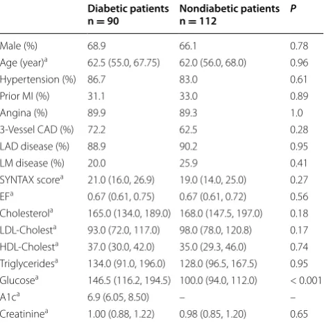
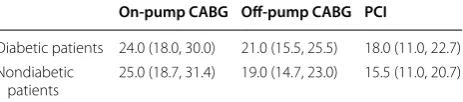
Between May 2012 and March 2014, 326 patients with multivessel CAD were analyzed for study enrollment. From these, 219 were included in this prospective, single-center, nonrandomized trial and were assigned to PCI, on-pump or off-pump CABG. Seventeen patients were excluded after the procedures (11 due to claustrophobia during CMR, four due to cerebrovascular accidents, and two due to septicemia). Thus, 202 patients were analyzed in this study. Of these, 66 were assigned to PCI, 67 to off-pump, and 69 to on-pump CABG. Ninety patients (44.5%) had diabetes mellitus at study entry. The baseline charac-teristics of diabetic and nondiabetic patients are presented in Table 1. Both groups were statistically similar regard-ing main demographic characteristics, despite glycemic levels. Table 2 shows the SYNTAX scores of patients with and without diabetes, stratified by treatment groups.
Biomarkers and CMR assessment
The study protocol assessed troponin and CK-MB at baseline, 6, 12, 24, 36, and 48 h after PCI and in the same periods up to 72 h after on-pump and off-pump CABG in 202 CAD patients. A total of 2658 biomarker samples were analyzed in this study.
After PCI, we analyzed 386 troponin samples until 48 h (97.5% of all 396 possible measurements). Regarding
Table 1 Baseline demographic variables of diabetic and nondiabetic patients in the MASS V trial
MI myocardial infarction, CAD coronary artery disease, LAD left anterior descending coronary artery, LM left main coronary artery, EF ejection fraction,
LDL low-density lipoprotein, HDL high-density lipoprotein, A1c glycated hemoglobin
a Presented as median (25th, 75th percentile)
Diabetic patients
n = 90 Nondiabetic patientsn = 112 P
Male (%) 68.9 66.1 0.78 Age (year)a 62.5 (55.0, 67.75) 62.0 (56.0, 68.0) 0.96
Hypertension (%) 86.7 83.0 0.61 Prior MI (%) 31.1 33.0 0.89 Angina (%) 89.9 89.3 1.0 3-Vessel CAD (%) 72.2 62.5 0.28 LAD disease (%) 88.9 90.2 0.95 LM disease (%) 20.0 25.9 0.41 SYNTAX scorea 21.0 (16.0, 26.9) 19.0 (14.0, 25.0) 0.27
EFa 0.67 (0.61, 0.75) 0.67 (0.61, 0.72) 0.56
Cholesterola 165.0 (134.0, 189.0) 168.0 (147.5, 197.0) 0.18
LDL-Cholesta 93.0 (72.0, 117.0) 98.0 (78.0, 120.8) 0.17
HDL-Cholesta 37.0 (30.0, 42.0) 35.0 (29.3, 46.0) 0.74
Triglyceridesa 134.0 (91.0, 196.0) 128.0 (96.5, 167.5) 0.95
Glucosea 146.5 (116.2, 194.5) 100.0 (94.0, 112.0) < 0.001
A1ca 6.9 (6.05, 8.50) – –
CK-MB, we analyzed 383 samples (96.7% of all 396 pos-sible samples).
After on-pump and off-pump CABG, we analyzed 942 troponin samples until 72 h (98.9% of all 952 possi-ble measurements). Regarding CK-MB, we analyzed 947 samples (99.5% of all 952 possible samples).
All 202 patients underwent 2 CMRs with LGE, and thus 404 CMRs were analyzed regarding new areas of myocardial fibrosis and myocardial edema after coronary interventions.
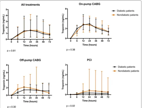
Troponin results
The group of diabetic patients had a mean peak troponin of 5.38 ng/mL with a standard deviation of 9.49 ng/mL, and a median and interquartile range (IQR) of 2.18 ng/ mL (0.47, 5.14). The group of nondiabetic patients had a mean peak troponin of 5.91 ng/mL with a standard devi-ation of 9.88 ng/mL, and median and IQR of 2.24 ng/mL (0.69, 5.42). The total area under the curve was also sta-tistically similar between both groups (P = 0.97).
We also compared troponin values at baseline and dur-ing each time point (6, 12, 24, 36, 48, and 72 h) between diabetic and nondiabetic patients and separately for each cardiac procedure (Fig. 1).
CK‑MB results
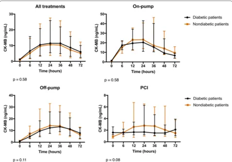
The group of diabetic patients had a mean peak CK-MB of 27.8 ng/mL with a standard deviation of 37.3 ng/mL, and a median and IQR of 14.1 ng/mL (6.8, 31.7). The group of nondiabetic patients had a mean peak CK-MB of 25.2 ng/mL with a standard deviation of 34.1 ng/mL, and median and IQR of 14.0 ng/mL (4.2, 29.8), P = 0.43. The total area under the curve was also statistically simi-lar in both groups (P = 0.56).
We also compared CK-MB values at baseline and dur-ing each time point (6, 12, 24, 36, 48, and 72 h) between diabetic and nondiabetic patients and separately for each cardiac procedure (Fig. 2).
CMR results
New LGE on CMR indicating new myocardial fibro-sis after interventions was present in 18.9% and 17.3%
(P = 0.91), and myocardial edema in 15.5% and 22.9% (P = 0.39) in diabetic and nondiabetic patients, respec-tively (Fig. 3).
Analyses of correlation
Peak CK-MB and peak troponin were plotted against fasting glycemic and A1c levels to look for any cor-relations among these variables. Glycemic levels and peak troponin had a very weak negative correlation (r = − 0.08, 95% CI − 0.22 to 0.07, P = 0.28). Glycemic levels and peak CK-MB also had a very weak correlation (r = 0.01, 95% CI − 0.13 to 0.16, P = 0.85).
The analysis of correlation of A1c levels from diabetic patients also showed a very weak negative correlation with peak troponin and CK-MB levels (r = − 0.18, 95% CI − 0.38 to 0.03, P = 0.08 for troponin; r = − 0.14, 95% CI − 0.34 to 0.08, P = 0.20 for CK-MB).
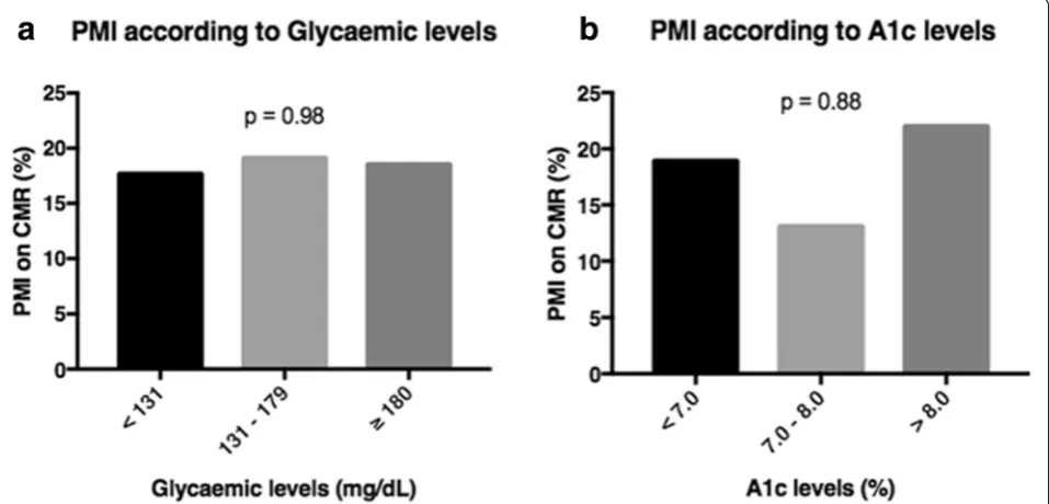
Periprocedural myocardial infarction on CMR according to tertiles of glycemia and A1c levels
Of 202 patients, 200 had their baseline glycemia meas-ured. These patients were grouped into tertiles of glyce-mia (< 131, n = 134; 131–179, n = 31; ≥ 180, n = 32). No difference was found regarding PMI on CMR (17.9% versus 19.3% versus 18.7%, P = 0.98). These results are shown in Fig. 4a.
From 90 diabetic patients, 89 had their baseline A1c levels measured. These patients were grouped into ter-tiles of A1c (< 7.0, n = 47; 7.0–8.0, n = 15; > 8.0, n = 27). No difference was also found regarding PMI on CMR (19.1% versus 13.3% versus 22.2%, P = 0.88). These results are shown in Fig. 4b.
Discussion
The analysis of the data from this study shows that patients with diabetes have similar myocardial sensitiv-ity to that of nondiabetic patients after revascularization interventions. The evidence of similar release of both cardiac biomarkers (troponin and CK-MB) after three distinct revascularization strategies, PCI, on-pump, or off-pump CABG, was confirmed by the findings of a high-sensitive and specific imaging technique to detect cardiac ischemic damage, the cardiac magnetic reso-nance, by the analysis of necrosis as well as myocardial edema.
Although the release of markers of myocardial necrosis after interventions are multifactorial and do not neces-sarily represent ischemic insults, the findings of myocar-dial necrosis and edema at CMR are mostly related to ischemic events during cardiac procedures and can be considered as strong evidence to confirm these findings in our sample. In fact, the strength of the present study lies in the assessment of multiple time points of both Table 2 SYNTAX scores in patients with and without
dia-betes, in the three treatment groups
Data is presented as median (25th, 75th percentile)
CABG coronary artery bypass graft surgery, PCI percutaneous coronary intervention
On‑pump CABG Off‑pump CABG PCI
Diabetic patients 24.0 (18.0, 30.0) 21.0 (15.5, 25.5) 18.0 (11.0, 22.7) Nondiabetic
cardiac biomarkers collected in a standardized fashion, with a minimum loss of data, and by bringing the rel-evant information of CMR results, for both myocardial necrosis and edema. Thus, the analysis of the release of these biomarkers and the findings of cardiac resonance shows similar myocardial sensitivity between diabetic and nondiabetic patients in this sample.
An interesting study from Verdoia et al. evaluated the effect of diabetes in PMI and myonecrosis after PCI treat-ment [18]. Using the release of CK-MB and troponin to define PMI and myonecrosis, the authors found that diabe-tes is not associated with increased risk of cardiac damage after PCI therapy. Thus, their findings agree with the results from the present study. However, adding to the information from that study, the present one also assessed the release of biomarkers after on-pump and off-pump bypass surgery and the evaluation of cardiac magnetic resonance.
Other studies have assessed the release of cardiac bio-markers in the setting of acute coronary syndromes and also evaluated mortality rates in diabetic patients [13, 19–22]. Interestingly, although the majority of them have found higher mortality rates in the diabetic population, the release of cardiac biomarkers was similar to that in the nondiabetic population, the same finding seen in the present study after coronary interventions. Thus, these studies show that myocardial sensitivity is probably not influenced by diabetes status, and other factors might be associated with the higher morbidity and mortality rates observed in the diabetic population. Potential reasons for this observation are speculative and still under debate, but might include greater extent of coronary artery dis-ease [23], systolic dysfunction [24], diastolic dysfunction [8], and a theoretically pro-thrombotic state that might be associated with diabetes [25]. Although some argue
Fig. 2 CK-MB results at baseline and after 6, 12, 24, 36, 48, and 72 h after coronary interventions in diabetic and nondiabetic patients. Data are presented as medians and interquartile ranges. P values are provided by the comparison of both groups regarding all time points
that myocardial intrinsic properties such as ischemic pre-conditioning might be altered by diabetes [26], our group has recently shown similar findings between diabetic and nondiabetic patients who underwent sequential exercise tests, as part of a preconditioning protocol [27].
In agreement with the findings of this study, Alegria et al. [28] have also evaluated mortality rates at 6 months and infarct size by myocardial scintigraphy in patients who suffered an acute myocardial infarction treated by thrombolytic therapy. They have shown higher mortal-ity rates associated with diabetes that persisted after adjustment for confounding factors. However, this higher mortality could not be totally explained by very modest differences in myocardial infarct size and reduced ejec-tion fracejec-tion in diabetic patients. Moreover, the authors could not completely rule out that these very small differ-ences could not still be due to the remaining influence of confounders.
Furthermore, a recent study by Grodzinsky et al. [29] has also assessed angina symptoms over 1 year after PCI treatment. In this study, angina prevalence and severity were similar between patients with and without diabetes. These findings also confirm the findings of the present study: ischemia assessed by the presence of angina and its effects in the myocardium of patients after coronary interventions occur independently of diabetes status.
Interestingly, the analysis of correlation between gly-cemic and A1c levels and peak troponin and CK-MB did
not show any correlation among these variables, although graphically it seems that the higher levels of troponin and CK-MB were found in the lower levels of glycemia. These findings might probably be because the higher peak lev-els of cardiac markers occurred within the most frequent observations of glycemic levels. In addition, the correla-tion coefficients do not support any further conclusions. These results are also confirmed by the analysis of car-diac damage assessed by magnetic resonance that did not show any association of PMI with glycemic and A1c level groups at baseline.
The results from this study deserve some consideration. First, it is possible that the small sample size could limit the study power to detect differences between the two populations. On the other hand, the multiple measure-ments of cardiac biomarkers in addition to the findings of cardiac resonance make this possibility less reasonable. Second, the well-controlled glycemic status, noted by the well-controlled levels of fasting glycemia and A1c in the diabetic population might have influenced study findings. Different findings could be observed in a diabetic popula-tion with poor controlled glycemic levels.
Conclusions
In this study, type 2 diabetes mellitus did not add risk to the development of myocardial injury after percutaneous coronary intervention or bypass surgery in patients with multivessel coronary artery disease.
Abbreviations
A1c: glycated hemoglobin; CABG: coronary artery bypass graft; CAD: coronary artery disease; CMR: cardiac magnetic resonance; IQR: interquartile range; LGE: late gadolinium enhancement; MASS V: medicine, angioplasty, or surgery study V; PCI: percutaneous coronary intervention; PMI: periprocedural myocar-dial infarction; TnI: troponin I.
Authors’ contributions
PCR researched the data, performed the statistical analysis, and wrote the manuscript. RMR researched the data and reviewed the manuscript. TLS, DFCA, and CLG reviewed the manuscript and made important suggestions to the analysis of the data. CAWS reviewed the manuscript and the statistical analysis. WH conceived the study, contributed to the discussion, and reviewed the manuscript. JAFR and RKF reviewed/edited the manuscript. All authors read and approved the final manuscript.
Acknowledgements
We thank all members of the MASS Research Team at the Heart Institute (InCor) of the University of São Paulo for the hard work in this project. Medical writing support was provided by Ann Conti Morcos of MorcosMedia during the preparation of the paper, supported by the Zerbini Foundation.
Competing interests
The authors declare that they have no competing interests.
Availability of data and materials
The datasets used and analyzed during the current study are available from the corresponding author on reasonable request.
Consent for publication Not applicable.
Ethics Approval and consent to participate
All patients provided written informed consent prior to inclusion in this study. The Ethics Committee of the Heart Institute of the University of São Paulo Medical School, São Paulo, Brazil, approved the trial. All procedures were performed in accordance with the Declaration of Helsinki.
Funding
Financial support was provided by a research grant from the Zerbini Founda-tion, São Paulo, Brazil.
Publisher’s Note
Springer Nature remains neutral with regard to jurisdictional claims in pub-lished maps and institutional affiliations.
Received: 19 July 2017 Accepted: 15 November 2017
References
1. Zhang Z, Kolm P, Boden WE, Hartigan PM, Maron DJ, Spertus JA, et al. The cost-effectiveness of percutaneous coronary intervention as a function of angina severity in patients with stable angina. Circ Cardiovasc Qual Outcomes. 2011;4(2):172–82.
2. Dagenais GR, Lu J, Faxon DP, Kent K, Lago RM, Lezama C, Bypass Angio-plasty Revascularization Investigation 2 Diabetes (BARI 2D) Study Group, et al. Effects of optimal medical treatment with or without coronary revascularization on angina and subsequent revascularizations in patients with type 2 diabetes mellitus and stable ischemic heart disease. Circulation. 2011;123(14):1492–500.
3. Domanski MJ, Mahaffey K, Hasselblad V, Brener SJ, Smith PK, Hillis G, et al. Association of myocardial enzyme elevation and survival following coronary artery bypass graft surgery. JAMA. 2011;305(6):585–91. 4. Thygesen K, Alpert JS, Jaffe AS, Simoons ML, Chaitman BR, White HD,
Joint ESC/ACCF/AHA/WHF Task Force for Universal Definition of Myocar-dial Infarction. Third universal definition of myocarMyocar-dial infarction. J Am Col Cardiol. 2012;60(16):1581–98.
5. Moussa ID, Klein LW, Shah B, Mehran R, Mack MJ, Brilakis ES, et al. Con-sideration of a new definition of clinically relevant myocardial infarction after coronary revascularization: an expert consensus document from the Society for Cardiovascular Angiography and Interventions (SCAI). J Am Col Cardiol. 2013;62(17):1563–70.
6. Barsotti A, Giannoni A, Di Napoli P, Emdin M. Energy metabolism in the normal and in the diabetic heart. Curr Pharm Des. 2009;15(8):836–40. 7. Paulson DJ. The diabetic heart is more sensitive to ischemic injury.
Cardio-vasc Res. 1997;34(1):104–12.
8. di Filippo C, Marfella R, Cuzzocrea S, Piegari E, Petronella P, Giugliano D, et al. Hyperglycemia in streptozotocin-induced diabetic rat increases infarct size associated with low levels of myocardial HO-1 during ischemia/reperfusion. Diabetes. 2005;54(3):803–10.
9. Matsumoto S, Cho S, Tosaka S, Ureshino H, Maekawa T, Hara T, et al. Pharmacological preconditioning in type 2 diabetic rat hearts: the roles of mitochondrial ATP-sensitive potassium channels and the phosphatidylinositol 3-kinase-Akt pathway. Cardiovasc Drugs Ther. 2009;23(4):263–70.
10. Feuvray D, Lopaschuk GD. Controversies on the sensitivity of the diabetic heart to ischemic injury: the sensitivity of the diabetic heart to ischemic injury is decreased. Cardiovasc Res. 1997;34(1):113–20.
11. Hadour G, Ferrera R, Sebbag L, Forrat R, Delaye J, Lorgeril M. Improved myocardial tolerance to ischaemia in the diabetic rabbit. J Mol Cell Cardiol. 1998;30(9):1869–75.
12. Korkmaz-Icöz S, Lehner A, Li S, Vater A, Radovits T, Hegedus P, et al. Mild type 2 diabetes mellitus reduces the susceptibility of the heart to ischemia/reperfusion injury: identification of underlying gene expression changes. J Diabetes Res. 2015;2015:396414.
13. Mukamal KJ, Nesto RW, Cohen MC, Muller JE, Maclure M, Sherwood JB, et al. Impact of diabetes on long-term survival after acute myocardial infarction: comparability of risk with prior myocardial infarction. Diabetes Care. 2001;24(8):1422–7.
14. Stone PH, Muller JE, Hartwell T, York BJ, Rutherford JD, Parker CB, The MILIS Study Group, et al. The effects of diabetes mellitus on prognosis and serial left ventricular function after acute myocardial infarction: con-tribution of both coronary disease and diastolic ventricular dysfunction to the adverse prognosis. J Am Coll Cardiol. 1989;14(1):49–57.
15. Hueb W, Gersh BJ, Alves da Costa LM, Costa Oikawa FT, Vieira de Melo RM, Rezende PC, et al. Accuracy of myocardial biomarkers in the diagnosis of myocardial infarction after revascularization as assessed by cardiac resonance: the medicine, angioplasty, surgery study V (MASS-V) trial. Ann Thorac Surg. 2016;101(6):2202–8.
16. Thygesen K, Mair J, Giannitsis E, Study Group on Biomarkers in Cardiol-ogy of ESC Working Group on Acute Cardiac Care, et al. How to use high-sensitivity cardiac troponins in acute cardiac care. Eur Heart J. 2012;33:2252–7.
17. American Diabetes Association. Standards of medical care in diabe-tes—2016. Diabetes Care. 2016;39(suppl 1):S1–106.
18. Verdoia M, Barbieri L, Schaffer A, Cassetti E, Di Giovine G, Narkin M, on behalf of the Novara Atherosclerosis Study Group (NAS), et al. Effect of diabetes mellitus on periprocedural myocardial infarction in patients undergoing coronary stent implantation. Diabetes Metab Res Rev. 2015;31(1):85–92.
19. Gwilt DJ, Petri M, Lewis PW, Nattrass M, Pentecost BL. Myocardial infarct size and mortality in diabetic patients. Br Heart J. 1985;54(5):466–72. 20. Lehto S, Pyörälä K, Miettinen H, Rönnemaa T, Palomäki P, Tuomilehto J,
et al. Myocardial infarct size and mortality in patients with noninsulin-dependent diabetes mellitus. J Intern Med. 1994;236:291–7.
21. Hasdai D, Granger CB, Srivatsa SS, Criger DA, Ellis SG, Califf RM, et al. Dia-betes mellitus and outcome after primary coronary angioplasty for acute myocardial infarction: lessons from the GUSTO-IIb angioplasty substudy. Global use of strategies to open occluded arteries in acute coronary syndromes. J Am Coll Cardiol. 2000;35:1502–12.
22. Murcia AM, Hennekens CH, Lamas GA, Jimenez-Navarro M, Rouleau JL, Flaker GC, et al. Impact of diabetes on mortality in patients with myocardial infarction and left ventricular dysfunction. Arch Intern Med. 2004;164:2273–9.
• We accept pre-submission inquiries
• Our selector tool helps you to find the most relevant journal
• We provide round the clock customer support
• Convenient online submission
• Thorough peer review
• Inclusion in PubMed and all major indexing services
• Maximum visibility for your research
Submit your manuscript at www.biomedcentral.com/submit
Submit your next manuscript to BioMed Central
and we will help you at every step:
24. Devereux RB, Roman MJ, Paranicas M, O’Grady MJ, Lee ET, Welty TK, et al. Impact of diabetes on cardiac structure and function: the strong heart study. Circulation. 2000;101:2271–6.
25. Nomura S. Dynamic role of microparticles in type 2 diabetes mellitus. Curr Diabetes Rev. 2009;5(4):245–51.
26. Miki T, Miura T, Hotta H, Tanno M, Yano T, Sato T, et al. Endoplasmic reticu-lum stress in diabetic hearts abolishes erythropoietin-induced myocardial protection by impairment of phosphoblycogen synthase kinase-3beta-mediated suppression of mitochondrial permeability transition. Diabetes. 2009;58:2863–72.
27. Rezende PC, Rahmi RM, Uchida AH, Alves da Costa LM, Scudeler TL, Garzillo CL, et al. Type 2 diabetes mellitus and myocardial ischemic
preconditioning in symptomatic coronary artery disease patients. Cardio-vasc Diabetol. 2015;14:66.
28. Alegria JR, Miller TD, Gibbons RJ, Yi QL, Yusuf S, for the Collaborative Organization of RheothRx Evaluation (CORE) Trial Investigators. Infarct size, ejection fraction, and mortality in diabetic patients with acute myocardial infarction treated with thrombolytic therapy. Am Heart J. 2007;154:743–50.