R E S E A R C H A R T I C L E
Open Access
Physical activity level in people with age
related white matter changes correlates to
better motor performance, lower
comorbidity and higher cognitive level
Anna F. Pettersson
1*, Lars-Olof Wahlund
2, Lena Bronge
3, Elisabeth Olsson
1, Kaarina Amberla
4,
Hansjoerg Baezner
5and Milita Crisby
2*Abstract
Background:Physical activity plays a pivotal role in the development of disability and may modify the negative effect of vascular risk factors on progression of both cardio and cerebrovascular disorders. The aim of this study was to evaluate the activity level in people with age-related white matter changes as identified on magnetic resonance imaging (MRI) in relation to motor performance, cognition and perceived health.
Methods:Data came from the first year follow up of one participating centers of the LADIS study. Fifty one subjects were first enrolled in the study. Complete first year follow up data was available for 41 subjects. Information on
comorbidity, physical activity level, physical function, cognition, level of white matter changes and perceived health was collected. Physical activity level was classified with a yes or no question and with the Frenchay Activities Index (FAI).
Results:Only 36% of the subjects in this study were physically active according to the yes/no question. 27.5% of the subjects were active according to the FAI score which evaluates the everyday activities. Being active discriminated subjects with better physical function. Subjects active according to the FAI score had a higher cognitive level (p≤0.01), lower comorbidity (p= 0.02) and performed better on all motor function tasks as assessed by walking speed (p≤0.01) and the Short Physical Performance battery (SPPB) (p≤0.01).
Conclusions:Being physically active seems to be a long term protective factor. In our study, the majority of subjects with Age Related White Mattter Changes (ARWMC) with no or mild Instrumental Activity of Daily Living (IADL) disability did not attain recommended level of activity at first year follow up. Whether or not increasing physical activity may slow down cognitive decline and lessen development of disability in physically inactive subjects with manifest ARWC remains to be studied. Trial registration: not applicable.
Keywords:Cognition, Gait, Health, Motor function, Physical activity
Background
Cognitive decline is among the most feared and eco-nomically costly sequela of ageing [1, 2]. Cognitive decline is associated with a reduction in the ability to perform everyday tasks, make important decisions, and thus live independently [3, 4]. Ageing is a complex process and
appears to affect white matter integrity and functional connectivity in the brain [5]. Physical fitness and posses-sion of the APOE e4 allele have been shown to be incre-mentally associated with cognitive change at statistically significant level [6].
Higher levels of physical activity have been associated with larger gray-matter volumes [7]. Furthermore, cardio-respiratory fitness affects white-matter integrity [8, 9], which may consequently have positive effects on func-tional brain activity in old age [10].
* Correspondence:[email protected];[email protected]
1
Division of Physiotherapy, Karolinska Institutet, Neurobiology Care Sciences and Society, 23 100, 141 83 Huddinge, Sweden
2Division of Clinical Geriatrics, Karolinska Institutet, Neurobiology Care
Sciences and Society, Huddinge, Sweden
Full list of author information is available at the end of the article
Moderate physical activity for at least 30 min, no less than 3 times/week has been reported to maintain cardio-vascular fitness [11]. This level of exercise has shown to decrease vascular risk factors and the incidence of cor-onary heart disease [12]. In addition, low intensity phys-ical activity renders other health benefits such as maintaining physical function, a higher quality of life, and wellbeing [13]. Even low intensity physical activity has been shown to be associated with larger hippocam-pus volume in older adults [14]. Many of our daily activ-ities include activactiv-ities that are dependent on lower extremity function, which has been found to be able to predict short-term mortality and nursing home admis-sion in older adults [15–17]. Gait speed alone has been found to predict incident disability in previously non-disabled older people [18].
Age related brain white matter changes, (ARWMC), are hyperintense lesions observed on T2-weighted im-ages on brain magnetic resonance imaging (MRI). ARWMC are common findings in older people and have been associated with cognitive impairment as well as gait disturbances [19, 20]. Cerebral ischemia related to deep cerebral small vessels of the brain due to aging, arterial hypertension and other vascular risk factors has been suggested as possible causes of ARWMC [21, 22]. The severity of ARWMC on brain MRI has been described by Fazekas et al. as; grade I (punctuate changes), grade II (early confluent changes) and grade III (severe confluent changes) [23]. Gait disturbances may lead to limitations in activities and a restricted ability to participate in life situations and subsequently lead to an inactive life style and increased risk of falls [19, 24, 25]. This has import-ant implications since inactive older people with low physiologic reserves are more vulnerable to disease events and subsequent disability [26, 27]. The present study evaluates cross sectional data of activity level in people with ARWMC in relation to motor performance, cognition and perceived health.
Methods
Design
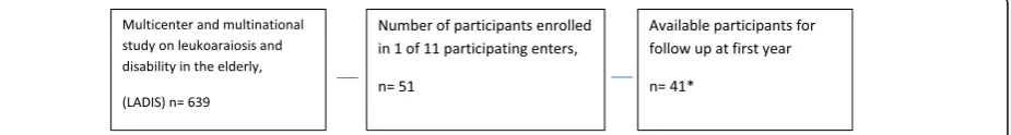
In this cross-sectional study data came from the first year follow up of one of the participating centres of the Leukoaraiosis and Disability study (LADIS) [28]. In brief the LADIS study is a multicentre and multinational longitudinal study with the aim to examine the net
contribution of various degrees of ARWMC on the de-velopment of disability in older people.
Subjects
LADIS participants were at entry between 65 and 84 years of age. They had no or mild disability as defined by the Instrumental Activities of Daily Living (IADL). All of them had a contactable informant and had agreed to sign an informed consent prior to participation in the study. Subjects were recruited between 2001 and 2002 through the radiology department where they had pre-sented with cerebral subcortical white matter changes on MRI of any degree. Two of the subjects had been re-ferred to MRI due to cognitive impairment; another five subjects had been referred to MRI due to minor stroke and the remaining subjects with ARWMC were inciden-tally detected. Subjects were excluded if they were judged likely to drop out due to any severe disease, un-able to give informed consent or inability to undergo further MRI scan. Based on the inclusion and exclusion criteria 51 subjects were enrolled in the study. At the first year follow up, data was available for 41 subjects, Fig. 1. The study was approved be the local ethics com-mittee, reference number 01–316.
Materials and procedure
Subjects underwent a thorough examination according to the LADIS protocol, which has been described in detail elsewhere [28]. In summary these examinations in-cluded medical history, social background and assess-ments of physical and cognitive function and brain MR. The LADIS study includes data on subjects from all European centers up to a 3 year follow-up period [28]. The present study is a sub-study that includes cross-sectional data assessed at the first year follow up at Karolinska University Hospital, Huddinge, Sweden. For the purpose of this study an additional evaluation of everyday activities was performed.
Medical history/co-morbidity
Information on comorbidity came from interviewing the patient and the informant, and from the medical records. Comorbidities included cardiovascular risk factors and age related comorbidities known as possible determinants of disability in the elderly. All of these were defined accord-ing to the currently most widely accepted criteria, selected
after a systematic literature search reported in detail else-where [28].
Medical history/physical activity
Physical activity level was classified with a yes or no question. Being physically active was defined according to the American Heart Association (AHA) Scientific def-inition, (i.e. at least 30 min of activity and at least 3 times/week) [11].
Additional assessment of every day physical activity
We were especially interested to look at lower intensities of physical activities since this also may have health benefits for older people. Consequently, for the purpose of the present study the activity level was further assessed with the Frenchay Activities Index (FAI) [29], a questionnaire reflecting lower intensity activities includ-ing questions about the frequency of domestic chores, leisure/work and outdoor activities. A high score indi-cates higher level of activity (max 45). The FAI has been validated in a general population aged 16 and over [30].
Assessment of cognitive function
General cognitive ability was assessed using the Mini Mental State Examination score (MMSE) [31].
Assessment of physical function
Physical function was assessed using the Short Physical Performance Battery (SPBB) that includes tests of stand-ing balance, walkstand-ing speed and time to rise from a chair five times. Subjects are scored 0–4 depending on time performance, with 0 representing the inability to complete the task and 4 representing the highest level of performance. Maximum score is 12.
In addition, walking speed (m/s) was recorded by meas-uring the free walking speed over an 8 m course [32].
Evaluation of perceived health
The subjects’perception of own health status was evalu-ated using the visual analogue rating scale (VAS) from the EuroQoul/EQ5D [33]. The EQ5D VAS is a vertical 20 cm visual analogue scale similar to a thermometer. It ranges from worst imaginable health state, 0, to best im-aginable health state, 100.
MRI scanning/rating of ARWMC
All subjects had been examined by MRI according to a standard protocol. The degree of ARWMC severity was rated by one of the co-authors, an experienced consult-ant in neuroradiology (LB), using the visual scale of Fazekas as grade 1 (punctuate), grade 2 (early confluent), grade 3 (confluent) [23]. ARWMC of grade one was de-noted as mild, grade 2 as moderate and grade 3 as severe
[28]. Details on the MRI procedure and rating of ARMC are published elsewhere [29].
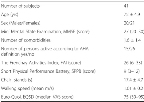
Subjects characteristics are shown in Table 1.
Statistical analysis
The subject sample was divided in subjects who were physically active as defined according to the AHA scien-tific definition [11] and less physically active respectively. In addition, we aimed to study those who were defined active at lower intensities using the FAI score that cap-tures domestic chores, leisure/work and outdoor activ-ities. The maximum score of FAI is 45 which indicate a higher level of such activities. For the purpose of this study we wanted to discriminate between high per-formers and low perper-formers thus subjects who had a FAI score≥the median of the whole group were consid-ered active and those subjects below the median were consequently considered inactive. In the analysis, the FAI Score was dichotomised taking the value zero for values below the median/mean and one for values equal to and above the median/mean. The Mann-Whitney U test was used to look at differences between physically active and physically inactive.
Differences among subjects with various degrees of ARWMC was assessed using Kruskal-Wallis ANOVA.
The variables FAI, SPPB, EQ5D, MMSE, age and num-ber of comorbidities were dichotomised, taking the value zero for values below the median/mean and one for values equal to and above the median/mean. Stepwise logistic and stepwise linear regression analysis were then used to evaluate to what extent physical function (SPPB, walking speed) and health (EQ5D) could be explained by the activity level, degree of ARWMC, age, sex, cognition (MMSE score) and co-morbidity.
Table 1Characteristics, all subjects (n= 41). Median (range), Mean ± SD
Number of subjects 41 Age (yrs) 75 ± 4.9 Sex (Males/Females) 20/21 Mini Mental State Examination, MMSE (score) 27 (20–30) Number of comorbidities 1.6 ± 1.4 Number of persons active according to AHA
definition yes/no
Results
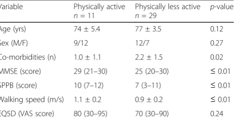
According to the AHA scientific definition 36% (n= 15) were physically active while 27.5% (n = 11) were active according to the FAI score (every day activities). Subjects being physically active according to the AHA scientific definition had significantly better SPPB-scores, (p-value 0.01). Subjects active according to the FAI score had a higher cognitive level (p = ≤ 0.01), less comorbidity (p = 0.02) and performed better on all motor function tasks such as the walking speed (p≤0.01) and the SPPB score (p≤0.01) as shown in Table 2.
Degree of ARWMC
There were no significant differences regarding age, sex, cognitive level, activity level, motor performance or per-ceived health between subjects with different degrees of ARWMC.
Tests of physical function
SPPB
Stepwise logistic regression analyses showed that cognitive level as measured with MMSE and the number of co-morbidities was significantly related to the performance on the SPPB score (p-value 0.01 and 0.02 respectively).
The OR for having a SPPB score equal to or above the median was 1.4 times higher for those with a higher cognitive level as measured with MMSE, (95% confi-dence interval, 1.07–1.85).
Subjects with co-morbidities equal to or above the mean for the whole group were less likely to perform better on SPPB (OR 0.48, 95% confidence interval 0.25–0.91).
Walking speed
Walking speed was significantly related to the subjects’ cognitive level as assessed by the MMSE (p< 0,005). In addition, the walking speed correlated to the number of comorbidities and moderate degree of ARWMC (p-value 0.02 and 0.04 respectively).
The MMSE scores, number of co-morbidities and de-gree of ARWMC appeared to be three independent
variables which explained 34% of the variation in the walking speed.
Perceived health
EQ5D
Only co-morbidities were significantly related to per-ceived health as assessed with the EQ5D (p < 0,005). Subjects with a number of co-morbidities equal to or above the mean (m = 1.6 ± 1.4) were less likely to score high on the EQ5D, (OR = 0.39; 95% confidence interval: 0.21–0.73).
Discussion
The majority of the subjects in this study were not active at the suggested level according to the AHA statement. There was a clear association between the activity level of the subjects according to the AHA’s definition and to better physical function such as motor performance and walking speed. A higher degree of physical activity has been shown to attenuate age-related myelin declines in portions of the corpus callosum that interconnect hom-ologous premotor cortex regions involved in motor planning [8]. A previous 3-year prospective study has shown that both sports and non-sports activities (walk-ing and household) have been found to be a determinant of change in mobility performance. Indeed, this effect was independent of presence of chronic disease. Further-more, the study showed that the subjects who started being active during follow up had a lesser degree of decline in mobility performance compared to those who remained at the same activity level [34].
We also found that the activity level of the subjects correlated to the degree of ARWMC. The positive cor-relation seen between physical activity and motor per-formance, cognition and walking speed remained significant even after correction for the demographic variables and were independent of the reported gait disturbances and previous falls. Studies have shown a clear association between white matter integrity and functional connectivity in the brain [35] and being phys-ically active appears to be protective for maintaining white matter integrity [36]. These protective effects could be partly due to the positive effects seen in the regulation of blood pressure and improvements of the glucose metabolism [37, 38].
A major limitation of this study is its’ small sample size. The cross sectional design also makes it difficult to determine the temporal relationship between exposure and outcome and does not allow to draw conclusions on causality.However, it is reasonable to consider the possi-bility that comorbidity, physical activity, physical func-tion and cognifunc-tion are interrelated. Co-morbidities may incorporate factors that are perceived as barriers for tak-ing part in physical activity [39]. Such factors may be Table 2Differences in age, sex, co-morbidity, cognitive and
physical function and health in active/inactive subjects accord-ing to FAI (n= 40) Median (range), Mean ± SD
Variable Physically active
n= 11
Physically less active
n= 29 p
tiredness, feelings of discomfort or pain. This may lead to a downward spiral of decreasing activity level eventually causing motor disability. Moreover, many of the co-morbidities were cardiovascular risk factors that are thought to play a role in the development of ischemic events and subsequent ARWMC and impaired cognitive function [21, 22]. Executive functions are often impaired in subjects with ARWMC, decrements in executive func-tion may cause less initiative to physical activity [22].
Physical activity has previously been linked to physical function [40, 41] and cognition [42]. In a recent publica-tion from the whole cohort included in the LADIS study, being physically active was associated with re-duced risk of cognitive impairment and rere-duced in more than half, the risk for vascular dementia. However, phys-ical activity did not influence evolution of Alzheimer de-mentia [43].
Conclusions
Being physically active seems to be a long term protect-ive factor for cognitprotect-ive function. In our study, the major-ity of subjects with ARWMC with no or mild IADL disability did not attain recommended level of activity at first year follow up. Whether or not increasing physical activity may slow down cognitive decline and lessen de-velopment of disability in physically inactive subjects with manifest ARWC remains to be studied.
Abbreviations
AHA:American Heart Association; ARWMC: Age Related White matters Changes; FAI: Frenchay Activities Index; IADL: Instrumental Activities of Daily Living; LADIS: Leukoaraiosis and Disability study; MMSE: Mini Mental State Examination score; MRI: Magnetic Resonance Imaging; SPBB: Short Physical Performance Battery; VAS: Visual Analogue rating Scale
Acknowledgements
Not applicable.
Funding
We’d like to thank Dr. Carl Herlitz och Mer Håkan Erlandsson for providing research resourses for Milita Crisby during the drafting of the manuscript and Gun & Bertil Stohnes foundation for providing research resources for Anna Pettersson.
Availability of data and materials
The data that support the findings of this study are available from an electronic case record but restrictions apply to the availability of these data, which were used under license for the current study, and so are not publicly available. Data are however available from the authors upon reasonable request.
Authors’contributions
AFP has been involved in the design of the study and assessments for the motor function and SPPB tests as well as the statistical analysis and the drafting of the manuscript. LOW has been involved in the design of the study. LB been responsible for the acquisition, selection, analysis and interpretation of the MRI scans.. KA has Performed and analysed the cognitive tests. EO has been involved in the design of the study. HBZ has been involved in the design of the study. MC has recruited the patients in the study, contributed to the analysis and the drafting of the manuscript. All authors made critical revisions and approved the submission.
Ethics approval and consent to participate
The study was approved be the local ethics committee, reference number 01–316. Participant’s signed an informed consent prior to participation in the study.
Consent for publication
Not applicable.
Competing interests
The authors declare that they have no competing interests.
Publisher’s Note
Springer Nature remains neutral with regard to jurisdictional claims in published maps and institutional affiliations.
Author details
1Division of Physiotherapy, Karolinska Institutet, Neurobiology Care Sciences
and Society, 23 100, 141 83 Huddinge, Sweden.2Division of Clinical
Geriatrics, Karolinska Institutet, Neurobiology Care Sciences and Society, Huddinge, Sweden.3Division of Radiology, CLINTEC Department, Karolinska Institutet, Huddinge, Sweden.4Department of Clinical Psychology, Helsinki
University, Helsinki, Finland.5Department of Neurology, Universitätsklinikum
Mannheim, University of Heidelberg, Heidelberg, Germany.
Received: 9 March 2017 Accepted: 4 July 2017
References
1. Wimo L, Jönsson J, Bond M, Prince B, Winblad B. The worldwide impact of dementia 2010. Alzheimers Dement. 2013;9:1–11. e3
2. Harper S. Economic and social implications of aging societies. Science. 2014; 346:587–91.
3. Tucker-Drob EM. Neurocognitive functions and everyday functions change together in old age. Neuropsychology. 2011;25:368–77.
4. Boyle PA, Yu L, Wilson RS, Gamble K, Buchman AS, Bennett DA. Poor decision making is a consequence of cognitive decline among older persons without Alzheimer's disease or mild cognitive impairment. PLoS One. 2012; doi:10.1371/journal.pone.0043647.
5. Yang AC, Tsai SJ, Liu ME, Huang CC, Lin CP. The Association of Aging with white matter integrity and functional connectivity hubs. Front Aging Neurosci. 2016;8:143.
6. Ritchie SJ, Tucker-Drob EM, Cox SR, Corley J, Dykiert D, et al. Predictors of ageing-related decline across multiple cognitive functions. Intelligence. 2016;59:115–26.
7. Papenberg G, Ferencz B, Mangialasche F, Mecocci P, Cecchetti R, et al. Physical activity and inflammation: effects on gray-matter volume and cognitive decline in aging. Hum Brain Mapp. 2016;37:3462–73.
8. Johnson NF, Kim C, Clasey JL, Bailey A, Gold BT. Cardiorespiratory fitness is positively correlated with cerebral white matter integrity in healthy seniors. NeuroImage. 2012;59:1514–23.
9. Voss MW, Heo S, Prakash RS, Erickson KI, Alves H, et al. The influence of aerobic fitness on cerebral white matter integrity and cognitive function in older adults: results of a one-year exercise intervention. Hum Brain Mapp. 2012;34:2972–85.
10. Suo C, Singh MF, Gates N, Wen W, Sachdev P, et al. Therapeutically relevant structural and functional mechanisms triggered by physical and cognitive exercise. Mol Psychiatry. 2016;21:1633–42.
11. Garber CE, Blissmer B, Deschenes MR, Franklin BA, Lamonte MJ, Lee IM, Nieman DC, Swain DP. American College of Sports Medicine position stand. Quantity and quality of exercise for developing and maintaining cardiorespiratory, musculoskeletal, and neuromotor fitness in apparently healthy adults: guidance for prescribing exercise. Med Sci Sports Exerc. 2011; doi:10.1249/MSS.0b013e318213fefb.
12. Villeneuve PJ, Morrison HI, Craig CL, Schaubel DE. Physical activity, physical fitness, and risk of dying. Epidemiology. 1998;9(6):626–31.
13. Spirduso WW, Cronin DL. Exercise dose-response effects on quality of life and independent living in older adults. Med Sci Sports Exerc. 2001; 33(Suppl):598–608.
15. Landi F, Cesari M, Onder G, Lattanzio F, Gravina EM, Bernabei R. Physical activity and mortality in frail, community-living elderly patients. J Gerontol A Biol Sci Med Sci. 2004;59:833–7.
16. Kokkinos P, Myers J, Faselis C, Panagiotakos DB, Doumas M, Pittaras A, Manolis A, Kokkinos JP, Karasik P, Greenberg M, Papademetriou V, Fletcher R. Exercise capacity and mortality in older men: a 20-year follow-up study. Circulation. 2010;122:790–7.
17. Guralnik JM, Simonsick EM, Ferrucci L, Glynn RJ, Berkman LF, Blazer DG, et al. A short physical performance battery assessing lower extremity function: association with self-reported disability and prediction of mortality and nursing home admission. J Gerontol. 1994;49:M85–94.
18. Guralnik JM, Ferrucci L, Pieper CF, Leveille SG, Markides KS, Ostir GV, et al. Lower extremity function and subsequent disability: consistency across studies, predictive models, and value of gait speed alone compared with the short physical performance battery. J Gerontol A Biol Sci Med Sci. 2000;55:M221–31. 19. Longstreth WT Jr, Arnold AM, Manolio TA, Burke GL, Bryan N, Jungreis CA,
et al. Clinical correlates of ventricular and sulcal size on cranial magnetic resonance imaging of 3,301 elderly people. The cardiovascular health study. Collaborative research group. Neuroepidemiology. 2000;19:30–42. 20. Baezner H, Blahak C, Poggesi A, Pantoni L, Inzitari D, Chabriat H, et al.
Association of gait and balance disorders with age-related white matter changes: the LADIS study. Neurology. 2008;70:935–42.
21. Pantoni L, Garcia JH. Pathogenesis of leukoaraiosis: a review. Stroke. 1997;28:652–9.
22. Pugh KG, Lipsitz LA. The microvascular frontal-subcortical syndrome of aging. Neurobiol Aging. 2002;23:421–31.
23. Fazekas F, Barkhof F, Wahlund LO, Pantoni L, Erkinjuntti T, Scheltens P, et al. CT and MRI rating of white matter lesions. Cerebrovasc Dis. 2002; 13(Suppl 2):31–6.
24. Baloh RW, Yue Q, Socotch TM, Jacobson KM. White matter lesions and disequilibrium in older people. I Case-control comparison. Arch Neurol. 1995;52:970–4.
25. Briley DP, Wasay M, Sergent S, Thomas S. Cerebral white matter changes (leukoaraiosis), stroke, and gait disturbance. J Am Geriatr Soc. 1997;45:1434–8. 26. Rantanen T, Guralnik JM, Ferrucci L, Penninx BW, Leveille S, Sipila S, et al.
Coimpairments as predictors of severe walking disability in older women. J Am Geriatr Soc. 2001;49:21–7.
27. Gill TM, Allore H, Holford TR, Guo Z. The development of insidious disability in activities of daily living among community-living older persons. Am J Med. 2004;117:484–91.
28. Pantoni L, Basile AM, Pracucci G, Asplund K, Bogousslavsky J, Chabriat H, et al. Impact of age-related cerebral white matter changes on the transition to disability–the LADIS study: rationale, design and methodology.
Neuroepidemiology. 2005;24:51–62.
29. Wade DT, Legh-Smith J, Langton HR. Social activities after stroke: measurement and natural history using the Frenchay activities index. Int Rehabil Med. 1985;7:176–81.
30. Turnbull JC, Kersten P, Habib M, McLellan L, Mullee MA, George S. Validation of the Frenchay activities index in a general population aged 16 years and older. Arch Phys Med Rehabil. 2000;81:1034–8.
31. Folstein MF, Folstein SE, McHugh PR.“mini-mental state”. A practical method for grading the cognitive state of patients for the clinician. J Psychiatr Res. 1975;12:189–98.
32. Guttmann CR, Benson R, Warfield SK, Wei X, Anderson MC, Hall CB, et al. White matter abnormalities in mobility-impaired older persons. Neurology. 2000;54:1277–83.
33. Brooks R. EuroQol: the current state of play. Health Policy. 1996;37:53–72. 34. Visser M, Pluijm SM, Stel VS, Bosscher RJ, Deeg DJ. Physical activity as a
determinant of change in mobility performance: the longitudinal aging study Amsterdam. J Am Geriatr Soc. 2002;50:1774–81.
35. Yang AC, Tsai SJ, Liu ME, Huang CC, Lin CP. The Association of Aging with white matter integrity and functional connectivity hubs. Front Aging Neurosci. 2016;15(8):143.
36. Brain and White Matter Hyperintensity Volumes After 10 Years of Random Assignment to Lifestyle Intervention. Espeland MA, Erickson K, Neiberg RH, Jakicic JM, Wadden TA, Wing RR, Desiderio L, et al. Diabetes Care. 2016; doi: 10.2337/dc15-2230.
37. Whelton SP, Chin A, Xin X, He J. Effect of aerobic exercise on blood pressure: a meta-analysis of randomized, controlled trials. Ann Intern Med. 2002;136:493–503.
38. Tuomilehto J, Lindström J, Eriksson JG, Valle TT, Hämäläinen H, Ilanne-Parikka P, et al. Prevention of type 2 diabetes mellitus by changes in lifestyle among subjects with impaired glucose tolerance. N Engl J Med. 2001;344:1343–50. 39. Ferrucci L, Bandinelli S, Benvenuti E, Di Iorio A, Macchi C, Harris TB, et al. Subsystems contributing to the decline in ability to walk: bridging the gap between epidemiology and geriatric practice in the InCHIANTI study. J Am Geriatr Soc. 2000;48:1618–25.
40. Wong CH, Wong SF, Pang WS, Azizah MY, Dass MJ. Habitual walking and its correlation to better physical function: implications for prevention of physical disability in older persons. J Gerontol A Biol Sci Med Sci. 2003;58:555–60. 41. Newman AB, Haggerty CL, Kritchevsky SB, Nevitt MC, Simonsick EM. Walking
performance and cardiovascular response: associations with age and morbidity–the health, aging and body composition study. J Gerontol A Biol Sci Med Sci. 2003;58:715–20.
42. Laurin D, Verreault R, Lindsay J, MacPherson K, Rockwood K. Physical activity and risk of cognitive impairment and dementia in elderly persons. Arch Neurol. 2001;58:498–504.
43. Verdelho A, Madureira S, Ferro JM, Baezner H, Blahak C, et al. LADIS study. Physical activity prevents progression for cognitive impairment and vascular dementia: results from the LADIS (Leukoaraiosis and disability) study. Stroke. 2012;43:3331–5.
• We accept pre-submission inquiries
• Our selector tool helps you to find the most relevant journal • We provide round the clock customer support
• Convenient online submission • Thorough peer review
• Inclusion in PubMed and all major indexing services • Maximum visibility for your research
Submit your manuscript at www.biomedcentral.com/submit