R E S E A R C H A R T I C L E
Open Access
Parathyroid cysts: experience of a rare
phenomenon at a single institution
Peipei Xu
1, Xiaotian Xia
1, Meifang Li
2, Minggao Guo
1*and Zhili Yang
1*Abstract
Background:Parathyroid cysts are relatively uncommon lesions and are often misdiagnosed. We evaluate our
experience in the diagnosis of and therapy to correct parathyroid cystic lesions.
Methods:We retrospectively reviewed a series of 32 patients with parathyroid cysts who were admitted to our department between July 2011 and November 2016. Clinical pathological features of the patients, including age, gender, location, size, ultrasonography, histopathology, surgery, and follow-up, were analyzed.
Results:There were 22 female and 10 male participants with a median age of 46.7 years old (27–76 years old). Only two cysts were found in the superior mediastinum. The rest were located under the lower pole of the thyroid. All of the patients underwent ultrasonography scans and serum parathyroid hormone (PTH) assays. Three patients had elevated serum PTH levels, and they were further scanned with Tc99m sestamibi as functional cysts. In 29 cases of nonfunctional cysts, 3 cases were preoperatively diagnosed by cystic aspiration with PTH detection. The rest were diagnosed by postoperative immunopathology. All of the patients underwent cystectomy, and 24 patients also underwent thyroidectomy. There was a significant difference in cyst diameter size between the cystectomy alone and cystectomy with thyroidectomy groups (4.0 ± 2.0 vs 1.5 ± 1.0 cm;p< 0.05). No participant experienced recurrence during the median 36 months of follow-up.
Conclusions:Cystic lesions located under the lower pole of the thyroid gland should be considered to have originated at the parathyroid gland. Cystic aspiration with PTH detection or postoperative immunopathology can lead to a definitive diagnosis. Cystectomy is still a commonly used and effective treatment.
Keywords:Parathyroid cyst, Parathyroid hormone, Thyroidectomy, Cystectomy
Background
Parathyroid cysts (PCs) are a rare entity, and only 300 cases have been reported worldwide [1]. Clinically, they are defined as either functional or non-functional based on the presence or absence of hyperparathyroidism [2]. Most PCs are non-functional and commonly present as asymptomatic nodular cervical lesions [3]. They are often mistaken for other neck masses, such as thyroid cysts. There are three options for treatment of PC: aspiration, percutaneous injec-tion of sclerosing agents, and surgical resecinjec-tion [4, 5]. At present, the diagnosis and treatment of PC is limited to a few small case reports. We reviewed 32 cases of PC
in our institution, summarized their clinical features, and highlighted the diagnosis and treatment procedures.
Methods
We retrospectively reviewed a series of cases of PCs in patients who had been admitted to our department between July 2011 and November 2016. Age, gender, cyst location and size, laboratory and imaging results, treatment, and pathology were recorded. All of the pa-tients underwent a histological examination for their final diagnosis. In this cohort, the indications for thy-roidectomy include nodules with suspected malignant sonographic features, nodules confirmed as malignant by fine needle aspiration cytology (FNAC), and benign nodules that are larger than 3 cm in diameter.
The surgical procedure involves a cystectomy alone or combined with thyroidectomy through a middle-access
* Correspondence:guominggao203@163.com;yangzhili2011@live.com
Peipei Xu and Xiaotian Xia are equal first authors Meifang Li is secondary author
1Department of General Surgery, Sixth People’s Hospital Affiliated Shanghai
Jiao Tong University, 600 Yi-Shan Road, Shanghai 200233, China Full list of author information is available at the end of the article
incision under the effect of general anesthesia. Briefly, the patient is positioned with the neck extended, and a 3- to 5-cm collar-type incision is made; two skin flaps are created by dissecting them away from the strap muscles, and the thyroid capsule is approached by splitting the strap muscles along the midline. According to the preoperative location and by exposing the lower pole of the thyroid and dissect-ing the superficial fascia, the PC will be found. To avoid in-jury of the recurrent laryngeal nerve, the surrounding fascia was dissociated along the cyst wall, the vessels were cut with an ultrasonic scalpel, and the cyst was removed and sent off for pathological examination. Two mediastinal PCs were removed using videothoracoscopy. The corresponding conventional thyroidectomy was performed for the con-comitant thyroid diseases (e.g., malignant or benign nod-ules). Intraoperative serum PTH (IOPTH) was measured 10 min after the excision of 3 functional PCs.
Serum calcium was measured in the postoperative 24 h for all of the cases and at 48 h, 72 h and 1 w for FPC cases. Calcium was administered intravenously in postoperative hypocalcemia functional parathyroid cyst (FPC) cases, and 2–3 days later, it was changed to oral calcium for 3 months. For nonfunctional parathyroid cysts (NFPCs), it is generally not routine to use calcium supplementation after surgery. All of the patients with PC underwent reex-amination of the neck ultrasound, blood PTH and serum calcium once a year except for the FPC patients, whose blood PTH and serum calcium were examined in the postoperative 3 month period. All of the enrolled patients were followed up by making an appointment and dialing the reserved phone number. The diameters of all PCs are reported as the means ± standard error and were com-pared between the cystectomy alone group and the cystec-tomy combined with thyroideccystec-tomy group using Student’s ttest.P-values < .05 were regarded as statistically signifi-cant. Statistical analysis was performed with SPSS 20.0 (SPSS Inc., Chicago, Illinois).
This retrospective study was endorsed by our hos-pital ethics committee, and all patients signed informed consent forms.
Results
We identified 32 patients (22 female and 10 male) with a diagnosis of PC. Their median age was 46.7 years (27–76 years). A total of 30 PCs were located under the lower pole of the thyroid gland, and 2 were in the su-perior mediastinum; their diameters ranged from 1 to 6 cm, and the diameters of only 6 cysts were more than 2.5 cm. All of the patients were asymptomatic, and only 5 complained of an anterior cervical mass (Table1).
Two cysts were found incidentally in the superior medi-astinum by enhanced computer tomography. Each was a large cyst that was later confirmed to be a PC by postoper-ative immunopathology (Fig. 1). Ultrasonography scans
and PTH assays in the serum were performed preopera-tively in all patients. Ultrasonography scans reported that the area under the lower pole of the thyroid was free of echoes (Fig. 2). Only three patients with elevated serum PTH levels (350 pg/ml, 324 pg/ml, and 224 pg/ml) were further examined with a Tc99m sestamibi scan, which presented a delayed imaging of the parathyroid. Cystic as-piration with PTH detection (540 pg/ml, 326 pg/ml, and 370 pg/ml) was conducted in 3 of the 29 patients with normal serum PTH levels, and the diagnosis of PC was confirmed by postoperative immunopathology (Fig.3).
All of the patients underwent cystectomy (Fig. 4). Of these patients, 8, including 3 patients with high serum PTH levels, underwent cystectomy alone, and the remaining patients also underwent thyroidectomy for other thyroid-related conditions. The median hospital stay was 5 days (range: 3–8 days). There was a significant dif-ference in cyst diameter size between the cystectomy alone and cystectomy with the thyroidectomy groups (4.0 ± 2.0 vs 1.5 ± 1.0 cm; p < 0.05). Only 2 cases of FPCs had transient hypocalcemia in the postoperative Table 1Clinical characteristics of PC patients in our institution
Parameter Value
Gender (female/male) (No.) 22/10
Age (<40/>40 years) 12/20
Size (diameter < 2.5/>2.5 cm) 26/6
Serum PTH (high/normal pg/ml) 3/29
Location (superior mediastinum/under the inferior pole of the thyroid gland) (No.)
2/30
Neck ultrasonigrapy (yes/no) (No.) 32/0
Cyst aspiration PTH measure (yes/no) (No.) 3/29
Surgery (simple cyst resection/thyroidectomy and cystomy) (No.) 8/24
Flollow-up (recurrence/no recurrence) (No.) 0/32
Fig. 1Representative case with enhanced computer tomography
24 h and were remitted in the postoperative 1 w period. There were no other complications in any of the surgical cases. No participant suffered recurrence during the me-dian 36 months of follow-up (12–60 months), as indicated by ultrasonography scans and their serum calcium and PTH levels.
Discussion
PC is a rare entity in clinical practice, accounting for 0.8– 3.41% of all parathyroid lesions and affecting 0.075% of the unselected population [6]. The first report of surgical re-section of PC was made in 1905 by Goris [7]. In 1953, Crile diagnosed PC for the first time by FNA [8]. Cur-rently, only approximately 300 cases have been reported worldwide.
In this series of cases, PCs were more common in women, and patients had a median age of 46.7 years. Al-most all of the lesions are located under the lower pole of
the thyroid, and the diameters ranged from 1 cm to 6 cm. Based on these demographic characteristics, the pathogen-esis of PCs may be related to the following factors. This cyst may have been caused by a vestigial remnant of the third or fourth branch or the persistence of the Kürsteiner canals [3]. The PC may have been formed from infarct and degen-eration of paralytic adenoma or by the development of mul-tiple microcapsules in normal parathyroid tissue [9]. There are no reports on the exact mechanism of the evolution of parathyroid cysts with hyperplasia and adenoma formation.
Ultrasound is faster, cheaper, and more widely available than other imaging techniques, and it is highly accurate in detecting neck masses and evaluating the local extent of disease [10]. Although it cannot distinguish between PC and other cervical diseases, ultrasonography may reveal a nonspecific cystic structure and establish its proximity to the thyroid gland [4, 11]. In this review, ultrasonographic scans were performed for all of the patients. Here, 30 were positioned under the lower pole of the thyroid, and 2 were ectopic. In this way, the diagnosis of a suspected PC can be established by ultrasonography and based on the location of cystic masses, which were under the lower pole of the thyroid. As a complement, a CT of the head and neck may indicate the cystic components of these lesions and help es-tablish the relationship to adjacent tissues [12]. This may be particularly helpful in the presence of substernal extension or compressive symptoms. Although most of the PCs are accompanied with thyroid nodules in this cohort, it may be difficult to speculate that the occurrence of PC is associated with thyroid disease. To date, there have been eight intrathyroidal PC cases reported in the literature [13]. No other reports of possible relationships between PC and thyroid disease have been described.
PCs can be classified as functional (FPC) or nonfunc-tional (NFPC) according to the presence or absence of
Fig. 2Representative case with PC ultrasonography scans. Cystic lesion
(2.5×2×0.8 cm) in right lobe of thyroid gland. PC, parathyroid cyst
Fig. 3Representative case with immunopathology of parathyroid
cysts. Cytoplasmically stained blown cells with antibody against PTH are parathyroid cells (arrow); cyst lumen (black asterisk) (40×)
Fig. 4Representative parathyroid cysts as seen intra-operatively. PC,
hyperparathyroidism. In our series of cases, 90% were NFPCs (29/32), and 10% were FPCs (3/32), which sug-gested that non-functional cysts are more common. NFPCs generally manifested as asymptomatic masses, which are hard to diagnose and often misdiagnosed. Among our 29 NFPCs, 26 were mistaken for other cer-vical diseases such as thyroid cysts, thyroglossal duct cysts, bronchial cleft cysts, and even mediastinal tumors. The remaining 3 cysts were diagnosed because of ele-vated PTH levels of cyst aspiration. PCs located in the mediastinum are even rarer and easier to misdiagnose as mediastinum tumors [14]. In our report, 2 cysts found incidentally in the superior mediastinum by enhanced computer tomography were misdiagnosed as tumors and confirmed by postoperative immunopathology.
The PTH assay for cystic aspiration is an effective method of diagnosing PCs, especially NFPCs [15]. There were 3 NFPCs diagnosed by cystic aspiration and elevated intact parathyroid hormone (iPTH) levels. The diagnosis was confirmed with postoperative immunopathology in this series of cases. It has been reported that the tissue synthesizes large amounts of PTH with degenerated carboxy-terminal parathyroid hormone (C-PTH), which renders it inactive in the NFPC [16]. Current laboratory practice is to perform an iPTH assay, which may under-estimate the PTH levels of cyst fluid. It is therefore recom-mended that C-PTH in cystic fluid should be measured instead of iPTH [17]. Even so, the presence of any iPTH in the cystic fluid, regardless of the level, can suggest a cyst of parathyroid origin. For this reason, as we reported in the present cases, iPTH testing may not affect the diag-nosis of PC. High-performance liquid chromatography may be a suitable means of accurately measuring the con-centration of PTH in fluid taken from cysts.
A high level of serum PTH is one of the most important parameters in the diagnosis of functional parathyroid
cysts. Meanwhile, ultrasound and 99 m Tc-MIBI parathy-roid imaging can be used for positional and functional diagnosis [18]. The results have shown that serum PTH was significantly higher than normal, and ultrasound and 99 m Tc-MIBI were used to precisely determine the loca-tion of the lesion within the parathyroid in our 3 cases of FPC. In this way, if the serum PTH is significantly higher than normal, the cystic lesions under the lower pole of the thyroid can be diagnosed easily using FPC. The possibility of rapidly developing adenocarcinoma should also be considered in FPC patients [19–22].
In this cohort, we realized from the following three aspects that the diagnosis of NFPC can be improved. First, from the imaging features of the lesions, almost all were free echo (ultrasound) or low density (CT), and there were no parenchymal tissue features. Second, most of the lesions are located under the lower pole of the thyroid gland. Third, in the laboratory examination, iPTH was detected in the ultrasound-guided puncture solution.
The best treatment for FPC is surgical removal of the lesion, but for NFPC, surgical treatment is optional. There are three options for the treatment of NFPC in-cluding simple aspiration, percutaneous sclerosing agent injections and surgical resection [4, 5]. Ultrasound-guided aspiration can be used alone as an initial treat-ment for symptomatic NFPCs, and it can immediately resolve compression symptoms. However, existing litera-ture has shown that aspiration alone is common in cases of small NFPCs with diameters less than 2.5 cm. Even so, there are still reports of recurrence during follow-up. Some cases even repeatedly relapse after multiple aspira-tions and are finally treated by surgical resection or ethanol ablation [23–26]. These reports suggest that the larger the cyst is, the more likely it is to relapse after the procedure. Sclerosing agent injections are not recom-mended because they can flow out from the capsule and
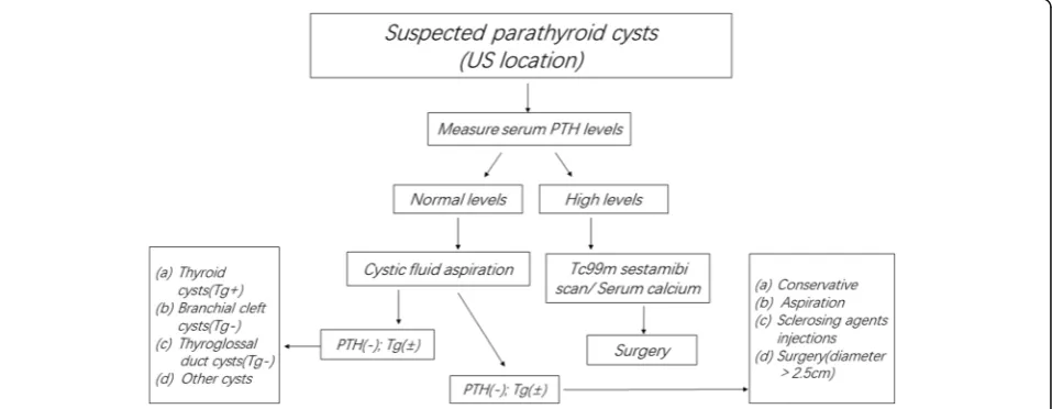
cause serious complications such as fiber degeneration or damage to the recurrent laryngeal nerve, resulting in vocal cord paralysis [26, 27]. It has been suggested that the optimal treatment for symptomatic NFPCs is surgi-cal resection [1, 28]. In the present report, 5 patients with NFPC (diameter greater than 2.5 cm) underwent cyst resection alone. Another 24 patients underwent thyroidectomy with parathyroid cystomy (diameter under 2.5 cm) due to a thyroid-related disease without any postoperative complications, and there was no re-currence during the median 36 months of follow-up. Our experience has shown that surgical resection is an effective and safe treatment for NFPCs with diameters greater than 2.5 cm. Figure 5 shows a synthesis of our cumulative experience with previously reported elegant data. We propose an algorithm to guide the diagnosis and management of PC.
Conclusion
Cystic lesions located under the lower pole of the thy-roid gland should be considered to have originated at the parathyroid gland. Cystic aspiration with PTH detec-tion or postoperative immunopathology can be defini-tively diagnosed. Resection of the cyst is still a common and effective treatment.
Abbreviations
C-PTH:Carboxy-terminal parathyroid hormone; CT: Computed tomography; FNAC: Fine needle aspiration cytology; FPC: Functional parathyroid cyst; IOPTH: Intraoperative serum parathyroid hormone; iPTH: Intact parathyroid hormone; NFPC: Nonfunctional parathyroid cyst; PCs: Parathyroid cysts; PTH: Parathyroid hormone
Acknowledgements
The authors thank all the medical staff who contributed to the maintenance of the medical record database.
Funding
This work was supported by the national natural science foundation of China (grant. no. 81502316) and the Shanghai Science and Technology
Commission Funded Project (grant. no. 14411964100)
Availability of data and materials
The datasets generated and analysed during the current study are available from the corresponding author on reasonable request.
Authors’contributions
PPX, MGG and ZLY carried out the main manuscript writing and design. XXX and MFL performed the operations. PPX and XXX collected and analyzed the data. PPX wrote the manuscript and MGG and ZLY assisted in drafting the manuscript and final revision of the article. All authors read and approved the final manuscript.
Ethics approval and consent to participate
The need for ethics approval was waived by the ethics committee of Shanghai Jiao Tong University Affiliated Sixth People’s Hospital due to the retrospective nature of the study.
Consent for publication
Written consent was given by the patients and their relatives to use their information in a research study and publish it.
Competing interests
The authors declare that they have no competing interests.
Publisher’s note
Springer Nature remains neutral with regard to jurisdictional claims in published maps and institutional affiliations.
Author details
1
Department of General Surgery, Sixth People’s Hospital Affiliated Shanghai Jiao Tong University, 600 Yi-Shan Road, Shanghai 200233, China.
2
Department of Emergency, Sixth People’s Hospital Affiliated Shanghai Jiao Tong University, 600 Yi-Shan Road, Shanghai 200233, China.
Received: 3 August 2017 Accepted: 22 January 2018
References
1. Roman-Gonzalez A, Aristizabal N, Aguilar C, Palacios K, Perez JC, Velez-Hoyos A, Duque CS, Sanabria A. Parathyroid cysts: the Latin-American experience. Gland Surg. 2016;5(6):559–64.https://doi.org/10.21037/gs.2016.12.08. 2. Rosenberg J, Orlando R 3rd, Ludwig M, Pyrtek LJ. Parathyroid cysts. Am J
Surg. 1982;143(4):473–80.https://doi.org/10.1016/0002-9610(82)90198-2. 3. Rossi ED, Revelli L, Giustozzi E, Straccia P, Stigliano E, Lombardi CP, Pontecorvi
A, Fadda G. Large non-functioning parathyroid cysts: our institutional experience of a rare entity and a possible pitfall in thyroid cytology. Cytopathology. 2015;26(2):114–21.https://doi.org/10.1111/cyt.12153. 4. Sung JY. Parathyroid ultrasonography: the evolving role of the
radiologist. Ultrasonography (Seoul, Korea). 2015;34(4):268–74.https:// doi.org/10.14366/usg.14071.
5. Arduc A, Tutuncu YA, Dogan BA, Arikan Ileri AB, Tuna MM, Ozcan HN, Isik S, Berker D, Guler S. Parathyroid cysts. Am Surg. 2015;81(4):E163–5. 6. Cappelli C, Rotondi M, Pirola I, De Martino E, Leporati P, Magri F, Rosei EA,
Chiovato L, Castellano M. Prevalence of parathyroid cysts by neck ultrasound scan in unselected patients. J Endocrinol Investig. 2009;32(4): 357–9.https://doi.org/10.1007/BF03345727.
7. Goris D. Extirpation de trios lobules parathyroidens kystiques. Ann Soc Belge Chir. 1905;5:394–400.
8. Crile G Jr, Perryman RG. Parathyroid cysts; report of five cases. Surgery. 1953; 34(1):151–4.
9. Pontikides N, Karras S, Kaprara A, Cheva A, Doumas A, Botsios D, Moschidis A, Efthimiou E, Wass J, Krassas G. Diagnostic and therapeutic review of cystic parathyroid lesions. Hormones (Athens, Greece). 2012;11(4):410–8. 10. Collins B, Stoner JA, Digoy GP. Benefits of ultrasound vs. computed
tomography in the diagnosis of pediatric lateral neck abscesses. Int J Pediatr Otorhinolaryngol. 2014;78(3):423–6.https://doi.org/10.1016/j.ijporl.2013.11.034. 11. Ujiki M, Sturgeon C, Nayar R, Angelos P. Parathyroid cyst: often
mistaken for a thyroid cyst. World J Surg. 2008;32(6):1234.https://doi. org/10.1007/s00268-008-9570-4.
12. McCoy KL, Yim JH, Zuckerbraun BS, Ogilvie JB, Peel RL, Carty SE. Cystic parathyroid lesions: functional and nonfunctional parathyroid cysts. Arch Surg. 2009;144(1):52–6.https://doi.org/10.1001/archsurg.2008.531. 13. Ahmad MM, Almohaya M, Almalki MH, Aljohani N. Intrathyroidal parathyroid
cyst: an unusual neck mass. Clin Med Insights Endocrinol Diabetes. 2017;10: 1179551417698135.https://doi.org/10.1177/1179551417698135.
14. Hoskuldsdottir A, Kristvinsson H, Gudjonsson H, Geirsson A, Gudbjartsson T. Mediastinal parathyroid cyst - a case report. Laeknabladid. 2014;100(9):453–5. 15. Caleo A, Vitale M, Valvano L, Siano M, Angrisani B, Forlenza M, Massari A,
Puzziello A, Salzano F, Zeppa P. Fine needle cytology pre-surgical differentiation of parathyroid neoplasms: is it reliable? Cytopathology. 2017; 28(4):273–9.https://doi.org/10.1111/cyt.12413.
16. Goomany A, Rafferty A, Smith I. An unusual neck mass: a case of a parathyroid cyst and review of the literature. Case Rep Surg. 2015;2015: 243527.https://doi.org/10.1155/2015/243527.
17. Lee SL. Parathyroid cyst fluid: discrepancy between C-terminal and intact parathyroid hormone assays. Thyroid. 2000;10(12):1125–6.https://doi.org/10. 1089/thy.2000.10.1125.
19. Khan A, Khan Y, Raza S, Akbar G, Khan M, Diwan N, Rizvi W. Functional parathyroid cyst: a rare cause of malignant hypercalcemia with primary hyperparathyroidism-a case report and review of the literature. Case Rep Med. 2012;2012:851941.https://doi.org/10.1155/2012/851941.
20. Sumana BS, Sabaretnam M, Sarathi V, Savith A. Functional parathyroid cystic adenoma: a rare cause of hypercalcemic crisis with primary
hyperparathyroidism. Indian J Pathol Microbiol. 2015;58(4):487–90.https:// doi.org/10.4103/0377-4929.168847.
21. Pirundini P, Zarif A, Wihbey JG. A rare manifestation of parathyroid carcinoma presenting as a cystic neck mass. Conn Med. 1998;62(4):195–7. 22. Kaplanoglu V, Kaplanoglu H, Ciliz DS, Duran S. A rare cystic lesion of
the neck: parathyroid cyst. BMJ Case Rep. 2013;2013https://doi.org/10. 1136/bcr-2013-200813.
23. Prinz RA, Peters JR, Kane JM, Wood J. Needle aspiration of nonfunctioning parathyroid cysts. Am Surg. 1990;56(7):420–2.
24. Okamura K, Ikenoue H, Sato K, Yoshinari M, Nakagawa M, Kuroda T. Sclerotherapy for benign parathyroid cysts. Am J Surg. 1992;163(3):344–5. 25. Shi B, Guo H, Tang N. Treatment of parathyroid cysts with fine-needle
aspiration. Ann Intern Med. 1999;131(10):797–8.
26. Sung JY, Baek JH, Kim KS, Lee D, Ha EJ, Lee JH. Symptomatic nonfunctioning parathyroid cysts: role of simple aspiration and ethanol ablation. Eur J Radiol. 2013;82(2):316–20.https://doi.org/10.1016/j.ejrad.2012.10.009.
27. Lorenzo J, Fernandez G, Iglesias B, Boente R, Sas M. Recurrent parathyroid cyst: a clinical case. An Med Interna. 2008;25(5):231–3.
28. Fustar Preradovic L, Danic D, Dzodic R. Small nonfunctional parathyroid cysts: single institution experience. Endocr J. 2017;64(2):151–6.https:// doi.org/10.1507/endocrj.EJ16-0398.
• We accept pre-submission inquiries
• Our selector tool helps you to find the most relevant journal
• We provide round the clock customer support
• Convenient online submission
• Thorough peer review
• Inclusion in PubMed and all major indexing services
• Maximum visibility for your research
Submit your manuscript at www.biomedcentral.com/submit