INTRODUCTION
I3C, DIM, and Estrogen-Enhanced Cancers
Indole-3-carbinol (I3C) and its chief condensation product, diindolyl-methane (DIM), are naturally occurring phytochemicals from cruciferous veg-etables that stimulate a number of cel-lular responses that are proapoptotic, anti-proliferative, and anti-estrogenic, i.e., processes incompatible with tumor development (1-3). Conversely, estro-gen-initiated activity can lead to in-creased replication (4-7) and inhibition of apoptosis (8), processes that are amenable with the development of tu-mors. Many animal studies indicate that I3C has anti-tumor efficacy for
breast, cervical, and endometrial can-cers (9-12), indicating that I3C holds promise for the prevention of estrogen-enhanced cancers. Both I3C and DIM abrogate estrogen-enhanced cell prolif-eration (1,2), and the amount of apopto-sis depends on the relative amount of I3C versus stradiol (E2) (13). Studies indicate that I3C and DIM affect estro-gen, for example, by inducing enzymes that modulate estrogen metabolism (14-16). I3C induces the expression of the tumor suppressor gene BRCA1 (17), which inhibits ER ␣-regulated gene ex-pression (18). I3C and DIM bind to es-trogen receptors (ER) (19) and can com-pete with estrogen in reporter gene assays to inhibit estrogen dependent gene expression (20).
Estrogen, I3C, DIM, and Gene Expression
Estrogen, I3C, and DIM regulate multi-ple genes, and this regulation is not al-ways mutually exclusive. 17β-Estradiol (E2) regulates genes by binding to either ER-αor ER-β, forming a complex, which binds the estrogen-responsive elements (ERE) in the promoter of estrogen-dependent genes (21,22). I3C or DIM binding to the aryl hydrocarbon receptor (AhR) activates it, resulting in nuclear translocation and complex formation with the basic helix loop helix region of the aryl hydrocarbon nuclear translocator protein (ARNT). The AhR/ARNT com-plex serves as a transcriptional unit, bind-ing to highly conserved enhancer se-quences termed xenobiotic response elements in promoters of relevant genes (23). However, I3C and DIM can also bind the ER acting as agonist (18,19) as well as competing with E2 for this binding (24). DIM-dependent AhR/ARNT-complex as-sociates with and co-activates unliganded ER, modulating the estrogen-driven tran-scriptional signature, and thus, may act to
Diindolylmethane in Breast Cancer Cell Lines
Address correspondence and reprint requests toLeslie Goodwin, Feinstein Institute for Medical Research, BoasMarks Biomedical Science Research Building, 350 Community Drive, Manhasset, NY 11030, USA. Phone: 516-319-4287; Fax: 516-562-1022; E-mail: log01@optonline.net.
Submitted May 22, 2006; Accepted for publication December 12, 2006.
Laura Mulvey,
1Alamelu Chandrasekaran,
1Kai Liu,
1Sarah Lombardi,
1Xue-Ping Wang,
1Karen J Auborn,
1,2,3,and Leslie Goodwin
11Feinstein Institute for Medical Research, Manhasset, New York, USA; 2Department of Otolaryngology,
Long Island Jewish Medical Center, The Long Island Campus of Albert Einstein College of Medicine, New Hyde Park, New York, USA; and 3Department of Microbiology and Immunology, Albert Einstein College of Medicine, Bronx, New York, USA
Diindolylmethane (DIM), a biologically active congener of indole-3-carbinol (I3C) derived from cruciferous vegetables, is a promising agent for the prevention of estrogen-sensitive cancers. Both DIM and estrogen affect transcription of genes by bind-ing receptors, such as aryl hydrocarbon receptor (AhR) or estrogen receptors (ER). Gene regulation by DIM and estradiol (E2) can be very complex. While DIM typically binds the AhR, this complex can directly associate with the ER, recruit co-activators that bind to estrogen-responsive promoters, and activate transcription. Alternately, DIM can bind the ER directly. In this study, we have analyzed gene expression using microarray profiling and quantitative real time–polymerase chain reaction in MCF7 breast can-cer cells treated with E2 (1 nM) or DIM (25 μM) alone or in combination for 16 h. The interplay of E2 and DIM was reflected in the expression of a subset of genes (<90) in which the combination of E2 and DIM acted either additively or antagonistically to alter gene expression.
enhance estrogenic activity (25). In con-trast, DIM or its precursor I3C, have been found to inhibit estrogen-induced genes (26), carcinogen-induced rat mammary tumor formation, as well as the growth of estrogen-dependent tumors in a mouse xenograft model (27,28,11).
In this paper, we report the interplay between DIM and E2 upon the gene ex-pression profile of the E2-responsive breast cancer cell line MCF-7.
MATERIALS AND METHODS
Reagents
17β-Estradiol and dimethyl sulfoxide (DMSO) were purchased from Sigma Chemical (St. Louis, MO, USA). DIM was a gift from Dr. M. Zeligs, BioResponse, Boulder, CO, USA.
Cell Lines and Cultures
The breast cancer cell line MCF-7 was purchased from the American Type Cul-ture Collection (Manassas, VA, USA). All cells were maintained as monolayer cul-tures at 37°C in 5% CO2and were grown in Dulbecco’s modified Eagle’s medium (DMEM) that contained 4.5 g glucose and bicarbonate/L (GIBCO-BRL, Gaithersburg, MD, USA) supplemented with 110 mg of sodium pyruvate/L, 200 mmol glutamine/L, 100 mL of fetal bovine serum/L, and 100,000 U each of penicillin and streptomycin/L.
Microarray
The experiment was performed on es-trogen responsive breast cancer MCF7 cells treated with combinations of DIM and E2 for 16 h. Four different sets of cul-ture conditions were used on 1 × 10⁵ MCF7 cells, and the cultures were done in triplicate. Cells grown in culture supple-mented with 1 nM E2, cells supplesupple-mented with 25 μM DIM, and cells supplemented with 1 nM E2 and 25 μM DIM combina-tion. As E2 and DIM were solubilized with DMSO, it served as vehicle control.
Microarray profiling
Total RNA was prepared from MCF-7 cells treated in the 4 culture conditions
using the Qiagen RNeasy Kit according to manufacturer’s instructions (Qiagen, Valencia, CA, USA). Total RNA quality was assessed using the Agilent 2100 Bio-analyzer (Agilent Technologies, Palo Alto, CA, USA) and the 260/280 ratio was determined using the BioSpec-1601 Spectrophotometer (Shimadzu, Colum-bia, MD, USA). cDNA and cRNA were processed following the Ambion mes-sage AMP cRNA kit (Ambion, Austin, TX, USA) employing a T-7oligo-dT primer according to the manufacturer’s instructions. Biotinylated 16-UTP (Roche-Boehringer-Mannheim, Palo Alto, USA) and 11-CTP (Perkin Elmer, Wellesley, MA, USA) were used during the in vitro transcription process, and the cRNA was fragmented and hy-bridized to Affymetrix Human Genome U133A chips (Affymetrix, Santa Clara, CA, USA), which contains over 20,000 known genes. The hybridized chips were stained with phycoerythrin-streptavidin and washed to remove nonspecific signal according to the Affymetrix protocol. The chips were scanned using a laser confocal scanner manufactured by Hewlett Packard. The expression levels were calculated by Affymetrix’s Microarray Suite 5.0, and the overall intensity values were all scaled to the same value, 1500.
Statistical Analysis
Microarray data analysis with Gene-spring (Agilent Technologies) was done by importing Affymetrix MAS5.0 data. Principle component analysis with the different treatment types indicated two predominant patterns showing additive up- and down-regulation of genes in cells treated with E2 + DIM (E + D) com-pared with E2 or DIM alone. Genes of these two expression patterns were fil-tered on flags and on confidence with a Pvalue of less than 0.05. This list was further filtered with a fold change cut off of 1.5 for E + D group for both up- and down-regulation. This gave a final list of 32 additively up-regulated genes and 46 additively down-regulated genes for E + D group. Important genes from these
two lists were confirmed by quantitative polymerase chain reaction (QPCR).
Pathway Analysis
Pathway analysis was conducted using Pathway Studio Central version 1.1 (Ariadne Genomics, MD, USA). We im-ported the Genespring list of up- or down-regulated genes to initiate database mining. The software retrieves the most relevant networks that are differentially perturbed in a disease or provides in-sights into the common regulatory mech-anisms of the set of genes. The networks are graphically displayed and allow vali-dation by referral to the original abstracts or articles the facts were drawn from. The database can be queried for all known in-teraction or pathways that involve a spe-cific protein or target molecules as well as for common relationships among a group of proteins. The software uses ResNet, a comprehensive molecular network data-base compiled by MedScan containing more than 500,000 events of regulation, interaction, and modification among thousands of proteins, cell processes, and small molecules. This is displayed as a global network of molecular interactions with pathways being sub-networks. This allows the building of individual as well as interplays among pathways.
probes for PGDH, LDLR, CXCR4, and IFIP were 500 nm and 200 nm, respec-tively. The concentration of B-actin primers and probe, served as the internal housekeeping gene control were 500 nm and 100 nm, respectively. Fifty nanograms of total RNA were used per 25 μL reaction with all samples run in duplicate. The thermal cycler conditions were 48°C for 30 min, 95°C for 10 min, and 45 cycles of 95°C for 0.15 min, and 60°C for 1 min. Data was analyzed using Sequence Detec-tion System (SDS) software version 1.9.1. Results were obtained as Ct (threshold cycle) values. Ct is inversely proportional to the starting template copy number. Rel-ative expression in all samples was calcu-lated in comparison with untreated con-trol samples using delta delta Ct method. Results were expressed as change with re-spect to the experimental control.
Primers and Probes
Gene ASNS
FP 5′TGGTTAAATATCATCACTGT CGGG
RP 5′AAC CTG GAA AGA GTT TCT CCA CAT
Probe 5′TGA ACC CCT GCA CGC CCT CTA TGA-TAMRA
Gene CXCR 4
FP 5′TGAGAAGCATGACGG
ACAAGTAC
RP 5′GGGAAGCGTGATGAC
AAAGAG
Probe 5′CTGCACCTGTCAGTG CCGACCT
Gene PGDH
FP 5′CCTGAAGAATGCTGG
GAACTG
RP 5′ACATCCGCCTGCTTGGAA
Probe 5′CTAAGCCCCGCAGTCATTGT CG
Gene CYP1A1
FP 5′AGCGGAAGTGTATCG
GTGAGA
RP 5′AATTCCACCCGTTGCAGC Probe 5′CATTGCCCGCTGGGAGGTCT
TTCT Gene CYP1B1
FP 5′TTTCGGCTGCCGCTACA
RP 5′CGAACTCTTCGTTGTGGCTG Probe 5′CGACGACCCCGAGTTCCGTG
AG
Gene H-SCD (solute carrier family 7) FP 5′GAGTACCGCTGGCACATCAA
RP 5′ATGGCGGCCTTGGAGACT
Probe 5′CCGCCCTCGGTCTGGCCTATG
Gene P8 (P8 protein-candidate of metastasis)
FP 5′CGCTGAGACAGAGCT
GGAGAT
RP 5′CTCCGCAGTCCCGTCTCTATT Probe 5′AGGCCAGACCATGGACACTA
CACCCA
Gene TGFb1 (Camurati-Engelmann disease)
FP 5′CCCTGCCCCTACATTTGGA RP 5′GCCCGGGTTATGCTGGTT Probe 5′ACACGCAGTACAGCAAGGTC
CTGGC
RESULTS
Microarray Analysis of Cell Lines
MAS5 analysis.We were interested in
genes whose expression was modulated by treatment with E2 and DIM when compared with control treatment with DMSO. A list of genes was compiled, and these same genes were examined for additive increases or decreases in expres-sion levels (fold changes) when E2 and DIM were used in combination. We found four different sets of gene lists: (a) genes changed by E2, but not by DIM or the combination; (b) genes changed by DIM but not by E2 or the combination; (c) genes changed by E2 or DIM and the expression of the genes were enhanced by the combination of both E2 and DIM; (d) genes modulated by E2 or DIM and the expression damp-ened by the combined effect. We have fo-cused on genes where we found additive effects when E2 and DIM were present together in the same culture, as in sets three and four (data not shown).
We analyzed gene expression profiles from three replicates in which MCF-7 cells were treated for 16 h with concen-trations of E2 (1 nM) or DIM (25 μM) or
the combination of both E2 and DIM. We imported our data as a tab-delimited file into GeneSpring software, where we were able to filter for genes of interest, i.e., genes whose expression reflected in-terplay of E2 and DIM.
Genes Significantly Up- or
Down-regulated by Treatment with E2 and DIM but Not E2 or DIM Alone
Table 1 is a list of genes whose expres-sion was significantly increased in the E2 + DIM combination when compared with the DMSO control, but not signifi-cantly changed in E2 or DIM alone when compared with the DMSO control. The up-regulated genes were 32 in number, and the fold change ranged from 3.93 for NM_000104, Cytochrome P450, CYP1B1 to 1.5 for AF070587.1 deleted in liver cancer 1. The genes whose expression were significantly decreased by the com-bination of E2 + DIM but not by E2 or DIM alone are shown in Table 2. We identified 46 down-regulated genes whose fold changes ranged from 2.21 for NM_006156 NEL to about 2 to 1.5 for NM_015950 mitochondrial ribosomal protein L2.
Increased Gene Expression by Combination Treatment with E2 + DIM or Inhibition by E2 + DIM Confirmation by Quantitative Real Time-Reverse Transcriptase PCR (Q-RT-PCR)
demonstrated decreased expression when treated with the combination of E2 and DIM, CXCR4, TGFb-1, and BCLL-6. All genes chosen from the microarray experi-ment and subsequently confirmed by QPCR were analyzed for correlation be-tween the two gene expression technolo-gies. The correlation coefficient is shown in each figure for comparison. The results of this analysis are shown in Figure 1.
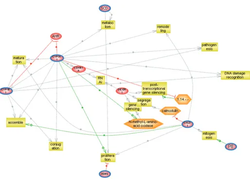
Pathway analysis
We imported our Genespring lists of ad-ditively up-regulated genes in cells treated with a combination of DIM and E2 into
PathwayStudio. We sought to analyze the relationships and biological interactions among the up-regulated genes identified by combination treatment. Figure two dis-plays the pathways retrieved from the PathwayStudio software when we put in three of our up-regulated genes.
In a like manner, we also analyzed the common regulators and/or biological in-teractions among our incrementally sup-pressed or down-regulated genes by querying the relationships between sev-eral of the genes showing the most sig-nificantly diminished gene expression, data not shown.
DISCUSSION
These results indicate that there are a subset of genes in MCF7 whose expres-sion can be modulated by the combina-tion of E2 and DIM. The expression of 32 genes was significantly up-regulated or enhanced by combination treatment with E2 and DIM. Expression of 46 genes was down-regulated by the combination of E2 and DIM in the MCF7 cultures. The subset of genes analyzed by QRT-PCR confirmed the results of microarray pro-filing. Clearly, the expression of many genes is interdependent on both E2 and DIM in cells that respond to estrogen.
Table 1.fold change of genes whose expression is increased by the combination of E2 + DIM
Gene title Genbank EvsC ± SE DvsC ± SE E+D ± SE Cytochrome P450 CYP1B1 NM_000104 1.389 ± 0.12 3.101 ± 0.22 3.93 ± 0.25 Cytochrome P450, CYP1B1 NM_000104.2 1.436 ± 0.15 2.738 ± 0.26 3.32 ±0.32 Cytochrome P450, CYP1B1 NM_000104.2 1.581 ± 0.13 2.617 ± 0.53 2.86 ± 0.66 Cytochrome P450 CYP1A1 NM_000499 0.773 ± 0.13 2.605 ± 0.61 2.84 ± 0.51 Stearoyl-CoA desaturase (delta-9-desaturase) AF116616.1 1.533 ± 0.14 1.96 ± 0.34 2.40 ± 0.28 Caldesmon 1 NM_018495 1.708 ± 0.32 1.75 ± 0.33 2.37 ± 0.31 Solute carrier family 7/ member 11 AB040875.1 1.444 ± 0.17 1.566 ± 0.19 1.90 ± 0.13 p8 protein (candidate of metastasis 1) AF135266.1 1.118 ± 0.12 1.395 ± 0.45 1.92 ± 0.27 Interferon-induced protein/ tetratricopeptide repeats 1 NM_001548 0.93 ± 0.17 1.438 ± 0.13 1.88 ± 0.20 Nucleophosmin/nucleoplasmin, 3 NM_006993 1.72 ± 0.22 1.775 ± 0.16 1.85 ± 0.16 Amyloid beta (A4) precursor protein (protease nexin-II, X06989.1 1.717 ± 0.39 1.624 ±0.31 1.84 ± 0.32
Alzheimer disease)
Zinc finger protein 557 NM_024341 1.324 ± 0.12 1.559 ± 0.21 1.83 ± 0.20 Like Bifunctional methylenetetrahydrofolate NM_025001 1.263 ± 0.13 1.055 ± 0.20 1.81 ± 0.23
dehydrogenase/cyclohydrolase, mitochondrial precursor
In general, the effect of the combina-tion treatment of E2 and DIM on MCF7 cells was antagonistic, reflected in the greater number of down-regulated genes observed in our microarray analysis. This finding is consistent with previous
reports that showed interactions that were primarily inhibitory in studies both in vitro and in vivo (27,28). Many of the changes are subtle, but show an effect over a wide variety of affected genes, in-cluding transcription factors as well as
numerous metabolic gene products whose expression is diminished in com-parison to the findings of treatment with E2, or for that matter DIM alone. Where the changes in gene expression are mod-est, it is important to note that, while
in-Table 2.Fold change of genes whose expression is diminished by the combination of E2 + DIM
triguing, these gene modulations are only possibilities and will need addi-tional studies to determine whether or not they significantly impact the behav-ior of the cell culture.
Previous studies by Chen et al. (26) using subtractive hybridization indicated that DIM could decrease the expression of a number of genes up-regulated by E2 and hypothesized cross-talk between AhR and ER. Our studies support these findings, but additionally showed that expression of a number of genes up-reg-ulated in the presence of 25 μM DIM could be modulated in the presence of 1 nM E2, with expression either dampened by E2 or enhanced by E2. Alternately,
some genes whose expression was up-regulated by E2 could be further en-hanced by DIM. Finally, the expression of some genes was only detectable when the cells were exposed to the combina-tion of E2 and DIM. These observacombina-tions dramatically increase the complexity of the interplay by E2 and DIM and how they might affect the microenvironment of a cell. The study raises questions as to imbalances of the relative amount of these compounds. It is clear that the risk of breast cancer is inversely proportional to the amount of cruciferous vegetables in diet (the natural source of DIM) (29), and that more estrogen increases risk of breast cancer (30). Relative amounts of
these compounds in combination may prove crucial to the protective or preven-tative benefits of DIM (or cruciferous vegetables).
As one example, DIM induces the ex-pression of CYP1A1 and CYP1B1 mem-bers of the P450 superfamily. This study indicated that a further enhancement of expression of these genes occurred in the presence of E2. CYP1A1, a phase I en-zyme, can be the first step in the detoxifi-cation of number of carcinogens. Con-versely, it is known to convert many procarcinogens to carcinogens. Impor-tantly, CYP1A1 increases 2-hydroxylation of estrone leading to 2-hydroxyestrone (not estrogenic), and which is rapidly
O-methylated into compounds that are proliferative, pro-apototic, and anti-angiogenic. However, induction of CYP-1B1 shifts metabolism toward 4-hydroxyestrone, which can be carcino-genic. An imbalance of estrogen metabo-lism is indicated in breast (31), cervical (32), and endometrial (unpublished re-sults) cancers, which are all estrogen-enhanced cancers where I3C and DIM appear to be preventative. Systemic lupus erythematosis and rheumatoid arthritis, diseases predominantly affect-ing women, also have abnormal estrogen metabolism (33). An animal study indi-cated that a diet rich in I3C ameliorated the lupus disease and changed estrogen metabolism (34).
An interesting aspect of altered estro-gen metabolism is found in the up-regulation of aldo-keto reductase family 1, member 3 (AKR1C3), an isomer of the AKR superfamily that is found most prominently in prostate and mammary glands, in samples treated with the com-bination of E2 and DIM. This enzyme has the ability to interconvert testos-terone with 4-androstene-3-17-dione but inactivate 5a-DHT and, therefore, elimi-nate active androgens from the prostate. In the mammary gland, AKR1C3 may function predominantly as a reductase to produce testosterone from 4-androstene-3-17-dione in an intracrine manner and to reduce estrone to estradiol (35). Re-cently, it has been shown that levels of these steroid-metabolizing genes are di-agnostic of tumor versus normal breast tissue (36). Levels of AKR1C3 are re-duced in tumor versus normal tissue, which reflect our findings in E2 treat-ment alone compared with treattreat-ment with both E2 and DIM. The induction of this gene, which has been correlated well with its enzyme activity, suggests a re-balancing to normal steroid-metabolism homeostasis.
One can predict numerous effects of how genes affected by DIM and E2 change the microenvironment of a cell. For instance, the DEAD box polypeptide 4 is a member of a family of genes char-acterized by the conserved motif
Asp-Glu-Ala-Asp (DEAD). These proteins are putative RNA helicases that mediate nu-cleoside triphosphate-dependent un-winding of double-stranded RNA. They are thought to be involved in a variety of cellular processes that involve modifica-tion of RNA secondary structure, i.e., translation initiation, nuclear and mito-chondrial splicing, ribosome and spliceo-some assembly. Members of this family are alleged to be involved in embryogen-esis, spermatogenembryogen-esis, and cellular growth and division.
It is of interest that the combination of DIM and E2 leads to a decrease in ex-pression of genes such as Bcl-6, a zinc finger nuclear phosphoprotein, normally expressed in the germinal center B cells and some intrafollicular T cells. This gene codes for a DNA-binding transcrip-tional repressor that exerts an important role in the development of normal ger-minal centers. Its constitutive expression has been associated with suppression of p53 expression as well as phenotypic changes in germinal center cells by af-fecting differentiation and/or apoptosis. Mature germinal center B cells that leave the germinal center environment gener-ally down-regulate Bcl-6 expression. A block in normal down-regulation of Bcl-6 has been postulated to cause genetic in-stability in the germinal center cells, and subsequently leads to neoplastic trans-formation (37). Additionally the down regulation of Bcl3 in cells treated in com-bination with E2 and DIM disrupts a sig-naling pattern observed in MCF7 cell (38) where Bcl3 complexes with phos-phorylated Bcl10 and translocates to the nucleus, where it alters transcription.
The mixture of E2 and DIM treatment dampens the expression of cytoplasmic FMR1 interacting protein 2, which asso-ciates with FMRP (Fragile X mental retar-dation protein) as well as FMRP-related proteins FXRIP and FXR2P. The protein is cytoplasmically colocalized with FMRP and ribosomes, and is thought to interact with RAC1. RAC1 is a small GTPase that stimulates actin polymeriza-tion toward lamellopodia formapolymeriza-tion, whose overexpression in tumor cells has
been associated with invasion and metastasis in human tumor cells (39,40).
Intriguingly, CXCR4 is also down-regulated by the combination treatment with E2 and DIM. CXCR4 appears to be necessary for breast cancer metastasis (41). This interesting gene was signifi-cantly suppressed in one gene list (data not shown), but not in the other, yet bears mention. CXCR4, a cytokine, is the cognate receptor for stromal cell derived factor 1, and the expression of this com-plex in breast cancer cells is associated with significant increases in invasiveness and faster migration of these cells to the lymph nodes (42). Silencing CXCR4 gene expression with siRNA blocks in vitro in-vasion and in vivo metastasis of breast cancer cells in animal models (41,43). Also of note is the down-regulation of CCR4-NOT complex 3 in the presence of both E2 and DIM, suggesting a suppres-sive effect on global transcription through regulation of transcription factor TFIID. As a master switch, dampening the expression of this factor would result in myriad gene effects both positive and negative (44).
Many of the noted gene changes and effects are subtle in form, with regulation modulated rather than radically altered. These findings suggest interesting per-turbations in a biological system in the presence of physiologic concentrations of hormone and low concentrations of bioactive chemicals present in the envi-ronment. As much of the effects of DIM and E2 shift metabolism toward a proapoptotic, moderated, proliferative state, our results are consistent with DIM offering an effective preventive adjuvant in a healthy nutritional regime.
on both lists. The six overlapping genes are among the largest fold changes on the dChip list while they are found throughout the list with GeneSpring. However, the list does completely over-lap with the clustering data identifying enhanced gene expression when treated with the combination of E2 + DIM (data not shown). In a similar manner, we identified 14 genes whose expression was suppressed or inhibited by treat-ment with E2 + DIM using dChip and the same filtering criterion as for gene in-duction, contrasted with 46 genes identi-fied with GeneSpring. Between these two groups were six genes in common.
The pathway analysis is consistent with our in vitro observations as well as our expression analysis of treatment of MCF7 cells with E2 or DIM or both. Among the up-regulated gene pathway we found interrelated cell processes, which contain other genes found in our up-regulated list but not included in our query, such as nuclear receptors, cyclin dependent kinase inhibitor, and dicer (a ribonuclease essential for RNA inter-ference of small temporal RNA pathways which represses gene expression). These genes are in keeping with a program fo-cused on increasing cell differentiation, through cytoskeletal development and assembly, detection of DNA damage and cell cycle arrest, as well as apoptosis, post translation, and gene silencing. These results suggest there may be com-mon regulators or signal transduction pathways among these genes.
When we examined cell systems af-fected by the expression of BCL-6 and TGF-b, the expression of both is down-regulated in treatment of cells with E2 and DIM, we find a multitude of processes that are common targets of these molecules but not necessarily over-lapping in their modulation. In fact, there are almost an equal number of processes, which are coordinately regu-lated by these genes as those that receive opposite and antagonistic signals from these powerful molecules. The net result may be due to the relative concentrations of each protein.
We noted common regulators between TGF-b and BCL6. These include MAPK1 and 3, ABL-1, FOXO 3a (belonging to the forkhead family of transcription factors, which may function as a trigger for apoptosis), tumor suppressor gene EP300, a cyclin D–related transcription factor, as well as cyclin D itself. In many ways, the paradoxical effects of the regu-lated genes we found with the E2 + DIM–treated cells mirror the complicated physiologic findings of estrogens and ex-posure to toxins such as TCDD and dioxin.
Joint effects of DIM and E2 are compli-cated. One possibility is coordinate gene regulation due to the binding of DIM to its cognate AhR receptor as well as unli-ganded ER complex. The binding of AhR/ARNT complex to its response ele-ments functions as a cis-acting enhancer in the regulatory domains of its target genes, a representative group identified as the AhR gene battery (47). The pres-ence of proximal regulatory elements to AhR binding sites within discrete chro-mosomal locals suggests a mechanism by which this complex can activate a num-ber of other transcription factors (not AhR targets) and clusters of genes as a secondary effect of binding (48). The jux-taposition of ligands dependent AhR-ER binding may have a concerted effect on the activation of additional transcription factors with both induction as well as re-pression of gene exre-pression. The tration of receptors as well as the concen-tration of DIM and E2 would matter and determine how much E2 or DIM bind to their cognate receptors (DIM binding to AhR and E2 binding to ERs) or compete for binding to the opposing receptors.
This work focused on the combined activity of E2 and DIM on gene expres-sion, i.e., how the combination would be different than individual effects. It is well established that most activities of I3C and DIM are not related to estrogen per se. Nonetheless, this study confirms that there is a strong interplay of estrogen and DIM, which is reflected in gene ex-pression changes when both compounds are present.
Note added in proof: A recent paper by Liu et al. (49) showed AhR agonists directly activate ERa in MCF-7 breast cancer cells.
ACKNOWLEDGMENTS
Laura Mulvey and Alamelu Chan-drasekaran contributed equally to this work. This work was supported by RO1-CA733850 to KJA from the National In-stitutes of Health.
REFERENCES
1. Ge X, Fares FA, Yannai S. 1999. Induction of apoptosis in MCF-7 cells by indole-3-carbinol is independent of p53 and bax. Anticancer Res. 19:3199-3203.
2. Chen DZ, Qi M, Auborn KJ, Carter TH. 2001. In-dole-3-carbinol and diindolylmethane induce apoptosis of human cervical cells and in murine HPV-16 transgenic preneoplastic cervical epithe-lium. J. Nutr. 131:3294-3302.
3. Bjeldanes LF, Kim JY, Grose KR, Bartholomew JC, Bradfield CA. 1991. Aromatic hydrocarbon responsiveness-receptor agonists generated from indole-3-carbinol in vitro and in vivo: compar-isons with 2,3,7,8-tetrachlorodibenzo-p-dioxin. Proc. Natl. Acad. Sci. U.S.A. 88:9543-9547. 4. Edwards DP, Adams DJ, McGuire WI. 1981.
Es-trogen regulation of growth and specific protein synthesis in human breast cancer cells in tissue culture. Adv. Exp. Biol. 138:133-149.
5. Newfield L, Bradlow HL, Sepkovic DW, Auborn K. 1998. Estrogen metabolism and the malignant potential of human papillomavirus immortalized keratinocytes. Exp. Biol. Med. 217:322-326. 6. Prall OWJ, Sarcevic B, Musgrove EA, Watts CK,
Sutherland RL. 1997. Estrogen-induced activation of Cdk4 and Cdk2 during G1-S phase progres-sion is accompanied by increased cyclin D1 ex-pression and decreased cyclin-dependent kinase inhibitor association with cyclin E-Cdk2. J. Biol. Chem. 272:10882-10894.
7. Lobenhofer EK, Lee Bennet P, Cable L, Li L, Bushel PR, Afshara CA. 2002. Regulation of DNA replication fork genes by 17b-estradiol. Mol. En-docrinol. 16:1215-1229.
8. Perillo B, Sasso A, Abbondanza C, Palumbo G. 2000. 17β-estradiol inhibits apoptosis in MCF-7 cells, inducing bcl-2 expression via 2 estrogen-responsive elements present in the coding se-quence. Mol. Cell. Biol. 8:2890-2901.
9. Jin L et al. 1999. Indole-3-carbinol prevents cervi-cal cancer in human papilloma virus type 16 (HPV16) transgenic mice. Cancer Res. 59:3991-3997.
10. Bell MC et al. 2000. Placebo-controlled trial of indole-3-carbinol in the treatment of CIN. Gynecol. Oncol. 78:123-129.
estradiol metabolism and spontaneous mam-mary tumors in mice. Carcinogenesis 12:1571-1574.
12. Kojima T, Tanaka T, Mori H. 1994. Chemopreven-tion of spontaneous endometrial cancer in female donyru rats by indole-3-carbinol. Cancer Res. 54:1446-1449.
13. Chen D-Z, Carter TH, Auborn KJ. 2004. Apopto-sis in cervical cancer cells: Implications for ad-junct anti-estrogen therapy for cervical cancer. Anticancer Res. 24:2649-2656.
14. Chen I, Safe S, Bjeldanes L. 1996. Indole-3-carbinol and diindolylmethane as aryl hydrocar-bon (Ah) receptor agonists and antagonists in T47D human breast cancer cells. Biochem. Phar-macol. 51:1069-1076.
15. Bradlow HL, Telang NT, Sepkovik DW, Osborn MP. 1996. 2-Hydroxyestrone: the “good” estro-gen. J. Endrocrinol. 150:S259-265.
16. LaVallee TM et al. 2002. 2-Methoxyestradiol in-hibits proliferation and induces apoptosis inde-pendently of estrogen receptors αand beta. Can-cer Res. 62:3691-3697.
17. Meng Q et al. 2000. Suppression of breast cancer invasion and migration by indole-3-carbinol: associated with up-regulation of BRCA1 and E-cadherin/catenin complexes. J. Mol. Med. 78:155-165.
18. Fan S et al. 1999. BRCA1 inhibition of estrogen receptor signaling in transfected cells. Science 284:1354-1356.
19. Liu H, Wormke M, Safe SH, Bjeldanes LF. 1994. Indolo[3,2-b]carbazole: a dietary-derived factor that exhibits both antiestrogenic and estrogenic activity. J. Natl. Cancer Inst. 86:1758-1765. 20. Auborn KJ et al. 2003. Indole-3-carbinol is a
neg-ative regulator of estrogen. J. Nutr. 133:2470s-2475s.
21. Evans RM. 1998. The steroid and thyroid hor-mone receptor superfamily. Science 240:889-895. 22. Katzenellenobogen BS. 1996. Estrogen receptors: bioactivities and interactions with cell signaling pathways. Biol. Reprod. 54:287-293.
23. Safe S, Krishnan V. 1995. Cellular and molecular biology of aryl hydrocarbon (Ah) receptor-mediated gene expression. Arch. Toxicol. Suppl. 17:99-115.
24. Riby J, Chang G, Firestone G, Bjeldanes L. 2000. Ligand-independent activation of estrogen recep-tor function by 3,3′diindolylmethane in human breast cancer cells. Biochem. Pharmacol. 60:167-177.
25. Ohtake F et al. 2003. Modulation of oestrogen re-ceptor signaling by association with the activated dioxin receptor. Nature 423:545-550.
26. Chen I, Hsieh T, Thomas T, Safe S. 2001. Identifi-cation of estrogen-induced genes downregulated by AhR agonists in MCF-7 breast cancer cells using suppression subtractive hybridization. Gene 262:207-214.
27. Chen I, McDougal A, Wang F, Safe S. 1998. Aryl hydrocarbon receptor-mediated antiestrogenic
and antitumorigenic activity of diindolyl-methane. Carcinogenisis 19:1631-1639. 28. Chang X et al. 2005. 3,3′-Diindolylmethane
in-hibits angiogenesis and the growth of trans-plantable human breast carcinoma in athymic mice. Carcinogenesis 26:771-778.
29. Terry P, Wolk A, Persson I, Magnusson C. 2001. Brassica vegetables and breast cancer risk. J. Am. Med. Assoc. 285:2975-2977.
30. Russo J, Hasan Lareef M, Balogh G, Russo IH. 2003. Estrogen and its metabolites are carcino-genic agents in human breast epithelial cells. Steroid Biochem. Mol. Biol. 87:1-25.
31. Rogan EG et al. 2003. Relative imbalances in es-trogen metabolism and conjugation in breast tis-sue of women with carcinoma: potential bio-markers of susceptibility to cancer. Carcinogenesis 24:697-702.
32. Jin L et al. 1999. Indole-3-carbinol prevents cervi-cal cancer in human papilloma virus type 16 (HPV16) transgenic mice. Cancer Res. 59:3991-3997.
33. Lahita RG, Bradlow HL, Kunkel HG, Fishman J. 1979. Alterations of estrogen metabolism in sys-temic lupus erythematosus. Arthritis Rheum. 22:1195-1198.
34. Auborn KJ et al. 2003. Lifespan is prolonged in autoimmune-prone (NZB/NZW) F1 mice fed a diet supplemented with indole-3-carbinol. J. Nutr. 133:3610-3613.
35. Penning TM et al. 2000. Human 3 α-hydroxysteroid dehydrogenase isoforms (AKR1C1-AKR1C4) of the aldo-keto reductase superfamily: functional plastic-ity and tissue distribution reveals roles in the in-activation and formation of male and female sex hormones. Biochem J. 351:67-77.
36. Lewis MJ, Wiebe JP, Heathcote JG. 2004. Expres-sion of progesterone metabolizing enzyme genes (AKR1C1, AKR1C2, AKR1C3, SRD5A1, SRD5A2) is altered in human breast carcinoma. BMC Can-cer 4:27-38.
37. Phan RT, Dalla-Favera R. 2004. The BCL6 proto-oncogene suppresses p53 expression in germinal-center B cells. Nature 432:635-639.
38. Yeh PY et al 2006. A pathway for tumor necrosis factor-a-induced Bcl10 nuclear translocation. J. Biol. Chem. 281:167-175.
39. Bouzahzah B et al. 2001. Rho family GTPases regulate mammary epithelium cell growth and metastasis through distinguishable pathways. Mol. Med. 7:816-830.
40. Baugher PJ, Krishnamoorthy L, Price JE, Dhar-mawardhane SF. 2005. Rac1 and Rac3 isoform ac-tivation is involved in the invasive and metasta-tic phenotype of human breast cancer cells. Breast Cancer Res. 7:R965-R974.
41. Liang Z et al. 2005. Silencing of CXCR4 blocks breast cancer metastasis. Cancer Res. 65:967-971. 42. Kang H et al. 2005. Stromal cell derived factor-1: its influence on invasiveness and migration of breast cancer cells in vitro, and its association with prognosis and survival in human breast cancer. Breast Cancer Res. 7:R402-R410.
43. Li YM et al. 2004. Upregulation of CXCR4 is es-sential for HER2-mediated tumor metastasis. Cancer Cell 6:459-430.
44. Collert MA, Struhl K. 1994. NOT1(CDC39), NOT2(CDC36), NOT3, and NOT4 encode a global-negative regulator of transcription that differentially affects TATA-element utilization. Genes Dev. 8:525-537.
45. Cheng L, Wing HW. (2001a). Model-based analy-sis of oligonucleotide arrays: Expression index computation and outlier detection. Proc. Natl. Acad. Sci. 98:31-36.
46. Cheng L, Wing HW. (2001b). Model-based analy-sis of oligonucleotide arrays: model validation, design issues and standard error application. Ge-nome Biol. 2:research0032.1-0032.11
47. Nebert DW et al. 2000. Role of the aromatic hy-drocarbon receptor and [Ah] gene battery in the oxidative stress response cell cycle control and apoptosis. Biochem. Pharmacol. 59:65-85. 48. Reymann S, Borlak J. 2006. Transcriptome
profil-ing of human hepatocytes treated with Aroclor 1254 reveals transcription factor regulatory net-works and clusters of regulated genes. BMC Ge-nomics 7:217-235.


![{2,2′ [Ethylenebis(nitrilomethylidyne)]diphenolato κ4O,N,N′,O′}oxidovanadium(IV)](data:image/gif;base64,R0lGODlhAQABAIAAAP///wAAACH5BAEAAAAALAAAAAABAAEAAAICRAEAOw==)