INTRODUCTION
Viral and nonviral vectors can be ma-nipulated to increase the efficiency and specificity for vascular cell transduction (1,2). This can be achieved either by modifying the cell binding properties of the vectors or by the use of cell-selective promoters (3,4). Altering the tropism of viral vectors can be achieved by genetic modification of the viral envelope (5-8) or by the use of proteins derived from other enveloped viruses (9,10). Alterna-tively, targeting molecules can be derived from nonretroviral proteins expressed on the packaging cell line (11-13). A further alternative is to use adaptor molecules that retarget the virus to specific cell-sur-face molecules (14-16). These approaches can be time consuming and often affect the production of the virus. In addition, although in some cases they offer high
specificity, they can result in poor trans-duction efficiency.
In this report, we describe an alterna-tive targeting strategy using immunovi-rosomes generated by mixing mildly aggregated monoclonal antibodies, lipo-somes, and viral particles. This strategy is based on the ability of cationic lipo-somes to form stable complexes with viral vectors (17,18). This interaction has been reported for Moloney murine leukemia virus (MMLV). Here, we have shown that immunovirosomes carrying monoclonal antibodies against endothe-lial markers can target activated vascular endothelium. The resulting gene transfer efficiency is enhanced if endocytic recep-tors are chosen. Importantly, our results demonstrate that changing viral tropism can alter the ability of viral transduction to result in long-term expression. These
findings will have profound conse-quences on future viral vector design with respect to improving specificity and efficiency.
MATERIALS AND METHODS
Reagents
RPMI-1640, CD hybridoma medium, hybridoma serum-free medium (SFM), human endothelial basal growth-SFM, L-glutamine, penicillin, streptomycin, and trypsin-EDTA were purchased from Invitrogen (Paisley, UK), and fetal calf serum (FCS) was purchased from Globepharm (Esher, UK). The synthetic liposome Tfx-50 was purchased from Promega (Southampton, UK). Lipofectin and LipofectAMINE were obtained from Invitrogen. Other reagents were pur-chased from Sigma (Poole, UK), unless stated otherwise.
Preparation of Monoclonal Antibodies Hybridomas producing the anti-human transferrin receptor (CD71) mAb, OKT9; anti-E/P-selectin (CD62E/P),
Stability of Gene Expression: Implications for Viral Vector
Design
Address correspondence and reprint requests to PH Tan, Nuffield Department of Surgery, John Radcliffe Hospital, Headley Way, Oxford, OX3 9DU, UK. Fax: +44 (0)208 846 8081; E-mail: [email protected]
*Contributed equally to this work.
Submitted July 2, 2006; Accepted for publication February 5, 2007.
Peng H Tan,*
1,2Shao-An Xue,*
3Bin Wei,
2,4Angelika Holler,
3Ralf-Holger Voss,
5and Andrew JT George
11Department of Immunology, Division of Medicine, 4Immunology Section, Division of Investigative Science, Faculty of Medicine,
Imperial College London, Hammersmith Hospital, London, UK; 2Nuffield Department of Surgery, University of Oxford, John
Rad-cliffe Hospital, Oxford; 3Department of Immunology and Molecular Pathology, Royal Free and University College Medical School,
Royal Free Hospital, London, UK; 5Department of Hematology and Oncology, Johannes Gutenberg University, Mainz, Germany
Many strategies for redirecting the tropism of murine Moloney leukemia virus (MMLV) have been described. Preformed virion-liposome complexes, termed virosomes, have been reported to be relatively stable. Virosomes mediate envelope-independent transduction that allows efficient superinfection of resistant cell lines; however, virosome-mediated transduction behaves in a non–target-specific manner. We developed a novel method using antibodies to direct MMLV to vascular endothelium. We have given the term immunovirosomes to the complexes formed between viruses, liposomes, and antibodies. These immunovirosomes improve the transduction efficiency of the viruses and alter their tropism. We have shown improved transduction when im-munovirosomes were targeted at the endocytic receptors CD71 and CD62E/P and rather less good delivery when targeted at CD106. The enhancement of the transduction efficiency was transient, however, suggesting that rerouting the entry pathway of viruses alters the expression properties of the viruses.
1.2B6 (dual-specificity), and anti-VCAM-1 (CDanti-VCAM-106), anti-VCAM-1.4C3, were grown and puri-fied by protein G chromatography, as described (19).
Immunoliposome Preparation Immunoliposomes were prepared as described (19,20). In brief, heat-aggregated Ab [optimal mAb concen-trations; OKT9 (60 µg/mL), 1.2B6 (30µg/mL), and 1.4C3 (60 µg/mL)] was mixed with liposomes (Tfx-50, Lipo-fectin, or LipofectAMINE), for 30 min at room temperature (in total volume of 250µL in Opti-MEM). For plasmid-medi-ated transfection, the immunoliposomes were then incubated with DNA at a ratio recommended by the manufacturer for the liposome-DNA complex. The result-ing transfection complexes were added to endothelial cells (ECs). As in the case of the immunovirosomes, the retroviral supernatants were mixed with immuno-liposomes at equal volumes for 30 min at room temperature before the addition to the cells (1 ×10⁵). In some cases, the
retroviral supernatant was treated with DNAse before transduction.
EC Isolation and Culture
ECs were isolated from human saphe-nous veins and maintained in culture as described (21,22). Briefly, ECs were cul-tured in EBM-2 (BioWhittaker, Cam-bridge, UK) and human endothelial-SFM basal growth medium supplemented with 10% heat-inactivated FCS, 2 mM L-glutamine, 100 U/mL penicillin, 100µg/mL streptomycin, 25 µg/mL fungizone, 0.03 mg/mL endothelial cell growth supplement (ECGS), and 100 U/mL heparin. All cells were used between the 3rd and 4th passages. Human saphenous veins were obtained from patients undergoing varicose vein or coronary artery bypass surgery. Se-lected patients were either healthy young adults or with coronary artery disease, but without other comorbidity. Patients with diabetes or other autoimmune dis-eases were excluded from the study. In-formed consent was obtained from all pa-tients and approval obtained from the
local research ethics committee. To inhibit de novo protein synthesis, the cells were treated with 50 µg/mL cycloheximide.
Vascular Samples
Vascular tissues were obtained from the Cardiothoracic Department of Hammer-smith Hospital, UK. The tissues were ob-tained as a surplus product from coronary bypass surgery, after local research ethics approval, of patients ages 45 to 85 years.
MMLV Production and Titration: Retroviral Vectors and Packaging Cell Lines
The plasmids—pHIT60 containing gag-polgenes (23), pCL-Eco env containing ecotropic envelope (24), pCOLT-GALV env containing gibbon ape leukemia virus envelope (GALV env) protein (25), or pRV67 env containing vesicular stom-atitis virus G envelope (VSV-G env) protein (22,26)—and the retroviral con-structs—pBullet-EGFP (22,26,27), LZRS-EGFP (22,26,28,29), pFB-rhGFP (Strata-gene, Cambridge, UK) (22,26), or Pinco-EGFP (30)—were used to generate retro-viral particles as described below.
Phoenix ecotropic (30) and ampho-tropic (23) packaging lines (kind gifts from Dr Gary Nolan’s laboratory, Stan-ford University) and 293T cells (22,26) were routinely maintained in Dulbecco’s modified Eagle’s medium (DMEM) sup-plemented with 10% FCS.
Ecotropic MMLV Production The ecotropic MMLV particles were generated as described (26). Briefly, the Phoenix ecotropic cells and 293T cells were cocultured in DMEM at a ratio of 1:1 (1.5 ×10⁶cells seeded in 25-cm2flask
on the day before transfection). Twenty-four hours later, the retroviral-GFP construct (Pinco-EGFP) together with pHIT60 and pCL-Eco were cotransfected into the seeded packaging cells using a calcium phosphate transfection kit (Invit-rogen) according to the manufacturer’s instructions. Sixteen hours after transfec-tion, cells were washed twice with warm PBS and the DMEM was replaced with RPMI-1640. Forty-eight hours after
trans-fection, the viral supernatants were har-vested and filtered through a 0.45-µm pore size filter and stored at –80°C. Retroviral titers of the PINCO vector were determined on NIH 3T3 cells as de-scribed (31). The cells were analyzed by flow cytometry to determine the propor-tion of NIH 3T3 cells expressing EGFP.
VSV-G–Pseudotyped and GALV-Pseudotyped MMLV Production and Titration
The MMLV-based constructs were prop-agated by a 3-plasmid cotransfection tech-nique in 293T cells as described (22,26). Transfections were performed using poly-ethylenimine (PEI) (Sigma) on 9-cm tissue culture plates as described (26). In the case, of GALV-pseudotyping, 2 ×106293T
were seeded into a T25 tissue culture flask and 24 hours later transiently transfected with retroviral-EGFP constructs with pCOLT-GALV using a calcium phosphate transfection kit (Invitrogen) according to the manufacturer’s instructions.
MMLV were titred on either D17 cell (VSV-G pseudotyped) or 293 and/or Hela cells (GALV pseudotyped) as previ-ously described (26). In brief cells were seeded on 24 well plates at 3 x 10⁴cells
per well, infected with serial 1:10 dilu-tions of virus and analyzed for EGFP ex-pression at 3 to 5 days post-infection.
Amphotropic MMLV Production and Titration
The amphotropic MMLV particles were generated as described (23). Briefly, 2 ×10⁶amphotropic packaging cells were
cells with serial 1:10 dilutions of virus and analyzing for EGFP expression at 3 to 5 days postinfection.
Transfection with Virosomes and Immunovirosomes
For transfection, virosomes and im-munovirosomes were generated as de-scribed above and cultured with 105EC
cells. The MMLV [with or without poly-brene (PB)], virosomes, or immunoviro-somes were exposed to cells for 2 h at 37°C. To block targeted endothelial re-ceptors of ECs before transduction, ECs were preincubated with 200 µg/mL of appropriate antibody at 4°C, then trans-fected. In some cases, the viral particles were exposed to 50 kGy gamma irradiation.
Flow Cytometry
The phenotype of transfected or un-transfected ECs was assessed by flow cytometry 48 h after transfection. Cell staining was performed using mouse an-tibodies conjugated to APC or primary antibodies followed by goat anti-mouse-APC, as described (19,26). Flow cytomet-ric analysis of all cells was performed using the following mouse monoclonal antibodies: 1.2B6 (anti-CD62E/P) (21), 1.4C3 CD106) (21), and 6.5B5 (anti-CD54) (21). All antibodies raised from hybridomas were kindly given by Prof. D Haskard, Imperial College London, UK, unless stated otherwise (21).
Assessment of EGFP Reporter Gene Expression
After transduction, EGFP reporter gene expression was determined using flow cytometry or an inverted fluores-cent microscope, as described (19,32).
Labeling of YOYO-1 and Determination of Internalization
YOYO-1 stock solution (1 ×10⁻³M)
(Molecular Probe, Cambridge, UK) was initially diluted 7.5 times in DMSO to a concentration of 1.33 ×10⁻⁴M. The
work-ing concentration of YOYO-1 in all sam-ples was 6.3 ×10⁻⁶M. To prevent
photo-bleaching of YOYO-1, its solutions were
kept in the dark and all experiments were protected from light. The viral par-ticles were labelled with YOYO-1 and vi-sualisation of these labelled particles using flow cytometry was carried out as previously described (33, 35). Uninternal-ized and surface-bound particles were removed as described (32). Internaliza-tion of viral particles was assessed using flow cytometry. In some cases, the cells were treated with chloroquine as de-scribed (35).
RT-PCR and Southern Blots
RT-PCR analysis for EGFP gene ex-pression was carried out as described (36) with the following primers; 5′-GAT CTT GAA GTT CAC CTT GAT GCC (sense) and 5′-AGC TGA CCC TGA AGT TCA TCT GC (antisense) at 60°C anneal-ing temperature. To detect the level of in-tegration of EGFP, we carried out South-ern blot hybridization for EGFP DNA in the genomic DNA of transduced cells. Genomic DNA was extracted in accor-dance with manufacturer’s instructions (Qiagen, Crawley, UK). It was then di-gested with NcoI (position 4137) and NotI (position 4870) (pBullet-EGFP). Gel
elec-trophoresis and Southern blotting were performed as described (37). The blot was hybridized with [32P]-labeled probe
using EGFP fragment released from pBullet-EGFP vector. After 12 h hy-bridization at 68°C, the blot was washed 3 times in 0.3×SSC, 0.1% SDS at 68°C be-fore exposing to X-ray film at –80°C for 2 h. The intensity of Southern blot was analyzed as previously described (38).
Reproducibility and Statistical Analysis
For multiple comparisons, results were analyzed by ANOVA, whereas for com-parison of 2 means, results were analyzed by Student ttest where P < 0.05 was con-sidered statistically significant. All data shown are representative of at least three experiments, unless stated otherwise.
RESULTS
Expression of Adhesion Molecules by Activated Endothelial Cells
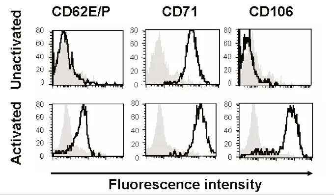
As previously reported (19), HSVECs constitutively express CD71 (Figure 1). However, after activation with proin-flammatory cytokines (80 ng/mL TNF-α,
Figure 1.Expression of surface receptors. HSVECs were cultured and the surface expres-sion of CD62E/P, CD71, and CD106 were analyzed using flow cytometry. Where indi-cated, the cells were stimulated with proinflammatory cytokines (80 ng/mL TNF-α, 80 ng/mL IFN-γ, and 80 ng/mL IL-1β) for 8 h. The gray backgrounds show staining with isotype-control antibody, and the dark black lines are specific antibodies against
80 ng/mL IFN-γ, and 80 ng/mL IL-1β) for 8 h, the cells upregulate the expres-sion of adheexpres-sion molecules (CD62E/P and CD106) and CD71 (21) (Figure 1).
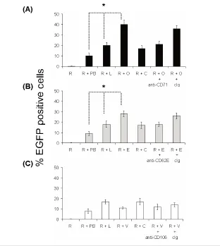
Immunoliposomes Enhance Gene Expression by MMLV and Alter Viral Tropism
In our experience, MMLV inefficiently transduces HSVECs, in both the presence
or absence of polybrene. The addition of liposomes to MMLV has been shown to enhance transduction efficiency (17,18). Using liposomes [Tfx-50 or lipofectin (data not shown) or lipofectAMINE (data not shown)] and amphotropic MMLV [LZRS-EGFP (data not shown), pFB-rhGFP (data not shown), or pBullet-EGFP (Figure 2 with Tfx50 liposomes)], we demonstrated an approximately
10-fold increase in cells expressing EGFP compared with MMLV alone. We investi-gated whether this could be further in-creased by incorporation of antibodies. An approximately two-fold increase in expression efficiency was seen with anti-CD71 immunovirosomes compared with virosomes (Figure 2). Similarly, anti-CD62E/P could increase transduction in stimulated HSVECs expressing CD62E/P (Figure 2b). However, as expected from a previous study with immunoliposomes (19), anti-CD106 (Figure 2c) showed only minor or no augmentation. The expres-sion levels of CD106 as determined by flow cytometry were more than
CD62E/P on the endothelial cells follow-ing 8-h activation (Figure 1) (21). In all cases, the enhanced expression efficiency mediated by an antibody could be blocked with free specific antibody, but not with control antibodies.
Similar augmentation of expression was seen whether the immunovirosomes were made with amphotropic, GALV, ecotropic, or VSV-G coat proteins. In all cases, the presence of liposomes and anti-CD71 antibody enhanced expression efficiency (Table 1). These effects were seen not only on human cells; a specific enhancement of expression efficiency in primary mouse cardiac ECs was also ob-served using transferrin (in nonactivated ECs) or anti-CD62E (in activated ECs) (data not shown).
Effect of Virosomes and
Immunovirosomes on Viral Particle Entry to Cells
We investigated the internalisation of viral particles in target cells by labelling the viruses with fluorescent dye and then following of internalisation of the virus by flow cytometry (using washing with NaOH to remove surface bound material). The data show that when im-munovirosomes are targeted at endo-cytic receptors, such as CD71, there is more rapid uptake than virus alone or incorporated into a virosome (Figure 3a). Chloroquine has been shown to in-crease gene expression in a number of vector systems, by inhibiting acidifica-Figure 2. Transducibility of ECs by retroviruses. pBullet-EGFP was used to generate
tion of the endolysosomal pathways and thus increasing the amount of cDNA reaching the nucleus (35). We showed that treatment with this endo-somolytic agent can increase cellular entry of MMLV particles in the presence and absence of polybrene. However, less impressive improvement in cellular entry was seen when the particles were packaged by liposomes and immunoli-posomes (Figure 3a).
Transduction of Virosomes and Immunovirosomes After an Endosomolytic Treatment
As observed in Figure 2, transduction of HSVECs with MMLV alone and MMLV in the presence of polybrene re-sulted in poor transduction efficiency. When comparing MMLV transduction of HSVECs in the absence of polybrene with virosomes and immunovirosomes, we saw enhancement in transduction ef-ficiency of ~100-fold and ~250-fold, re-spectively (Figure 3b). On the other hand, when MMLV transduction in the presence of polybrene was compared to virosomes and immunovirosomes, only ~two-fold and ~five-fold increases, re-spectively, was observed (Figure 3b).
Cellular treatment with chloroquine can improve transduction efficiency of MMLV alone or MMLV in the presence of polybrene by ~25-fold and ~two-fold, respectively (Figure 3b). However, in the case of virosome and immunovirosome transduction, the addition of chloroquine resulted in only marginal increase in transduction efficiencies (Figure 3b) (al-beit statistically significant but probably with limited any biological significance).
Effect of Virosomes and
Immunovirosomes on Kinetics of Gene Expression
The time-course of expression after gene transfer with virus, virosomes, or immunovirosomes was compared. Fol-lowing amphotropic MMLV, virosome, or immunovirosome transduction/transfec-tion, the early expression of EGFP (less than 24 h) indicated that there was a slow increase in EGFP-positive cells and mean
Table 1.Gene transfer with different pseudotyped MMLV, virosomes, and immunovirosomes.
Viral coat proteins
Amphotropic Ecotropic GALV VSV-G
Unstimulated HSVECs
MMLV 0.3 ± 0.3 0.1 ± 0.1 0.7 ± 0.5 0.4 ± 0.8
MMLV + polybrene 9.2 ± 1.7 3.1 ± 2.9 10.2 ± 2.8 10.1 ± 2.3
Virosomes 19.1 ± 3.2 20.8 ± 2.4 22.8 ± 3.1 21.8 ± 1.7
Anti-CD71 immunovirosomes 39.3 ± 3.4 40.8 ± 1.7 41.9 ± 6.9 42.7 ± 3.5 Stimulated HSVECs
MMLV 0.2 ± 0.1 0.3 ± 0.2 0.3 ± 0.1 0.4 ± 0.3
MMLV + polybrene 10.8 ± 0.3 2.8 ± 3.9 10.7 ± 3.4 10.6 ± 4.9
Virosomes 22.2 ± 8.1 22.7 ± 3.2 22.3 ± 4.5 24.1 ± 3.8
Anti-CD62E/P immunovirosomes 35.2 ± 1.8 36.9 ± 3.3 37.1 ± 5.2 38.1 ± 7.1
HSVECs (either nonactivated or activated) were treated with amphotropic, ecotropic, GALV- and VSV-G-pseudotyped MMLV. These pseudotyped MMLV were then used to generated virosomes [Tfx-50, lipofectin (data not shown) or LipofectAMINE (data not shown)] and anti-CD71 containing immunovirosomes for nonactivated ECs (upper panel) or anti-CD62E/P containing immunovirosomes for cytokine-activated ECs (lower panel). All results are expressed as mean ± SD of transfection efficiency of triplicate wells and all experiments were carried out at least three times.
fluorescence intensity of cells (Figure 4) in all methods of transduction/transfection.
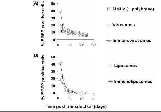
After 24 h following amphotropic MMLV gene transfer, the expression of EGFP was stable with only a slight de-crease over time in the proportion of cells expressing EGFP (Figure 5). How-ever, in cells treated with either viro-somes or immunoviroviro-somes made with anti-CD71 (Figure 5) or anti-CD62E/P (data not shown), there was a decrease over two weeks in the proportion of cells expressing EGFP, at which time expres-sion was equivalent to that seen follow-ing retroviral transduction. This reduction in the proportion of EGFP-expressing cells showed a similar kinet-ics to the reduction in expression seen following transfection with plasmid DNA and liposomes or immunolipo-somes alone (with the exception that with these transient nonviral vectors, ex-pression fell to background levels over two weeks) (Figure 5a, lower panel).
We compared the kinetics of mRNA expression following treatment with amphotropic MMLV (Figure 6, pBullet-EGFP) (LZRS-EGFP, data not shown), virosomes, or immunovirosomes. As can be seen in Figure 6, upper panels, expression of EGFP mRNA was similar at all times following retroviral gene transfer. However, following treatment with either virosomes or
immunoviro-somes, EGFP mRNA expression was higher at early time-points but de-creased over time to reach levels compa-rable to retroviral-transduced levels [shown for CD71 monoclonal anti-bodies for activated EC (Figure 6, upper panels), but similar data (not shown) were seen with anti-CD71 for
nonvated EC and anti-CD62E/P for acti-vated EC].
To assess the persistence of EGFP gene integrated into the EC genome, we car-ried out Southern blot analysis of ge-nomic DNA derived from ECs following gene transfer. Interestingly, the data showed that the number of copies of Figure 4.Early kinetics of gene expression. HSVECs that had been stimulated with 80 ng/mL TNF-α, 80 ng/mL IFN-γ, and 80 ng/mL IL-1β) for 8 h were treated with amphotropic MMLV-EGFP in combination with polybrene or amphotropic MMLV-EGFP containing virosomes or immunovirosomes (made with anti-CD71 mAb). After transduction/transfection, the cells were cultured for 24 h. Flow cytometric analysis was performed at 30, 60, 180, 360, 720, and 1440 min to determine the percentage of EGFP-positive cells (transfection efficiency) (A) and the mean fluorescence intensity of cell (B). All results are expressed as the mean (% transfection efficiency) ± SD of triplicates (A) and the mean fluorescence intensity (B).
EGFP integrated into EC genomic DNA remained stable at all times following treatment with viruses, virosomes, or im-munovirosomes (Figure 6, lower panels). To exclude the possibility that the aug-mentative effects of liposomes and im-munoliposomes were due merely to the transfer of fluorescent protein (produced in the packaging cell) by the viral parti-cles, the particles were irradiated (90 kGy) before use. As can be seen (Figure 7a), no expression of EGFP seen after irradiation, indicating that passive uptake of fluores-cent viral particles was not responsible.
This was further confirmed by treating HSVECs with cycloheximide. Again, no EGFP expression was seen, indicating that EGFP expression is due to the de novo synthesis rather than transfer of EGFP protein (Figure 7b).
To exclude the likelihood that the EGFP-containing plasmids used to make the retrovirus particles, which might still be present in the viral supernatants, were responsible, we treated the supernatant with DNAse before use. As can be seen Figure 7c, treatment of DNAse had mini-mal effect on gene expression, indicating
that carryover of EGFP plasmids were unlikely to be the cause.
DISCUSSION
cytic receptors can increase transfection efficiency transiently but has no effect on proviral integration.
Since the initial description of lipofec-tion as a mean of DNA transfeclipofec-tion (39), cationic liposomes have been widely used for gene delivery both in vitro and in vivo (40,41). We, like others, have used liposomes to package viral particles to increase the infectivity (17,18,42). Using immunoliposomes targeted to endothe-lial receptors, such as CD71 (nonacti-vated ECs) or acti(nonacti-vated endothelial re-ceptors, such as CD62E/P, we can enhance gene transfer of MMLV. We showed selectivity of viral tropism when viral particles were complexed with im-munoliposomes containing anti-CD71 antibodies. Although expression of CD71 is high on ECs, its ubiquitous expression restricts its use as a specific target for gene delivery. We have, therefore, devel-oped this approach with anti-CD62E/P. This allows a target-specific delivery of viral particles to activated ECs. On the other hand, other adhesion molecules, such as CD54 (data not shown) and CD106 expressed by activated ECs (21), did not show enhancement of transduc-tion efficiency. Our observatransduc-tions suggest that the cellular receptors chosen to be targeted are important in determining the success of the receptor-mediated ap-proach. Whereas some antigens may be attractive targets in terms of patterns of expression, they are poor at delivering the viral particles to the appropriate cel-lular compartment. For example, even though CD106 has been shown to inter-nalize through clathrin-coated vesicles (43), when the molecule is bound by an-tibody, it is shed rather than internalized (44). CD54, on the other hand, has a 10-fold lower endocytosis rate than E-selectin (19,45).
As MMLV on its own has poor trans-duction efficiency on HSVECs, we have therefore chosen this group of viruses as a model. On the basis of their host range, MMLVs have been classified into six sub-groups (ecotropic, amphotropic, poly-tropic, xenopoly-tropic, MDEV, and 10A1) (9). The envelope motifs that bind to the var-Figure 7.Expression of EGFP is dependent on de novo synthesis. To exclude the possibility
ious viral receptors and determine the in-fection specificity have been mapped within the N-terminal one-third of the surface protein SU (gp70), which bears the most variable regions of MMLV en-velope (46-48). Thus, ecotropic MMLV enters cells after binding to a cationic amino acid transporter (CAT-1 or REC1) (49), and amphotropic MMLV enters cells after binding to a sodium phosphate symporter (PIT-2 or GLVR1) (50,51). How-ever, the fine mechanism by which retro-viruses enter the cell is one of the most poorly understood aspects of the viral life cycle. We have shown that MMLV pseudotyped with various viral en-velopes [amphotropic, ecotropic, gibbon ape leukemia virus (GALV), vesicular stomatitis virus G (VSV-G) (Table 1)] can be targeted using immunoliposomes, in-dicating that the coat protein has little ef-fect on the interaction of the virus with liposomes or their subsequent delivery to cells. By generating immunovirosomes, there are three independent pathways for cell entry which likely will compete with each other. Our data suggest the most ef-ficient pathway of entry would be the predominant entry delivery mechanism in our mixed system. It is therefore not surprising that viral entry pathway me-diated by its pseudotyped envelope is negligible, as it represents the slowest entry pathway (Figure 3).
Using virosomes or immunovirosomes targeting at highly endocytic receptors, we could improve transduction efficiency of MMLV alone by ~100- and ~250-fold, respectively. Compared with MMLV transduction in the presence of polybrene, however, these increases are less impres-sive. These findings are not surprising, as the use of polybrene is a well-optimized method to improve transduction effi-ciency of MMLV in vitro. Whereas the use of polybrene to assist transduction of MMLV is effective in vitro, its application for in vivo transduction is limited. On the other hand, virosomes or, in our case, immunovirosomes may allow in vivo ap-plication (18).
Parameters that control the kinetics of transduction of viral vectors are poorly
understood. The first step is binding of the virus to its receptor on the cell sur-face. This is thought to be the rate-limit-ing step of the infection process (52). It has been argued that the Brownian mo-tion of viral particles in the medium im-poses a significant limitation for infection of adherent cells (53). However, the bind-ing kinetics of MMLV vectors to cells in suspension fits a bimolecular, noncooper-ative model, which rapidly reaches equi-librium at 37°C when virus is in excess (54). The association rate constant is sig-nificantly lower than the calculated limi-tation imposed by viral diffusion, sug-gesting that binding, rather than encounter, is the rate-limiting step (55). In our experiments, the cells were ex-posed to viral particles with or without liposomes or immunoliposomes for 2 h. Longer time exposure to viral particles often results in higher cellular toxicity (data not shown). The use of immunoli-posomes or liimmunoli-posomes may increase the rate of binding of MMLV.
Further studies have shown that MMLV infection follows internalization of intact virions by endocytosis, followed by a membrane fusion event releasing virion cores to the cytoplasm (56,57). To determine if MMLV transduction is en-docytosis dependent, we showed that transduction efficiency could be aug-mented by treating the cells with the en-dosomolytic agent chloroquine, as previ-ously reported (58). This suggests that once the complex is internalized, it is di-rected into the endolysosomal pathway. By inhibiting the lysosomal enzymes with the endosomolytic agents and thereby increasing endolysosomal es-cape, gene expression was increased. However, in the case of virosomes and immunovirosomes, treatment with the endosomolytic agents resulted in only a marginal improvement in the number of viral particles retained intracellularly and productive transduction. This may be because the efficient delivery of viral particles overwhelms the degradation ca-pacity of endolysosomal pathways. However, with a less efficient delivery system such as MMLV alone or in the
presence of polybrene, these pathways have more influence on the total number of particles remained intracellularly (Figure 3a) at the 4-h time-point and the productive transduction (Figure 3b).
The enhancement of expression with immunovirosomes or virosomes was tran-sient. Previous groups have also shown short-term expression after transduction with either lentivirus or MMLV. This could be due to transfer of protein by the viral particles (59,60) or, in the case of viruses that have been disabled in such as a way as to prevent either reverse tran-scription of RNA or integration of virally derived DNA into the host genome, as a result of direct translation from virally de-livered RNA (61) or transcription of episo-mal viral DNA (60,62). Such “pseudo-transduction” has been suggested both as a way of transient expression of toxic pro-teins (61) and as a safe, nonintegrating, viral vector for gene therapy (62).
transduction. We cannot exclude that the use of liposomes may interfere with the integration process.
Modifications of the structure of the virus capsid proteins and expression cas-sette to generate vectors that are highly selective and efficient for target cell bind-ing and entry have been described (6). For example, fusing a gene for a single chain antibody (sFv) to the region of the envelope gene encoding its N-terminal amino acids has been carried out (63). This technique has been attempted using different sFvs and other proteins capable of specifically attaching to cellular recep-tors (62). Most have resulted in very low infectivity, less than 3%, and poor virion stability (63). It is therefore difficult to compare the approach with our approach.
It may be possible to exploit the differ-ent pathways utilized by virus alone or virosomes/immunovirosomes to alter the behavior of retroviral vectors. Retroviral vectors are likely to be of greatest use in the context of requiring long-term expres-sion of transgene. However, nonviral vec-tors or adenovirus that resulted in tran-sient expression of transgene may be useful for clinical settings requiring tran-sient expression. In our system, we saw biphasic expression profile with higher and transient expression followed by low and sustained expression of transgene. This might be in particular useful for transplantation, as the initial expression of an immunomodulatory transgene would be required at higher levels to pre-vent acute rejection (3). Later and sus-tained expression of this transgene might be required to prevent chronic rejection or maintain tolerance (3).
The successful utilization of a new generation of viral vectors in the clinic may well pave the way for superior gene delivery systems in the future that specifically deliver the therapeutic gene to a particular target cell. Immunoviro-somes may offer an opportunity to ad-dress some of the current deficiency of viral vectors, in particular for transplan-tation (3), and to test different targets for gene transfer. They can be easily and
quickly assembled, avoiding complicated molecular engineering of the viral enve-lope. However, it is important to under-stand how these vectors alter long-term expression of genes in target cells. Our findings have significant implication for viral vector design and improved tropism.
ACKNOWLEDGMENTS
This research is supported by a Re-search Training Fellowship from the MRC and the Royal College of Surgeons Edinburgh 2001–2004 (PHT) and project and program grants from the BBSRC and the MRC (AJTG). AJTG was a BBSRC Re-search Development Fellow. The authors thank Prof. K Taylor and his team for supplying human vascular tissues for the work and the theatre nursing staff at Hammersmith and St. Mary’s Hospitals for collecting and preserving human vessels.
REFERENCES
1. Tan PH, Chan CL, Chan C, George AJT. (2005) The evolving role of gene-based treatment in sur-gery. Br. J. Surg.92:1466-80.
2. Liu Y, Deisseroth A. (2006) Tumor vascular tar-geting therapy with viral vectors. Blood 107:3027-3033.
3. Tan PH, Tan PL, George AJ, Chan CL. (2006) Gene therapy for transplantation with viral vec-tors: how much of the promise has been real-ized? Expert Opin. Biol. Ther.6:759-72.
4. Beck C, Uramoto H, Boren J, Akyurek LM. (2004) Tissue-specific targeting for cardiovascular gene transfer: potential vectors and future challenges.
Curr. Gene Ther.4:457-67.
5. Turunen MP et al. (2002) Peptide-retargeted ade-novirus encoding a tissue inhibitor of metallo-proteinase-1 decreases restenosis after intravas-cular gene transfer. Mol. Ther.6:306-12. 6. Kasahara N, Dozy AM, Kan YW. (1994)
Tissue-specific targeting of retroviral vectors through ligand-receptor interactions. Science266:1373-6. 7. Cosset FL, Morling FJ, Takeuchi Y, Weiss RA,
Collins MK, Russell SJ. (1995) Retroviral retarget-ing by envelopes expressretarget-ing an N-terminal bind-ing domain. J. Virol.69:6314-22.
8. Valsesia-Wittmann S, Morling FJ, Hatziioannou T, Russell SJ, Cosset FL. (1997) Receptor co-oper-ation in retrovirus entry: recruitment of an auxil-iary entry mechanism after retargeted binding.
EMBO J.16:1214-23.
9. Battini JL, Heard JM, Danos O. (1992) Receptor choice determinants in the envelope glycopro-teins of amphotropic, xenotropic, and polytropic
murine leukemia viruses. J. Virol.66:1468-75. 10. Schnierle BS et al. (1997) Pseudotyping of murine
leukemia virus with the envelope glycoproteins of HIV generates a retroviral vector with speci-ficity of infection for CD4-expressing cells. Proc. Natl. Acad. Sci. U. S. A.94:8640-5.
11. Hatziioannou T, Delahaye E, Martin F, Russell SJ, Cosset FL. (1999) Retroviral display of functional binding domains fused to the amino terminus of influenza hemagglutinin. Hum. Gene Ther.
10:1533-44.
12. Morizono K, Bristol G, Xie YM, Kung SK, Chen IS. (2001) Antibody-directed targeting of retrovi-ral vectors via cell surface antigens. J. Virol.
75:8016-20.
13. Sandrin V et al. (2002) Lentiviral vectors pseudo-typed with a modified RD114 envelope glycopro-tein show increased stability in sera and aug-mented transduction of primary lymphocytes and CD34+cells derived from human and non-human primates. Blood100:823-32.
14. Roux P, Jeanteur P, Piechaczyk M. (1989) A versa-tile and potentially general approach to the tar-geting of specific cell types by retroviruses: ap-plication to the infection of human cells by means of major histocompatibility complex class I and class II antigens by mouse ecotropic murine leukemia virus-derived viruses. Proc. Natl. Acad. Sci. U. S. A.86:9079-83.
15. Snitkovsky S, Young JA. (1998) Cell-specific viral targeting mediated by a soluble retroviral recep-tor-ligand fusion protein. Proc. Natl. Acad. Sci. U. S. A.95:7063-8.
16. Boerger AL, Snitkovsky S, Young JA. (1999) Retroviral vectors preloaded with a viral recep-tor-ligand bridge protein are targeted to specific cell types. Proc. Natl. Acad. Sci. U. S. A.96:9867-72. 17. Hodgson CP, Solaiman F. (1996) Virosomes:
cationic liposomes enhance retroviral transduc-tion. Nat. Biotechnol.14:339-42.
18. Porter CD, Lukacs KV, Box G, Takeuchi Y, Collins MK. (1998) Cationic liposomes enhance the rate of transduction by a recombinant retroviral vec-tor in vitro and in vivo. J. Virol.72:4832-40. 19. Tan PH et al. (2003) Antibody targeted gene
transfer to endothelium. J. Gene Med.5:311-23. 20. Tan PH et al. (2005) Immunolipoplexes: an
effi-cient, nonviral alternative for transfection of human dendritic cells with potential for clinical vaccination. Mol. Ther.11:790-800.
21. Tan PH et al. (2004) Phenotypic and functional differences between human saphenous vein (HSVEC) and umbilical vein (HUVEC) endothe-lial cells. Atherosclerosis173:171-83.
22. Tan PH et al. (2006) Effect of vectors on human endothelial cell signal transduction: implications for cardiovascular gene therapy. Arterioscler. Thromb. Vasc. Biol.26:462-7.
23. Xue SA et al. (2005) Elimination of human leukemia cells in NOD/SCID mice by WT1-TCR gene-transduced human T cells. Blood106:3062-7. 24. Morris EC, Tsallios A, Bendle GM, Xue SA,
receptor-transduced MHC class I-restricted helper T cells in tumor protection. Proc. Natl. Acad. Sci. U. S. A.102:7934-9.
25. Miller AD, Garcia JV, von Suhr N, Lynch CM, Wilson C, Eiden MV. (1991) Construction and properties of retrovirus packaging cells based on gibbon ape leukemia virus. J. Virol.65:2220-4. 26. Tan PH et al. (2005) Modulation of human
den-dritic-cell function following transduction with viral vectors: implications for gene therapy. Blood
105:3824-32.
27. Weijtens ME, Willemsen RA, Hart EH, Bolhuis RL. (1998) A retroviral vector system ‘STITCH’ in combination with an optimized single chain anti-body chimeric receptor gene structure allows ef-ficient gene transduction and expression in human T lymphocytes. Gene Ther.5:1195-203. 28. Dardalhon V et al. (1999) Green fluorescent
pro-tein as a selectable marker of fibronectin-facili-tated retroviral gene transfer in primary human T lymphocytes. Hum. Gene Ther.10:5-14. 29. Dardalhon V et al. (2000) Highly efficient gene
transfer in naive human T cells with a murine leukemia virus-based vector. Blood96:885-93. 30. Chandrashekran A, Gordon MY, Casimir C.
(2004) Targeted retroviral transduction of c-kit+ hematopoietic cells using novel ligand display technology. Blood104:2697-703.
31. Chandrashekran A, Gordon MY, Darling D, Farzaneh F, Casimir C. (2004) Growth factor dis-played on the surface of retroviral particles with-out manipulation of envelope proteins is biologi-cally active and can enhance transduction. J. Gene Med.6:1189-96.
32. Manunta M, Tan PH, Sagoo P, Kashefi K, George AJ. (2004) Gene delivery by dendrimers operates via a cholesterol dependent pathway. Nucleic Acids Res.32:2730-9.
33. Guindulain T, Comas J, Vives-Rego J. (1997) Use of nucleic acid dyes SYTO-13, TOTO-1, and YOYO-1 in the study of Escherichia coli and ma-rine prokaryotic populations by flow cytometry.
Appl Environ Microbiol63: 4608-4611.
34. Nermut MV, Fassati A. (2003) Structural analyses of purified human immunodeficiency virus type 1 intracellular reverse transcription complexes.
J Virol77: 8196-8206.
35. Xue SA, Lu QL, Poulsom R, Karran L, Jones MD, Griffin BE. (2000) Expression of two related viral early genes in Epstein-Barr virus-associated tu-mors. J. Virol.74:2793-803.
36. Xue SA, Jones MD, Lu QL, Middeldorp JM, Grif-fin BE. (2003) Genetic diversity: frameshift mech-anisms alter coding of a gene (Epstein-Barr virus LF3 gene) that contains multiple 102-base-pair di-rect sequence repeats. Mol. Cell. Biol.23:2192-201. 37. Tan PH et al. (2005) Inhibition of NF-κB and ox-idative pathways in human dendritic cells by an-tioxidative vitamins generates regulatory T cells.
J. Immunol.174:7633-44.
38. Felgner PL, Ringold GM. (1989) Cationic lipo-some-mediated transfection. Nature337:387-8. 39. Caplen NJ et al. (1995) Liposome-mediated CFTR
gene transfer to the nasal epithelium of patients with cystic fibrosis. Nat. Med.1:39-46.
40. Tan PH, Chan CL, George AJ. (2006) Strategies to improve non-viral vectors: potential applications in clinical transplantation. Expert Opin. Biol. Ther.
6:619-30.
41. Porter CD. (2002) Rescue of retroviral envelope fusion deficiencies by cationic liposomes. J. Gene Med.4:622-33.
42. Ricard I, Payet MD, Dupuis G. (1998) VCAM-1 is internalized by a clathrin-related pathway in human endothelial cells but its alpha 4 beta 1 in-tegrin counter-receptor remains associated with the plasma membrane in human T lymphocytes.
Eur. J. Immunol.28:1708-18.
43. Harrison AA et al. (1997) Expression of vascular cell adhesion molecule-1 by vascular endothelial cells in immune and nonimmune inflammatory reactions in the skin. J. Immunol.159:4546-54. 44. von Asmuth EJ, Smeets EF, Ginsel LA,
Onderwa-ter JJ, Leeuwenberg JF, Buurman WA. (1992) Evi-dence for endocytosis of E-selectin in human en-dothelial cells. Eur. J. Immunol.22:2519-26. 45. Battini JL, Danos O, Heard JM. (1995)
Receptor-binding domain of murine leukemia virus enve-lope glycoproteins. J. Virol.69:713-9.
46. Ott D, Rein A. (1992) Basis for receptor specificity of nonecotropic murine leukemia virus surface glycoprotein gp70SU. J. Virol.66:4632-8. 47. Morgan RA, Nussbaum O, Muenchau DD, Shu
L, Couture L, Anderson WF. (1993) Analysis of the functional and host range-determining re-gions of the murine ecotropic and amphotropic retrovirus envelope proteins. J. Virol.67:4712-21. 48. Kavanaugh MP et al. (1994) Control of cationic
amino acid transport and retroviral receptor functions in a membrane protein family. J. Biol. Chem.269:15445-50.
49. Miller DG, Edwards RH, Miller AD. (1994) Cloning of the cellular receptor for amphotropic murine retroviruses reveals homology to that for gibbon ape leukemia virus. Proc. Natl. Acad. Sci. U. S. A.91:78-82.
50. Miller DG, Miller AD. (1994) A family of retro-viruses that utilize related phosphate trans-porters for cell entry. J. Virol.68:8270-6. 51. Weiss RA, Tailor CS. (1995) Retrovirus receptors.
Cell82:531-533.
52. Chuck AS, Clarke MF, Palsson BO. (1996) Retro-viral infection is limited by Brownian motion.
Hum. Gene Ther.7:1527-34.
53. Yu H, Soong N, Anderson WF. (1995) Binding ki-netics of ecotropic (Moloney) murine leukemia retrovirus with NIH 3T3 cells. J. Virol.69:6557-62. 54. Andersen KB, Nexo BA. (1983) Entry of murine
retrovirus into mouse fibroblasts. Virology
125:85-98.
55. Aboud M, Shoor R, Salzberg S. (1979) Adsorp-tion, penetraAdsorp-tion, and uncoating of murine leukemia virus studied by using its reverse tran-scriptase. J. Virol.30:32-7.
56. Risco C, Menendez-Arias L, Copeland TD, Pinto da Silva P, Oroszlan S. (1995) Intracellular
trans-port of the murine leukemia virus during acute infection of NIH 3T3 cells: nuclear import of nu-cleocapsid protein and integrase. J. Cell Sci.
108:3039-50.
57. Zhong Q, Kolls JK, Schwarzenberger P. (2001) Retrovirus molecular conjugates: a novel, high transduction efficiency, potentially safety-im-proved, gene transfer system. J. Biol. Chem.
276:24601-7.
58. Nash KL, Lever AM. (2004) Green fluorescent protein: green cells do not always indicate gene expression. Gene Ther.11:882-3.
59. Haas DL, Case SS, Crooks GM, Kohn DB. (2000) Critical factors influencing stable transduction of human CD34+cells with HIV-1-derived lentiviral vectors. Mol. Ther.2:71-80.
60. Galla M, Will E, Kraunus J, Chen L, Baum C. (2004) Retroviral pseudotransduction for tar-geted cell manipulation. Mol. Cell16:309-15. 61. Vargas J Jr, Gusella GL, Najfeld V, Klotman ME,
Cara A. (2004) Novel integrase-defective lentivi-ral episomal vectors for gene transfer. Hum. Gene Ther.15:361-72.