activity
Rapicavoli
et al
.
R E S E A R C H A R T I C L E
Open Access
The long noncoding RNA
Six3OS
acts in
trans
to
regulate retinal development by modulating Six3
activity
Nicole A Rapicavoli
1,2, Erin M Poth
1, Heng Zhu
3and Seth Blackshaw
1*Abstract
Background:Thousands of different long non-coding RNAs are expressed during embryonic development, but the function of these molecules remains largely unexplored.
Results:Here we characterize the expression and function ofSix3OS, a long non-coding RNA that is transcribed from the distal promoter region of the gene encoding the homeodomain transcription factor Six3. Overexpression and knockdown analysis ofSix3OSreveals that it plays an essential role in regulating retinal cell specification. We further observe thatSix3OS regulates Six3 activity in developing retina, but does not do so by modulating Six3 expression. Finally, we show thatSix3OSbinds directly to Ezh2 and Eya family members, indicating thatSix3OS can act as a molecular scaffold to recruit histone modification enzymes to Six3 target genes.
Conclusions:Our findings demonstrate a novel mechanism by which promoter-associated long non-coding RNAs can modulate the activity of their associated protein coding genes, and highlight the importance of this diverse class of molecules in the control of neural development.
Background
It has recently become clear that long non-coding RNAs (lncRNAs) comprise a large fraction of the mammalian transcriptome [1]. Much effort has been focused on func-tional analysis of lncRNAs that are processed into short fragments, such as microRNAs, that regulate expression of protein coding genes via homologous base pairing. However, several thousand mammalian lncRNAs have been identified that span multiple kilobases in length, and in some cases show extensive conservation at the nucleotide level [2-4].
To date, only a small number of lncRNAs have been functionally characterized, although this list is growing rapidly. Some lncRNAs act via antisense base pairing to block gene expression [5-7], but many show no clear sequence overlap with the mRNAs of protein coding genes. Several of these lncRNAs are known instead to
regulate mRNA transcription, acting incisto regulate het-erochromatin formation at nearby genomic loci. TheXist/
Tsixtranscripts mediate X-inactivation in placental mam-mals [8], andKcnq1otis important for silencing of the Kcnq locus resulting from parental imprinting [9]. Other lncRNAs regulate transcription of genes that are located great distances away from their own genomic loci. One notable example of such a trans-acting lncRNA is
HOTAIR, which is transcribed from within specific Hox
gene clusters, but which regulates the expression of Hox genes located on different chromosomes [10,11].HOTAIR,
Kcnq1otandXistall mediate their effects by interacting
with the Polycomb-repressive complex 2 (PRC2) compo-nent Ezh2 (enhancer of zeste homolog 2 (Drosophila)) and modulating histone methylation [9,11,12]. Finally, a small number of lncRNAs also directly interact with transcrip-tion factors, and potentially functranscrip-tion as transcriptranscrip-tional coregulators [13-15]. Although the emerging picture sug-gests that lncRNAs may play an important and widespread role in regulating mammalian gene expression, a central and still unresolved question is how lncRNAs act intrans to regulate expression of specific target genes without the use of homologous base paring.
* Correspondence: [email protected]
1
Department of Neuroscience, Neurology and Ophthalmology, Center for High-Throughput Biology and Institute for Cell Engineering, Johns Hopkins University School of Medicine, 733 N. Broadway Avenue, Baltimore, MD 21287, USA
Full list of author information is available at the end of the article
A complex assortment of lncRNAs is expressed in the developing and mature mammalian central nervous system, with the cellular expression patterns of nearly 1, 000 different lncRNAs having been previously described [16-18]. Many show highly specific expression in specific brain regions and neuronal subtypes and it has been speculated that these lncRNAs may play a critical role in generating and maintaining the great cellular com-plexity found in the central nervous system [19,20]. Although a limited number of intergenic lncRNAs have been found to regulate neural development, their mode of action remains obscure [21,22]. Mechanistic insight into the function of one brain-expressed lncRNAs has come from analysis ofEvf-2, a lncRNA co-transcribed with the homeodomain factor Dlx6. Evf-2 modulates transcription ofDlx6by recruiting DLX2 and MECP2 to the ultraconservedeienhancer element that is also tran-scribed as part of Evf-2 itself. The transcribed domain containing the eisequence is essential for Evf-2to acti-vate expression of Dlx6, which has raised the possibility
thatEvf-2 might regulateDlx6transcription at least in
part through the formation of a RNA-DNA hybrid; this hybrid may in turn facilitate binding of theei sequence by DLX2 and MECP2 [14,15].
Recent studies have also begun to address the function of long non-coding opposite-strand transcripts (lncOSTs), which are divergently co-transcribed with a broad range of neuronally expressed genes. Over one-third of brain-expressed homeodomain genes possess an associated lncOST, which typically spans the promoter, but not the transcribed region, of the protein coding gene in question [23,24]. Since short promoter-associated ncRNAs can reg-ulate expression of nearby protein coding genes [25-27], this has raised the possibility that these lncOSTs might also act incisto selectively regulate the expression of their associated protein coding gene. However, although lncOSTs comprise a substantial fraction of all brain-expressed lncRNA species, their function has yet to be directly investigated.
In this study, we characterize the molecular function and mechanism of the lncOSTSix3OS.Six3OSis co-expressed with the homeodomain factor Six3, a homologue of the
Drosophila sine oculisgene [28,29]. Likesine oculis,Six3
plays a critical role in mammalian eye development, regu-lating both early eye formation and cell specification in the postnatal retina [30,31]. BothSix3andSix3OSare strongly and selectively expressed in the developing mouse retina and hypothalamus [23,32,33]. We use bothin vivo overex-pression and short hairpin RNA (shRNA)-mediated knockdown analysis to analyze whether gain or loss of function ofSix3OSresults in altered differentiation of spe-cific retinal cell subtypes. We also examine whether
Six3OSacts cooperatively with Six3 to regulate retinal
dif-ferentiation, but find thatSix3OSdoes not regulateSix3
expression levels. Finally, we provide evidence thatSix3OS can directly bind both to known transcriptional coregula-tors ofSix3and to histone modification enzymes, thereby functioning as an RNA-based transcriptional scaffold. These results demonstrate the mechanism by which this diverse class of molecules regulates cell specification dur-ing development.
Results
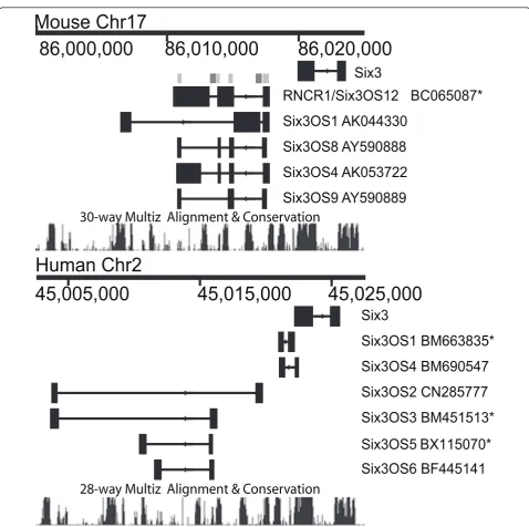
Genomic organization ofSix3andSix3OS
To determine which regions ofSix3OSto target for func-tional analysis, we first examined the genomic organiza-tion and evoluorganiza-tionary conservaorganiza-tion of this lncOST using publicly available cDNA and genomic sequence. As pre-viously reported, we found thatSix3OSis alternatively spliced in both mouse and human [23], althoughSix3OS -like transcripts were not identified in any other vertebrate species examined (data not shown). To determine which sequences to target for overexpression and knockdown, we aligned these sequences and observed that the puta-tive full-length mouse cDNA BC065087 contains the exon sequences shared by all alternative splice forms of
Six3OS(Figure 1, light grey bars). Two of these exons are
adjacent to alternatively spliced exons of the human
Six3OSorthologue, which lie within intronic regions of
the mouse transcript (Figure 1, dark grey bars). This cDNA corresponds to the most abundant isoform in neo-natal retina as measured by serial analysis of gene expres-sion (SAGE) tag abundance [32], and also matches the 4.5-kb isoform ofSix3OS, previously reported to be the most abundant isoform expressed in embryonic brain [33]. We therefore selected this cDNA for analysis by overexpression.
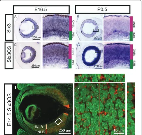
Cellular expression pattern ofSix3OS
To determine ifSix3andSix3OSare co-expressed in ret-inal progenitors, as had been previously suggested [23,32,33], we performed chromogenicin situ hybridiza-tion (ISH) on sechybridiza-tions of embryonic day (E)16.5 and postnatal day (P)0.5 retina (Figure 2A-H). We observed
that Six3and Six3OSare co-expressed in both retinal
fluorescent ISH (FISH) with a probe forSix3OSat E14.5, at which point prominent expression in progenitor cells of the outer neuroblastic layer is seen (Figure 2I). Inter-estingly, though mostSix3OSRNA is localized in the cytoplasm of retinal progenitors at this stage, some
Six3OSRNA is also associated with nuclear DNA (Figure
2I, J). This subcellular distribution has also been noted for the lncRNAEvf-2[14]. Using FISH in conjunction with immunostaining for the cytoplasmic ribosomal
protein S6, we observed that theSix3OStranscript is also found in the nucleus and cytoplasm when expressed in transfected HeLa cells (Additional file 1).
Overexpression and knockdown ofSix3andSix3OS
SinceSix3OSwas coexpressed withSix3in retinal pro-genitors, we hypothesized thatSix3OSmight regulate the expression and/or activity of Six3 in developing retina. To determine if this was indeed the case, we employed
RNCR1/Six3OS12 BC065087*
Six3OS1 AK044330
Six3OS4 AK053722
Six3OS8 AY590888
Six3OS9 AY590889
Six3
86,000,000 86,010,000
86,020,000
Mouse Chr17
30-way Multiz Alignment & Conservation
Six3
Six3OS1 BM663835*
Six3OS4 BM690547
Human Chr2
45,005,000 45,015,000 45,025,000
Six3OS2 CN285777
Six3OS3 BM451513*
Six3OS5 BX115070*
28-way Multiz Alignment & Conservation
Six3OS6 BF445141
in vivoelectroporation of neonatal (P0.5) mice to deter-mine whether overexpression and knockdown ofSix3OS phenocopied the effects of overexpression and knock-down ofSix3. For overexpression ofSix3OS, we used full-length cDNAs corresponding to BC065087 (Figure 1) cloned into the pCAG plasmid [34]. Upon transfection into HeLa cells, robustSix3OSexpression was confirmed by FISH (Additional file 1). To visualize electroporated cells, all constructs were co-electroporated with plasmids
expressing green fluorescent protein (GFP) from the same CAG promoter.
We observed that overexpression of 1 μg ofSix3OS resulted in no significant change in any major retinal cell type, and section immunohistochemistry revealed that electroporated cells showed grossly normal mor-phology (Figure 3B, C, white arrowheads). However, when dissociated cell preparations were examined, a significant decrease (P < 0.05) in the fraction of
J
I
INLB
ONLB
250
μ
m
10
μ
m
E14.5 Six3OS
GFP-positive cells expressing amacrine cell-specific mar-ker syntaxin was observed relative to cells electroporated with the GFP control vector at P21 (Figure 3A). Since
Six3OSoverexpression did not alter the fraction of
GFP-positive cells with amacrine-like morphology and lami-nar position, we conclude that Six3OSdoes not inhibit
amacrine cell specification. The reduction in syntaxin expression seen following CAG-Six3OS electropora-tion thus reflects a quantitative reducelectropora-tion in syntaxin levels or possibly reflects reduced adhesion ofSix3OS -expressing amacrine cells following dissociation for immunocytochemistry.
Figure 3Overexpression ofSix3OSin developing retina inhibits changes in cell fate and photoreceptor morphology observed followingSix3overexpression.(A)Electroporation of CAG-Six3led to an increase in the fraction of GFP-positive cells expressing the amacrine cell marker syntaxin and a decrease in cells expressing the rod bipolar marker PKCa, CAG-Six3OSled to a decrease in syntaxin-positive cells, and co-expression led to a cell composition that was indistinguishable from CAG-GFP controls. *P< 0.05.(B-E)Section immunohistochemistry of retinas electroporated with CAG-GFP, CAG-Six3OS, CAG-Six3or both CAG-Six3OSand CAG-Six3. White dashed lines define the outer third of the outer nuclear layer (OONL). Syntaxin (red) is co-immunostained with GFP (green). (C) No obvious difference is observed in either amacrine cell number or morphology (white arrowheads) in retinas electroporated with CAG-Six3OSrelative to CAG-GFP controls or in the fraction of the cells in the OONL. (D) An increase in amacrine cell number and in the number of cells in the OONL and a decrease in outer segment length is observed in the case ofSix3. (E) An increase in the number of cells in the OONL is observed, but no difference in amacrine cell number or outer segment length is observed from controls following co-electroporation of CAG-Six3and
CAG-Six3OS. GCL, ganglion cell layer; INL, inner nuclear layer; ONL, outer nuclear layer; OS, outer segment..(F)Laminar position of cells within
the OONL. Electroporation of CAG-Six3leads to a shift of rod photoreceptor cell bodies to the OONL, and this effect is not affected by co-electroporation of CAG-Six3OS(white dashed lines in B-E).(G)Rod photoreceptors electroporated with CAG-Six3show substantially
We next tested the effects of overexpression of
pCAG-Six3usingin vivoelectroporation. Upon electroporation, expression of Six3 was confirmed by section immuno-histochemistry. Cell fate specification in the postnatal retina is highly sensitive to Six3 dosage, and both gain and loss of function of Six3 have been reported to result in similar defects in bipolar cell and photoreceptor development [30]. We observed that electroporation of 1 μg of pCAG-Six3led to a reduction in the number of rod bipolar cells, and detected an increase in the frac-tion of GFP-positive amacrine cells by P21 (P < 0.05; Figure 3A, D). The length of the outer segments of GFP-positive rod photoreceptors was also decreased (P < 0.05; Figure 3D, G). Finally, we observed that the cell bodies of the Six3 electroporated photoreceptor cells were primarily located in the outer third of the outer nuclear layer (P < 0.05; Figure 3D, white dashed line, and 3F), in contrast to photoreceptors electropo-rated with control vector, which were distributed throughout the outer nuclear layer.
We then analyzed the effects of loss of function of
Six3OS and Six3. To this end, we first tested shRNAs
for their ability to reduce expression of endogenousSix3
andSix3OS expression byin vivoelectroporation of P0.5
retina. Reduction in expression of the target gene was determined by analyzing expression of either Six3 pro-tein or Six3OS RNA dissociated GFP-positive cells. For
both Six3 and Six3OS, individual shRNA constructs
were identified that resulted in a substantial reduction in the average fluorescence intensity in either Six3 pro-tein orSix3OS RNA in GFP-positive cells (Additional file 2).
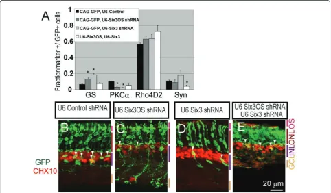
A significant decrease in the fraction of protein kinase C
a(PKCa) positive rod bipolar cells was observed at P21 following electroporation of either 1μg ofSix3shRNA construct or 1μg ofSix3OSshRNA (P< 0.05; Figure 4A), reminiscent of effects of retroviral overexpression of a dominant-negative mutant form ofSix3[30]. The fraction of glutamine synthetase-positive Muller glia was also increased (P< 0.05; Figure 4A). Section immunohisto-chemistry confirmed an increase in glutamine synthetase-positive cells with Muller glia-like morphology, at the expense of cells expressing the bipolar cell marker CHX10 and demonstrating bipolar-like morphology (Figure 4B-D,
Figure 4shRNA-mediated knockdown ofSix3andSix3OSin the developing retina alters bipolar cell and Muller glial development.(A)
Six3knockdown andSix3OSknockdown both lead to an increase in Muller glia and a decrease in rod bipolar cells as measured by
white arrowheads). To further confirm that knockdown experiments did indeed result in a selective loss of func-tion ofSix3OS, we also inhibitedSix3OSfunction by over-expression ofSix3OS-IRES-GFP. Fusion of IRES-GFP to lncRNAs results in a mislocalization of lncRNAs to the ribosome, and can produce dominant negative phenotypes when overexpressed [22]. When this is performed, we observed a reduction in rod bipolar markers and an increase in Muller markers, phenocopying the effects of
Six3OSknockdown (data not shown).
Simultaneous overexpression and knockdown ofSix3and
Six3OSdemonstrates non-additive effects on retinal differentiation
Having observed that loss of function ofSix3andSix3OS resulted in similar phenotypes in developing retina, we next investigated whether these two genes acted coopera-tively or independently to regulate retinal differentiation. We first usedin vivoelectroporation to overexpress 1μg of both theSix3andSix3OSconstructs simultaneously in P0.5 retina. We observed that co-expression ofSix3and
Six3OSresulted in a normal cell fate phenotype (Figure
3A). The length of GFP-positive rod photoreceptor outer segments was indistinguishable from controls (Figure 3B, E, G). However, the cell bodies of the electroporated photoreceptor cells were primarily located in the outer third of the outer nuclear layer (P< 0.05; Figure 3B, E, white dashed line, and 3F). We thus concluded that over-expression ofSix3OSwas largely able to reverse the cellu-lar phenotypes observed following overexpression ofSix3. As a follow up to these experiments, we simultaneously reduced expression ofSix3andSix3OSby co-electropor-ating 1 μg of theSix3 shRNA construct and 1 μg of
Six3OS shRNA construct. We observed that
simulta-neous knockdown of bothSix3andSix3OSresulted in a normal number of bipolar cells and Muller glia. However, we observed a significant decrease in the fraction of GFP-positive amacrine cells, which was not observed following electroporation of either the Six3 orSix3OS shRNA alone (P< 0.05; Figure 4A, E). These data show a non-additive effect of simultaneous loss of function of both
Six3andSix3OS, and show that these genes functionally
interact to regulate retinal differentiation.
Lastly, we directly investigated the functional relation-ship betweenSix3andSix3OS in vivoby simultaneously overexpressingSix3and knocking downSix3OS, andvice
versa. When we co-electroporated 1μg of CAG-Six3with
1μg ofSix3OSshRNA, we found thatSix3overexpression
fully rescued theSix3OSknockdown phenotype. Further-more, loss of function of Six3OS fully eliminated the effects ofSix3overexpression. No differences in the position of GFP-positive cells were observed when com-pared to controls (Figure 5A). Both the morphology and distribution of the cells within the retina were normal
(Figure 5C, D; Additional file 3A), and the rod photorecep-tor outer segment length was normal (Additional file 3B). We conclude that Six3OSis required for many of the effects on retinal differentiation that result from Six3 overexpression.
In contrast, when 1 μg of CAG-Six3OSwas co-elec-troporated with 1 μg of Six3 shRNA, we observed an increase in glutamine synthetase-positive cells with Mul-ler glia-like morphology (P < 0.05; Figure 5B) and a decrease in PKCa-positive and CHX10-positive bipolar cells (P< 0.05; Figure 5B, C, E) when compared to con-trols. Additionally, a decrease in cells expressing the amacrine cell-specific marker syntaxin was observed (P < 0.05; Figure 5B). These retinas, however, do not show any change in the number of GFP-positive cells with amacrine cell-like morphology. We therefore con-clude that, as is the case whenSix3OS alone was overex-pressed, Six3OS overexpression in conjunction with knockdown of Six3reduces syntaxin expression without otherwise affecting amacrine cell differentiation (Figure 5B, E). The phenotype seen here is the sum of the phe-notypes seen following Six3OSoverexpression and Six3 knockdown, and stands in contrast to the non-additive phenotype seen whenSix3OS andSix3are both simulta-neously knocked down (Additional file 4). We thus con-clude that the phenotype observed following Six3OS overexpression is not affected by Six3 loss of function, and that Six3OSmay also regulate syntaxin expression through aSix3-independent mechanism.
Six3OSdoes not directly regulate Six3 protein levels
(Additional file 6). Although 14 additional human and mouse cell lines were tested, noSix3OS-dependent effects on Six3-mediated autorepression could be detected (data not shown). We conclude that transcription of Six3 mRNA is not regulated bySix3OS. InsteadSix3OSRNA likely modulates the ability of Six3 protein to activate or repress expression of its target genes in developing retinal cells.
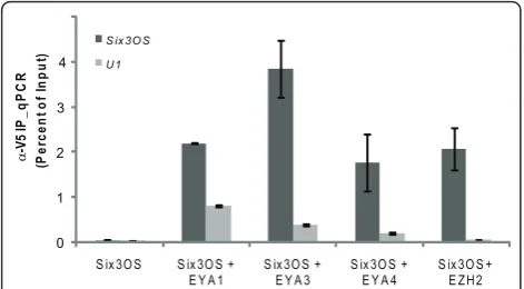
Ezh2 and Eya1/3/4 directly bindSix3OS
To identify proteins that directly bind Six3OS, Cy5-labeledSix3OSRNA was used to probe human protein microarrays [37]. MouseSix3OSand three human splice variants ofSix3OSwere transcribedin vitroand labeled with Cy5, and antisense cRNAs for these same tran-scripts were hybridized in parallel as negative controls. A total of five proteins that specifically and selectively interact with both the mouse and human forms of
Six3OSwere thus identified. We found that both human
and mouse Six3OS selectively interact with Eya1, encoded by a homologue of theeyes absentgene of
Dro-sophila. In addition, Six3OS directly binds to the
chromatin remodeling enzyme subunits Smarce1 and Ezh2, as well as Eno1 and Ppp5c (Additional file 7). Members of the Eya protein family have been previously shown to interact with Six family proteins [38], but Eya1 is not expressed in the mouse neuroretina [32]. There-fore, we also performed RNA immunoprecipitation (RIP) experiments on Eya3 and Eya4, which are both robustly expressed in embryonic retina.
To test whether Ezh2 and Eya family members inter-act with mouseSix3OS in vivo, co-immunoprecipitation experiments were performed in HEK 293T cells overex-pressing V5-tagged Eya1, Eya3, Eya4 and Ezh2. Expres-sion and immunoprecipitation of V5-tagged protein was verified by immunoblot (Additional file 8). We con-firmed that mouse Six3OS RNA selectively interacts with each of these proteins using RIP (Figure 6), and conclude that these proteins interact with Six3OS in transfected HEK 293T cells. To confirm that Six3OS interacts with Eya family members and Ezh2 specifically, we examined whether Six3OS interacts directly with Six3 protein by RIP and found thatSix3OSand Six3 do not interact in transfected HEK 293T cells (data not
Figure 5Six3overexpression rescues theSix3OSknockdown phenotype whereasSix3OSoverexpression is not sufficient to rescue the loss ofSix3.(A)Six3OSknockdown combined withSix3overexpression led to a cell type distribution indistinguishable from cells electroporated with control constructs when tested by immunostaining of dissociated electroporated retinal cells.(B)Six3knockdown combined withSix3OS
shown). This suggests thatSix3OSregulates Six3 func-tion by facilitating interacfunc-tion between Eya proteins and chromatin-modifying enzyme complexes.
Discussion
Six3OSand Six3 modulate retinal cell specification and differentiation
Our findings demonstrate that both gain and loss of function ofSix3OSandSix3affect retinal differentiation (Additional file 4). Knockdown ofSix3OSresulted in a decrease in the fraction of bipolar cells and an increase in Muller glia in electroporated cells. An identical pheno-type was observed following knockdown ofSix3(Figure 4). Knockdown of eitherSix3OSorSix3phenocopies the effect of retroviral overexpression of a dominant-negative form of Six3 in neonatal retina, where a substantial decrease in bipolar cells was also seen [30].
In contrast, overexpression of either Six3or Six3OS alone produced very different phenotypes.Six3OS over-expression resulted in a reduction in syntaxin staining in electroporated amacrine cells, but did not alter the morphology or laminar position of electroporated cells. On the other hand, Six3overexpression, like Six3loss of function, resulted in a dramatic reduction in the number of bipolar cells (Figure 3A), and also resulted in decreased rod photoreceptor outer segment length at P21 (Figure 3D, G). These effects were previously reported following retroviral-mediated overexpression of
Six3 [30]. Interestingly, we also observed phenotypes that were not reported in this previous study, such as an increase in amacrine cells. A number of technical differ-ences between these two experiments may account for these differences, most notably the fact that electropora-tion-mediated overexpression typically results in much higher levels of construct expression in developing retina than does retroviral transduction [34].
Six3OSregulates Six3 activity in developing retina, but does not regulateSix3expression levels
If Six3 and Six3OSact independently to control retinal cell specification, we would expect to observe purely additive phenotypes when their activities are both altered. Strikingly, when both Six3OS and Six3 were overexpressed simultaneously, we no longer observed the cellular phenotypes that resulted from overexpres-sion of Six3 alone. Simultaneous knockdown of both
Six3OSandSix3 also showed non-additive effects. The
loss of bipolar cells and increase in Muller glia, which was seen following knockdown of each gene individually, were not observed when expression of both Six3and
Six3OS was reduced. Instead, a decrease in amacrine
cells was observed, which was a phenotype not observed when each individual gene was knocked down. These non-additive phenotypes demonstrate an epistatic rela-tionship between Six3 andSix3OS, and imply that they functionally interact to regulate retinal differentiation.
Having demonstrated that Six3 andSix3OSinteract to regulate retinal differentiation, we next investigated whether defects in Six3 could be rescued by overexpres-sion ofSix3OS, andvice versa. WhenSix3is overexpressed
butSix3OSis knocked down, none of the phenotypes
nor-mally seen following eitherSix3overexpression alone or
Six3OSknockdown alone were observed (Figure 5A, C-D).
These data indicate that loss of function ofSix3OS sup-presses the effects of both gain and loss of function of
Six3. However, simultaneous overexpression ofSix3OS and knockdown ofSix3resulted in a purely additive phe-notype that was the sum of the effects ofSix3knockdown
andSix3OSoverexpression. These findings suggest that
Six3OSacts to regulate Six3 activity, since overexpression
ofSix3can rescue the effects ofSix3OSknockdown, but
notvice versa. AsSix3OSoverexpression leads to a
reduc-tion in syntaxin expression even followingSix3 knock-down, this implies thatSix3OSmay also regulate retinal differentiation through a Six3-independent mechanism, although this remains to be characterized further.
Since both promoter- and enhancer-associated ncRNAs have been previously thought to act incis to selectively regulate expression of nearby protein coding genes [36,39], our finding thatSix3OSdid not regulate Six3 expression came as something of a surprise. Our finding thatSix3OSacts intransto regulate retinal differ-entiation was supported by several lines of evidence. First, by the finding that the activity of Six3 expressed from the CAG promoter can be modulated by overex-pression or knockdown ofSix3OS(Figure 5). Second, we have directly demonstrated that neither overexpression nor knockdown ofSix3OShas any detectable effect on
Six3expression in retina, nor doesSix3OShave an effect on Six3-dependent autorepression when measured by
0 1 2
S ix3O S S ix3O S +
E Y A 1 S ix3O S + E Y A 3 S ix3O S + E Y A 4 S ix3O S + E ZH2
S ix3O S U1 3 4 (P er ce nt of Input ) α -V5 I P _q P C R
luciferase analysis in transfected tissue culture cells. These data imply thatSix3OSand perhaps other lncOSTs do not regulate transcription of their associated protein coding genes, but instead act intrans to regulate the activity directly via protein-RNA interactions. This mechanism may not hold true for all promoter-associated lncRNAs, however, and each will have to be characterized independently to determine its mechanism of action.
Six3OSselectively interacts with Eya family members and Ezh2
While these data demonstrate a functional relationship between Six3OSand Six3, they still leave the precise mechanism by whichSix3OSregulates Six3 activity unre-solved. Our finding that both human and mouse isoforms
ofSix3OSinteract with multiple different members of the
eyes absentprotein family demonstrates how this might
occurin vivo. Theeyes absent(eya) gene and its mamma-lian homologues encode protein tyrosine phosphatases that function as transcriptional coregulators.Eyabinds directly tosine oculis, and acts in conjunction withsine
oculisin controlling eye field specification inDrosophila
[28,29]. We also observe thatSix3OSdirectly binds the PRC2 subunit Ezh2, and also possibly the BAF57 subunit Smarce1. Additional confirmation thatSix3OSinteracts with Ezh2in vivocomes from a recent study of poly-comb-associated RNAs, which found thatSix3OSwas co-precipitated with PRC2 in embryonic stem cells [40]. We propose thatSix3OScan modulate the expression of Six3 target genes without necessarily regulating expression of
Six3itself by acting as a transcriptional scaffold. When Six3 directs the Six3-Eya complex to bind to specific genomic target sequences,Six3OSmay thus act as a tran-scriptional scaffold, recruiting histone modifying enzyme complexes to regulate expression of Six3 target genes. Althoughtrans-acting lncRNAs have previously been found to regulate gene expression through recruitment of the Ezh2-containing PRC2 histone methyltransferase complex, their action is restricted to a small subset of tar-get genes, and the mechanism by which this occurs is unknown [11]. Our findings point towards a plausible mechanism by which this could occur. However, in the absence of RNA-IP data that clearly demonstrate interac-tion ofSix3OSwith both Eya proteins and Six3 in devel-oping retina, other potential mechanisms of action of
Six3OSremain plausible.
Several unresolved questions remain, the most obvious being which Eya subtype is the most relevant target of
Six3OS in vivo. Eya1 is known to bind Six3 bothin vitro
and in transfected tissue culture cells [41], although the co-expression of Six3 and Eya1 is limited to the ciliary margin and lens [42]. Eya2, Eya3 and Eya4, however, are all co-expressed with Six3 andSix3OSin both the devel-oping retina and in other forebrain regions [42]. Eya4
and Six3 have been demonstrated to interact in a study of holoprosencephaly, indicating that Eya4 may be the functional bridge between Six3 andSix3OSin the devel-oping ventral forebrain [43]. Our findings indicate that
Six3OSmay also regulate retinal differentiation by
Six3-independent mechanisms. Since Eya family members can interact with other classes of transcription factors, including Hox and Tlx family members [44], this sug-gests thatSix3OSmight also modulate the expression of genes regulated by these transcription factors.
Many homeodomain transcription factors that are essential for central nervous system development possess an associated lncOST, including Pax6, Rax, Vax2 and Six6 [23,45]. Each of these proteins exhibits complex and dynamic expression during development, and their expres-sion patterns often diverge considerably from those of their associated lncOST. Although their target sequences are present in all cells, the genes that are directly regulated by each protein can vary considerably, both at different developmental stages and in different cell types. Models that explain how this context-dependent regulation of transcription factor activity occurs have typically empha-sized combinatorial regulation by other proteins, or cell-and stage-specific epigenetic modifications. Our finding that lncOSTs can modulate the activity of their associated transcription factors lends an additional layer of complex-ity to these models, and suggests that this diverse class of molecules may play a critical function in generation of cell subtype diversity within the developing central nervous system.
Materials and methods Animals
Pregnant CD-1 mice were purchased from Charles River Breeding Laboratories, Wilmington, MA, USA. Animal experiments were approved by the Johns Hopkins Uni-versity Institutional Animal Care and Use Committee.
In vivoelectroporation
In vivoelectroporation of mouse retina was performed as
described [34]. Retinas were electroporated at P0.5. Dis-sociated cells and section immunohistochemistry data shown here were performed at P21. Error bars represent ± standard error of the mean for at least three indepen-dent electroporated retinas. A two-tailed Stuindepen-dentst-test was performed to determine theP-value. Control and experimental constructs were co-electroporated with 0.2
μg of pCAG-GFP to readily visualize electroporated cells by GFP [34]. For overexpression experiments, 1μg of pCAG-GFP was injected with either 1μg of pCAG-Six3 or 1μg of CAG-Six3OS. For simultaneous overexpression
ofSix3andSix3OS, retinas were electroporated with 1μg
of pCAG-Six3and 1μg of CAG-Six3OS. For knockdown
either 1μg of U6-Six3or 1μg of U6-Six3OS. For simulta-neous knockdown, 1 μg of U6-Six3and 1 μg of
U6-Six3OSwere injected. For overexpression ofSix3in
com-bination with knockdown ofSix3OS, retinas were electro-porated with 1μg of pCAG-Six3and 1μg of U6-Six3OS were injected. Finally, for overexpression of Six3OS in combination with knockdown ofSix3, 1μg of
pCAG-Six3OS and 1 μg of U6-Six3 were injected and
electroporated.
In situhybridization
For cryosections, untimed E14.5 and E16.5 embryos and P0.5 neonates were dissected, fixed overnight in 4% par-aformaldehyde in PBS at 4°C and cryoprotected overnight in 30% sucrose/PBS at 4°C before being embedded in OCT compound (Sakura, Torrance, CA, USA) on dry ice. Cryosections (20μm) were cut on a cryostat. Section ISH methodology was as previously described [32] with the exception that probe BC065087 was used and a Tyramide Signal Amplification system (TSA Plus, Perkin Elmer, NEL 744, Waltham, MA, USA) combined with an antidi-goxigenin-HRP antibody (1:1, 000; Roche, Indianapolis, IN, USA). for fluorescent ISH. Fluorescent ISH sections were counterstained with DAPI. Fluorescent ISH samples were photographed on a Zeiss Meta 510 confocal micro-scope. Chromogenic images were visualized on a Zeiss Axioskop2 microscope.
Immunohistochemistry
For cryosections, electroporated eyes were harvested 21 days after electroporation, fixed for 1 hour in 4% parafor-maldehyde in PBS at 4°C and cryoprotected overnight in 30% sucrose/PBS at 4°C before being embedded in OCT compound (Sakura) on dry ice. Cryosections (20μm) were cut on a cryostat. Retinal cryosections were immunos-tained as described except that sections were post fixed in 4% paraformaldehyde for 5 minutes prior to blocking [21]. Samples were photographed on a Zeiss Meta 510 confocal microscope.
For cell marker analysis, dissociation and immunos-taining were performed as described [34] except that reti-nas were harvested at P21. Samples were visualized and quantified on a Zeiss Axioskop2 microscope. To quantify the effects ofSix3knockdown, retinas were electropo-rated withSix3shRNA, harvested at P4.5, dissociated, and immunostained as described above with anti-Six3 and detection with Alexa568 goat anti-guinea pig IgG and rabbit anti-GFP and detection with Alexa488 goat anti-rabbit. For Six3 quantification,Six3OS overexpres-sion andSixOSshRNA electroporated retinas were har-vested at P5.5, dissociated and immunostained as described above with anti-Six3 and detection with Alexa568 goat anti-guinea pig IgG and rabbit anti-GFP and detection with Alexa488 goat anti-rabbit. Samples
were visualized on a Zeiss Axioskop2 microscope and signal intensity was quantified with Velocity 4.0 (Perkin Elmer) software by calculating the average signal inten-sity per cell and normalized to cell size.
In situhybridization and immunocytochemistry
ForSix3OSknockdown quantification, retinas were
har-vested at P4.5 and dissociated as described above. FISH was performed as described using TSA Plus Cyanine3 kit (1:125; Perkin Elmer, NEL 744) followed by staining as described (with rabbit anti-GFP and detection with Alexa488 goat anti rabbit). Samples were visualized and quantified as above. For all dissociations, nuclear DNA was visualized with DAPI counterstaining. Cell counts were analyzed using the two-tailed Student’st-test. A minimum of three retinas were counted for each con-struct examined using dissociated immunocytochemis-try, with 100 to 300 GFP-positive cells per retina counted for each marker tested.
HeLa cell FISH followed by immunocytochemistry was performed as previously described [22] except that 20 cells were counted.
Luciferase reporter assays
NIH 3T3 cells were grown in DMEM supplemented with 10% FCS. Cells were transfected with Fugene6 (Roche) per the manufacturer’s instructions. Cells were transfected with 500 ng of luciferase reporter construct and 50 ng of the expression plasmids forSix3andSix3OS. Cells were harvested 2 days post-transfection. Luciferase was mea-sured per manufacturer’s instructions with the Dual-Luci-ferase Reporter System (Promega, E1910, Madison, WI, USA). pTK-Renilla (50 ng) was co-transfected to control for transfection efficiency.
Synthesis of RNA probes for protein microarray
BC065087 (5μg) was linearized with DraI, and 5 μg of BM663835, BX115070, and BM451513 were linearized with NotI. Linearized DNA was then phenol-chloroform extracted, ethanol precipitated and resuspended in 10μl water. Probes were in vitrotranscribed with T7 poly-merase per manufacturer’s instructions using the Ribop-robe Combination system kit (Promega, P1405), spiking the reaction with 1 μl of 5 mM Cy5 labeled CTP (GE Healthcare, 25-8010-87, Piscataway, NJ, USA). The RNA probe was ethanol precipitated with LiCl and resus-pended in 1 mM EDTA.
Protein microarray analysis
BSA for 1 hour at room temperature. RNA was hybri-dized at 250 nM concentration in binding buffer (PBS supplemented with 2 mM MgCl2, 2 mg ml-1 BSA) at
room temperature for 1 hour. The slides were washed four times in TBST, dried and scanned by a GenePix 400B scanner. Data were analyzed with GenePix Pro 6.10 as previously described [46].
RNA immunoprecipitation experiments
HEK 293T cells were grown in DMEM supplemented with 10% FCS. Cells were transfected with Fugene6 (Roche) per the manufacturer’s instructions. Ten million cells were transfected 24 hours post-plating with 5μg of pCAGIG-V5, pCAGIG-Six3-V5, pCAGIG Eya1-V5, pCAGIG Eya3-V5, pCAGIG Eya4-V5 or pCAGIG Ezh2-V5, and pCAG-Six3OS. Cells were harvested 2 days post-transfection, lysed and precipitated essentially as previously described [11], except that 5μg V5 anti-body was used (1:5, 000; Invitrogen, R96025, Carlsbad, CA, USA) with 50 μl protein A-agarose (Invitrogen, 15918-014) in each immunoprecipitation reaction. For each sample, 10% of total volume was set aside after lysis for RNA extraction and 5% set aside for immuno-blot analysis. After precipitation, 80% of the beads were resuspended in Trizol and 20% were resuspended in Laemmle buffer. RNA was Trizol extracted and resus-pended in 50 μl of nuclease free water. RNA was then DNAse treated with Turbo DNA-free (Ambion, AM1907, Carlsbad, CA, USA) per manufacturer’s instructions. RNA was quantified by quantitative RT-PCR using Brilliant II SYBR Green QRT_RT-PCR Master Mix (Agilent, 600825, Santa Clara, CA, USA) on a Roche LightCycler480. No-RT controls were simulta-neously performed to demonstrate that signal was not from DNA contamination.
Expression of V5-tagged protein was confirmed by western analysis using anti-V5 antibody (1:10, 000; Invi-trogen, R96025) dilution in 5% milk in PBST, and detected with horse radish peroxidase goat anti-mouse IgG (1:10, 000; Santa Cruz, sc-2031, Santa Cruz, CA, USA) and ECL Western Blotting Detection System (GE Healthcare, RPN2132 Piscataway, NJ, USA) per the man-ufacturers’instructions.
Full details of all plasmids and antibodies used in this study are included in Additional File 9.
Additional material
Additional file 1:Six3OSis localized equally in the nucleus and cytoplasm. HeLa cells were transfected withSix3OSconstructs and RNA location was analyzed by FISH followed by immunohistochemistry against the cytoplasmic S6 ribosomal protein. CytoplasmicSix3OSRNA was identified by localization with S6 protein. The relative proportion of nuclearSix3OS, defined as FISH signal that did not colocalize with S6 protein, is indicated. N = 20 cells.
Additional file 2: Confirmation of shRNA-mediated knockdown of endogenousSix3andSix3OSin developing retina. (A-E) A construct encoding either control hairpin,Six3targeted hairpin orSix3OStargeted hairpin was electroporatedin vivo, into P0.5 retina and harvested at P4.5, and dissociated, and immunocytochemistry against Six3 and GFP, or FISH againstSix3OSwas performed followed by immunostaining for GFP. GFP-positive cells were counted to analyze the fluorescence intensity for each cell that expressed (A) Six3 or (D)Six3OS. At least 100 GFP-positive cells from three different electroporated retinas were counted for each construct tested. Error bars represent standard error for at least three independent retinas. (A)P< 0.05; (D)P< 0.05. (B-E) Examples of dissociated cells positive for GFP and either Six3 protein orSix3OSRNA.
Additional file 3:Six3OSknockdown rescues changes in retinal cell morphology and position observed following overexpression of Six3. (A, B)Six3OSknockdown combined withSix3overexpression rescues the effects observed on rod photoreceptor cell body position (A) and on photoreceptor outer segment length observed withSix3
overexpression (B).
Additional file 4: Summary of overexpression and knockdown data forSix3andSix3OS. These results demonstrate that co-expression of
Six3OSandSix3rescues the phenotypes observed withSix3
overexpression except that the photoreceptors are displaced in the outer third of the outer nuclear layer. Simultaneous knockdown ofSix3OSand
Six3results in a novel phenotype, fewer amacrine cells. Additionally,
Six3OSoverexpression rescues the phenotype of knockdown ofSix3.
However, expression ofSix3combined with knockdown ofSix3OSresults in an additive phenotype.
Additional file 5: Cellular levels of Six3 protein are not affected by overexpression or knockdown ofSix3OS.(A)Overexpression ofSix3OS
at P0.5 does not affect Six3 protein levels when measured at P5.5.(B)
shRNA-mediated knockdown at P0.5 ofSix3OSdoes not affect Six3 protein levels when measured at P5.5
Additional file 6: Autorepression of Six3 is not affected bySix3OS. Transfection of CMV-Six3together with the Six3-pro luciferase reporter into NIH 3T3 led to > 70% reduction in luciferase expression. Co-transfection of CMV-Six3and CAG-Six3OSwith theSix3-pro luciferase reporter was not significantly different than CMV-Six3alone.
Additional file 7: List of proteins that interact with the mouse and human forms ofSix3OSfrom the transcription factor/RNA binding protein microarray.
Additional file 8: V5 expression constructs are expressed in HEK 293T cells.aV5 western analysis demonstrating that Eya1-V5, Eya3-V5, Eya4-V5 and Ezh2-V5 are expressed.
Additional file 9: Supporting Information. Includes plasmid information, shRNA sequences and antibodies used.
Abbreviations
BSA: bovine serum albumin; DMEM: Dulbecco’s modified Eagle’s medium; E: embryonic day; FCS: fetal calf serum; FISH: fluorescentin situhybridization; GFP: green fluorescent protein; ISH:in situhybridization; lncOST: long non-coding opposite-strand transcript; lncRNA: long non-non-coding RNA; ncRNA: non-coding RNA; P: postnatal day; PBS: phosphate-buffered saline; PKC: protein kinase C; PRC2: Polycomb-repressive complex 2; RIP: RNA immunoprecipitation; shRNA: short hairpin RNA; TBST: Tris- buffered saline with Tween-20.
Acknowledgements
The authors thank Shiming Chen, Tomomi Shimogori, Jeremy Nathans, Wendy Yap and members of the Blackshaw lab for comments on the paper. This work was supported by a grant from the Whitehall Foundation to SB and a VNTP training grant to EP. SB is a WM Keck Foundation Distinguished Young Scholar in Medical Research.
Author details
1
University School of Medicine, 733 N. Broadway Avenue, Baltimore, MD 21287, USA.2Howard Hughes Medical Institute and Program in Epithelial
Biology, Department of Dermatology, Stanford University School of Medicine, Stanford, CA 94305, USA.3Department of Pharmacology and
Center for High-Throughput Biology, Johns Hopkins University School of Medicine, 733 N. Broadway Avenue, Baltimore, MD 21287, USA.
Authors’contributions
NAR designed and performed experiments, analyzed the data and wrote the manuscript. EMP performed experiments. HZ analyzed the data. SB designed experiments, analyzed the data and wrote the manuscript. This manuscript has been seen and approved by all authors.
Competing interests
The authors declare that they have no competing interests. Received: 28 May 2011 Accepted: 21 September 2011 Published: 21 September 2011
References
1. ENCODE Project Consortium, Birney E, Stamatoyannopoulos JA, Dutta A, Guigó R, Gingeras TR, Margulies EH, Weng Z, Snyder M, Dermitzakis ET, Thurman RE, Kuehn MS, Taylor CM, Neph S, Koch CM, Asthana S, Malhotra A, Adzhubei I, Greenbaum JA, Andrews RM, Flicek P, Boyle PJ, Cao H, Carter NP, Clelland GK, Davis S, Day N, Dhami P, Dillon SC, Dorschner MO,et al:Identification and analysis of functional elements in 1% of the human genome by the ENCODE pilot project.Nature2007, 447:799-816.
2. Mattick JS, Makunin IV:Non-coding RNA.Hum Mol Genet2006,15(Spec No 1):R17-29.
3. Prasanth KV, Spector DL:Eukaryotic regulatory RNAs: an answer to the
‘genome complexity’conundrum.Genes Dev2007,21:11-42.
4. Guttman M, Amit I, Garber M, French C, Lin MF, Feldser D, Huarte M, Zuk O, Carey BW, Cassady JP, Cabili MN, Jaenisch R, Mikkelsen TS, Jacks T, Hacohen N, Bernstein BE, Kellis M, Regev A, Rinn JL, Lander ES:Chromatin signature reveals over a thousand highly conserved large non-coding RNAs in mammals.Nature2009,458:223-227.
5. Tochitani S, Hayashizaki Y:Nkx2.2 antisense RNA overexpression enhanced oligodendrocytic differentiation.Biochem Biophys Res Commun
2008,372:691-696.
6. Schwartz JC, Younger ST, Nguyen NB, Hardy DB, Monia BP, Corey DR, Janowski BA:Antisense transcripts are targets for activating small RNAs.
Nat Struct Mol Biol2008,15:842-848.
7. Yu W, Gius D, Onyango P, Muldoon-Jacobs K, Karp J, Feinberg AP, Cui H: Epigenetic silencing of tumour suppressor gene p15 by its antisense
RNA.Nature2008,451:202-206.
8. Plath K, Mlynarczyk-Evans S, Nusinow DA, Panning B:Xist RNA and the mechanism of X chromosome inactivation.Annu Rev Genet2002, 36:233-278.
9. Pandey RR, Mondal T, Mohammad F, Enroth S, Redrup L, Komorowski J, Nagano T, Mancini-Dinardo D, Kanduri C:Kcnq1ot1 antisense noncoding RNA mediates lineage-specific transcriptional silencing through chromatin-level regulation.Mol Cell2008,32:232-246. 10. Rinn JL, Kertesz M, Wang JK, Squazzo SL, Xu X, Brugmann SA,
Goodnough LH, Helms JA, Farnham PJ, Segal E, Chang HY:Functional demarcation of active and silent chromatin domains in human HOX loci by noncoding RNAs.Cell2007,129:1311-1323.
11. Tsai MC, Manor O, Wan Y, Mosammaparast N, Wang JK, Lan F, Shi Y, Segal E, Chang HY:Long noncoding RNA as modular scaffold of histone modification complexes.Science2010,329:689-693.
12. Zhao J, Sun BK, Erwin JA, Song JJ, Lee JT:Polycomb proteins targeted by a short repeat RNA to the mouse X chromosome.Science2008, 322:750-756.
13. Lanz RB, McKenna NJ, Onate SA, Albrecht U, Wong J, Tsai SY, Tsai MJ, O’Malley BW:A steroid receptor coactivator, SRA, functions as an RNA and is present in an SRC-1 complex.Cell1999,97:17-27.
14. Feng J, Bi C, Clark BS, Mady R, Shah P, Kohtz JD:The Evf-2 noncoding RNA is transcribed from the Dlx-5/6 ultraconserved region and functions as a Dlx-2 transcriptional coactivator.Genes Dev2006,20:1470-1484. 15. Bond AM, Vangompel MJ, Sametsky EA, Clark MF, Savage JC, Disterhoft JF,
Kohtz JD:Balanced gene regulation by an embryonic brain ncRNA is
critical for adult hippocampal GABA circuitry.Nat Neurosci2009, 12:1020-1027.
16. Cao X, Yeo G, Muotri AR, Kuwabara T, Gage FH:Noncoding RNAs in the mammalian central nervous system.Annu Rev Neurosci2006,29:77-103. 17. Dinger ME, Amaral PP, Mercer TR, Pang KC, Bruce SJ, Gardiner BB,
Askarian-Amiri ME, Ru K, Soldà G, Simons C, Sunkin SM, Crowe ML, Grimmond SM, Perkins AC, Mattick JS:Long noncoding RNAs in mouse embryonic stem cell pluripotency and differentiation.Genome Res2008,18:1433-1445. 18. Amaral PP, Dinger ME, Mercer TR, Mattick JS:The eukaryotic genome as
an RNA machine.Science2008,319:1787-1789.
19. Chodroff RA, Goodstadt L, Sirey TM, Oliver PL, Davies KE, Green ED, Molnár Z, Ponting CP:Long noncoding RNA genes: conservation of sequence and brain expression among diverse amniotes.Genome Biol
2010,11:R72.
20. Qureshi IA, Mattick JS, Mehler MF:Long non-coding RNAs in nervous system function and disease.Brain Res2010,1338:20-35.
21. Young TL, Matsuda T, Cepko CL:The noncoding RNA taurine upregulated gene 1 is required for differentiation of the murine retina.Curr Biol2005, 15:501-512.
22. Rapicavoli NA, Poth EM, Blackshaw S:The long noncoding RNA RNCR2 directs mouse retinal cell specification.BMC Dev Biol2010,10:49. 23. Alfano G, Vitiello C, Caccioppoli C, Caramico T, Carola A, Szego MJ,
McInnes RR, Auricchio A, Banfi S:Natural antisense transcripts associated with genes involved in eye development.Hum Mol Genet2005, 14:913-923.
24. Rapicavoli NA, Blackshaw S:New meaning in the message: noncoding RNAs and their role in retinal development.Dev Dyn2009,238:2103-2114. 25. Seila AC, Calabrese JM, Levine SS, Yeo GW, Rahl PB, Flynn RA, Young RA,
Sharp PA:Divergent transcription from active promoters.Science2008, 322:1849-1851.
26. Preker P, Nielsen J, Kammler S, Lykke-Andersen S, Christensen MS, Mapendano CK, Schierup MH, Jensen TH:RNA exosome depletion reveals transcription upstream of active human promoters.Science2008, 322:1851-1854.
27. Core LJ, Waterfall JJ, Lis JT:Nascent RNA sequencing reveals widespread pausing and divergent initiation at human promoters.Science2008, 322:1845-1848.
28. Bonini NM, Leiserson WM, Benzer S:The eyes absent gene: genetic control of cell survival and differentiation in the developingDrosophila eye.Cell1993,72:379-395.
29. Pignoni F, Hu B, Zavitz KH, Xiao J, Garrity PA, Zipursky SL:The eye-specification proteins So and Eya form a complex and regulate multiple steps inDrosophilaeye development.Cell1997,91:881-891.
30. Zhu CC, Dyer MA, Uchikawa M, Kondoh H, Lagutin OV, Oliver G: Six3-mediated auto repression and eye development requires its interaction with members of the Groucho-related family of co-repressors.
Development2002,129:2835-2849.
31. Lagutin OV, Zhu CC, Kobayashi D, Topczewski J, Shimamura K, Puelles L, Russell HR, McKinnon PJ, Solnica-Krezel L, Oliver G:Six3 repression of Wnt signaling in the anterior neuroectoderm is essential for vertebrate forebrain development.Genes Dev2003,17:368-379.
32. Blackshaw S, Harpavat S, Trimarchi J, Cai L, Huang H, Kuo WP, Weber G, Lee K, Fraioli RE, Cho SH, Yung R, Asch E, Ohno-Machado L, Wong WH, Cepko CL:Genomic analysis of mouse retinal development.PLoS Biol
2004,2:E247.
33. Geng X, Lavado A, Lagutin OV, Liu W, Oliver G:Expression of Six3 Opposite Strand (Six3OS) during mouse embryonic development.Gene
Expr Patterns2007,7:252-257.
34. Matsuda T, Cepko CL:Electroporation and RNA interference in the rodent retina in vivo and in vitro.Proc Natl Acad Sci USA2004,101:16-22. 35. Ørom UA, Derrien T, Beringer M, Gumireddy K, Gardini A, Bussotti G, Lai F,
Zytnicki M, Notredame C, Huang Q, Guigo R, Shiekhattar R:Long noncoding RNAs with enhancer-like function in human cells.Cell2010, 143:46-58.
36. Kim TK, Hemberg M, Gray JM, Costa AM, Bear DM, Wu J, Harmin DA, Laptewicz M, Barbara-Haley K, Kuersten S, Markenscoff-Papadimitriou E, Kuhl D, Bito H, Worley PF, Kreiman G, Greenberg ME:Widespread transcription at neuronal activity-regulated enhancers.Nature2010, 465:182-187.
human protein-DNA interactome reveals ERK2 as a transcriptional repressor of interferon signaling.Cell2009,139:610-622. 38. Jemc J, Rebay I:The eyes absent family of phosphotyrosine
phosphatases: properties and roles in developmental regulation of transcription.Annu Rev Biochem2007,76:513-538.
39. Ørom UA, Derrien T, Beringer M, Gumireddy K, Gardini A, Bussotti G, Lai F, Zytnicki M, Notredame C, Huang Q, Guigo R, Shiekhattar R:Long noncoding RNAs with enhancer-like function in human cells.Cell2010, 143:46-58.
40. Zhao J, Ohsumi TK, Kung JT, Ogawa Y, Grau DJ, Sarma K, Song JJ, Kingston RE, Borowsky M, Lee JT:Genome-wide identification of polycomb-associated RNAs by RIP-seq.Mol Cell2010,40:939-953. 41. Purcell P, Oliver G, Mardon G, Donner AL, Maas RL:Pax6-dependence of
Six3, Eya1 and Dach1 expression during lens and nasal placode induction.Gene Expr Patterns2005,6:110-118.
42. Xu PX, Woo I, Her H, Beier DR, Maas RL:Mouse Eya homologues of the
Drosophilaeyes absent gene require Pax6 for expression in lens and nasal placode.Development1997,124:219-231.
43. Abe Y, Oka A, Mizuguchi M, Igarashi T, Ishikawa S, Aburatani H,
Yokoyama S, Asahara H, Nagao K, Yamada M, Miyashita T:EYA4, deleted in a case with middle interhemispheric variant of holoprosencephaly, interacts with SIX3 both physically and functionally.Hum Mutat2009,30: E946-955.
44. Ravasi T, Suzuki H, Cannistraci CV, Katayama S, Bajic VB, Tan K, Akalin A, Schmeier S, Kanamori-Katayama M, Bertin N, Carninci P, Daub CO, Forrest AR, Gough J, Grimmond S, Han JH, Hashimoto T, Hide W, Hofmann O, Kamburov A, Kaur M, Kawaji H, Kubosaki A, Lassmann T, van Nimwegen E, MacPherson CR, Ogawa C, Radovanovic A, Schwartz A, Teasdale RD,et al:An atlas of combinatorial transcriptional regulation in mouse and man.Cell2010,140:744-752.
45. Mercer TR, Dinger ME, Sunkin SM, Mehler MF, Mattick JS:Specific expression of long noncoding RNAs in the mouse brain.Proc Natl Acad
Sci USA2008,105:716-721.
46. Zhu J, Gopinath K, Murali A, Yi G, Hayward SD, Zhu H, Kao C:RNA-binding proteins that inhibit RNA virus infection.Proc Natl Acad Sci USA2007, 104:3129-3134.
47. Siepel A, Bejerano G, Pedersen JS, Hinrichs AS, Hou M, Rosenbloom K, Clawson H, Spieth J, Hillier LW, Richards S, Weinstock GM, Wilson RK, Gibbs RA, Kent WJ, Miller W, Haussler D:Evolutionarily conserved elements in vertebrate, insect, worm, and yeast genomes.Genome Res
2005,15:1034-1050.
doi:10.1186/1749-8104-6-32
Cite this article as:Rapicavoliet al.:The long noncoding RNASix3OS
acts intransto regulate retinal development by modulating Six3
activity.Neural Development20116:32.
Submit your next manuscript to BioMed Central and take full advantage of:
• Convenient online submission
• Thorough peer review
• No space constraints or color figure charges
• Immediate publication on acceptance
• Inclusion in PubMed, CAS, Scopus and Google Scholar
• Research which is freely available for redistribution