R E V I E W
Open Access
Ingested engineered nanomaterials: state
of science in nanotoxicity testing and future
research needs
Ikjot Singh Sohal
1*, Kevin S. O
’
Fallon
2, Peter Gaines
3, Philip Demokritou
4and Dhimiter Bello
1,4,5*Abstract
Background:Engineered nanomaterials (ENM) are used extensively in food products to fulfill a number of roles, including enhancement of color and texture, for nutritional fortification, enhanced bioavailability, improved barrier properties of packaging, and enhanced food preservation. Safety assessment of ingested engineered nanomaterials (iENM) has gained interest in the nanotoxicology community in recent years. A variety of test systems and approaches have been used for such evaluations, with in vitro monoculture cell models being the most common test systems, owing to their low cost and ease-of-use. The goal of this review is to systematically assess the current state of science in toxicological testing of iENM, with particular emphasis on model test systems, their physiological relevance, methodological strengths and challenges, realistic doses (ranges and rates), and then to identify future research needs and priorities based on these assessments.
Methods:Extensive searches were conducted in Google Scholar, PubMed and Web of Science to identify peer-reviewed
literature on safety assessment of iENM over the last decade, using keywords such as“nanoparticle”,“food”,“toxicity”, and
combinations thereof. Relevant literature was assessed based on a set of criteria that included the relevance of nanomaterials tested; ENM physicochemical and morphological characterization; dispersion and dosimetry in an in vitro system; dose ranges employed, the rationale and dose realism; dissolution behavior of iENM; endpoints tested, and the main findings of each study. Observations were entered into an excel spreadsheet, transferred to Origin, from where summary statistics were calculated to assess patterns, trends, and research gaps.
Results:A total of 650 peer-reviewed publications were identified from 2007 to 2017, of which 39 were deemed relevant. Only 21% of the studies used food grade nanomaterials for testing; adequate physicochemical and morphological characterization was performed in 53% of the studies. All in vitro studies lacked dosimetry and 60% of them did not provide a rationale for the doses tested and their relevance. Only 12% of the studies attempted to consider the dissolution kinetics of nanomaterials. Moreover, only 1 study attempted to prepare and characterize standardized nanoparticle dispersions.
(Continued on next page)
* Correspondence:[email protected];
1Biomedical Engineering & Biotechnology Program, University of Massachusetts
Lowell, Lowell, MA 01854, USA
Full list of author information is available at the end of the article
(Continued from previous page)
Conclusion:We identified 5 clusters of factors deemed relevant to nanotoxicology of grade iENM: (i) using food-grade nanomaterials for toxicity testing; (ii) performing comprehensive physicochemical and morphological characterization of iENM in the dry state, (iii) establishing standard NP dispersions and their characterization in cell culture medium, (iv) employing realistic dose ranges and standardized in vitro dosimetry models, and (v) investigating dissolution kinetics and biotransformation behavior of iENM in synthetic media representative of the gastrointestinal (GI) tract fluids, including analyses in a fasted state and in the presence of a food matrix. We discussed how these factors, when not considered thoughtfully, could influence the results and generalizability of in vitro and in vivo testing. We conclude with a set of recommendations to guide future iENM toxicity studies and to develop/adopt more relevant in vitro model systems representative of in vivo animal and human iENM exposure scenarios.
Keywords:Ingested nanoparticles, Food grade, Gastrointestinal tract, Caco-2, Titanium dioxide E171, Zinc oxide
Background
Nanotechnology, a term first used by the late professor Norio Taniguchi in 1974, is the science of manipulating matter at the nanoscale, e.g. atomic, molecular and supra-molecular scale [1,2]. After more than 2 decades of exten-sive basic nanoscience research, nanotechnology and nano-enabled products have penetrated nearly every field of scientific and economic activity. One such important set of commercial and emerging applications of nanotech-nology involves the food industry. Engineered nanomater-ials (ENM) in the food industry are used as food additives, in food packaging, as antimicrobials for improving food preservation, for nutrient encapsulation and enhancing bioavailability, as well as in sensing applications for micro-organism detection and identification [3–5]. The main function of these ENM additives in the above-mentioned applications is to maintain and/or enhance food texture, flavor, color, consistency, food stability (or preservation), nutrient bioavailability, as well as consumers’ perception of food qualities [6]. We will refer to the ENM used intentionally in foods as ingested ENM, or iENM, to dis-tinguish them from other nanoparticles that may end up in food incidentally, such as those present in airborne pol-lutants that are deposited on fruits and vegetables, or nanoparticles in food and water that are taken up by or synthesized by plants, or ENM ingested as a result of clearance processes from the lungs following inhalation of airborne ENM.
It is important to note that the term food-grade engi-neered nanomaterial (referred to as iENM herein) has a particular definition, which we are briefly summarizing here for clarity. According to the European Food Safety author-ity (EFSA 2011, 2017), the term ENM refers to any intentionally produced material that has one or more di-mensions of the order of 100 nm or less or that is com-posed of discrete functional parts, either internally or at the surface, many of which have one or more dimensions of the order of 100 nm or less, including structures, agglomer-ates or aggregagglomer-ates, which may have a size above the order of 100 nm but retain properties that are characteristic of
the nanoscale [7]. This definition of ENM is consistent with the generally accepted definition of an ENM in nanos-ciences [8–10]. Within the context of this EFSA guidance document mentioned above, the term “engineered” is equivalent to the term “manufactured” and/or“processed” as used in other reports (e.g. SCENIHR, 2009, 2010 [8,11]). Only certain (nano) materials are authorized for use in food products and the list varies across countries. Some engineered materials may contain a broad particle size distribution, for which the nanoscale fraction may vary con-siderably. Engineered materials that contain < 50% nano-particles by number are not considered nano by certain regulatory agencies (e.g. EFSA), other agencies, such as FDA, do not have such specifications. The 50% cut-point of nanoparticles by number in the definition of an engineered nanomaterial is rather arbitrary and has no toxicological or physiological basis. The term iENM in these cases would strictly refer to only the nanoscale fraction of that material. TiO2 E171 is a good example of such a material, as dis-cussed in more detail in later sections.
as part of the powdered sugar coating on donuts, agreed to remove the potentially harmful nanomaterial in 2015 due to pressure from shareholders after an independent study from the San-Francisco-based As You Sow con-firmed their presence [15]. Chen et al. [16] found that greater than 96% of TiO2can be found in the sugar coat-ing of chewcoat-ing gums and only 0.15–0.38% can be attrib-uted to the gum base [17]. Moreover, the authors further estimated that chewing a sugar-coated gum for 10 min could lead to an intake of as much as 5.1 mg of TiO2 par-ticles. Significant variation in diets, food preferences, and the content of such iENM additives in different food prod-ucts produces a wide range of estimated daily human con-sumption of TiO2, silicon dioxide (SiO2), or any other iENM for that matter. In extreme cases, the use of specific products, such as salad dressing containing TiO2 as a whitening agent, can lead to more than a 40 fold increase in the daily average intake [17]. It should be mentioned that food-grade TiO2(E171) used in these products as a whitening agent contains less than 30% nanoscale parti-cles, with the remainder being in the 150–400 nm (nm) range [18]. Such widespread occurrence of iENM in com-mon food products necessitates studying their impact on the GI tract and human health in general.
The interest in evaluating nanotoxicity of iENM has been recently renewed, as reflected in the number of pub-lications on this topic (detailed in the Results and Discus-sion section). Various test models have been used, of which cell monocultures and certain co-cultures predom-inate. In vitro assays provide quick and inexpensive ap-proaches to testing toxicity of an ENM, including iENM, in a specific cell culture system. However, interpretation of in vitro results and their relevance to in vivo risk assess-ment is less straightforward [19, 20]. To date, little is known about the toxicokinetic and toxicodynamic pro-cesses, as well as in situ biotransformation kinetics follow-ing oral exposure, particularly in relation to follow-ingestion of iENM that are present in food. Several in vivo studies have been conducted in rats to determine the biodistribution, elimination and toxicity of iENM [21–25]. Recently, how-ever, it has been suggested that the impact of iENM on the GI tract should be re-evaluated because of significant differences in the physiology and nutrient uptake of the GI tract between humans and rats [26–29].
The main objective of this review is to summarize and critically evaluate the nanotoxicology literature on iENM, especially direct iENM food additives, in the context of test systems, methodological approaches, persistent chal-lenges, and critical research gaps that require addressing in the future. Although the findings of these studies have been summarized, the focus of our review is to critically assess how the findings may have been impacted by the selection of test materials, methodological issues such as dispersion and dosimetry, the dose ranges employed, and
iENM dissolution behavior. We conclude with recom-mendations for improving the design of future studies.
Methods
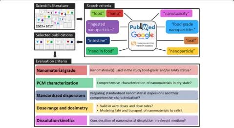
A combination of the terms“nano”,“nanoparticle”,“food”, “food grade nanoparticles”, “nano in food”, “toxicity”, “nanotoxicity”,“oral”,“intestine”, and“ingested nanoparti-cles” were used in Google Scholar, PubMed, and Web of Science search engines to identify scientific publications on toxicity of iENM (Fig.1). The search process focused on toxicity studies using nano/materials such as titanium dioxide, silicon dioxide, iron oxide and zinc oxide that were directly added to food products. Furthermore, we also used Google search engine to search for additional magazine articles related to identification of iENM in food products. Additional publications were traced from the references section of other articles and from our own col-lection of nano literature dating back to 2007. Only peer-reviewed scientific publications that were published in“English” were included in the analysis. A total of 650 peer-reviewed articles were identified, of which 39 met the inclusion criteria. Each scientific publication was thor-oughly reviewed, and information was extracted on 5 do-mains related to aspects of the methodology, as detailed in Fig. 1. Other published reviews have argued for the need to intensify research on the toxicology and risk assessment of iENM [30]. For publications that presented both in vitro and in vivo data, each component was evaluated separately.
Using food-grade nanomaterials
Each publication was evaluated based on whether the nanomaterials used in the study were certified as food-grade (i.e. allowed to be used as food additives), or not. Publications that did not specify nanomaterial type, or for which we were unable to collect that information based on the data reported in the manuscript, were de-noted as lacking that information (“not reported”).
Comprehensive physicochemical and morphological (PCM) characterization of ENM in dry state
Standardized nanoparticle dispersions and their characterization
Each publication was evaluated for whether the study used standardized protocols to disperse test nanomaterials and characterize the dispersions. A“standard dispersion proto-col”was defined as a protocol that follows best available sci-ence on nanoparticle dispersion, characterization, and fate and transport modeling [31–33]. Such protocols describe in detail information such as the make of the sonicator plus amplitude and power used, sample volume, dispersing media, measured energy density, and energy delivered to prepare stable nanoparticle dispersions, so that the protocol could be reproduced across labs. Characterization of nano-particle dispersions was considered minimally sufficient when size and charge distribution analysis data, obtained by DLS (dynamic light scattering) or NTA (Nanoparticle tracking analysis) and electron microscopy, were provided together with the polydispersity index (PdI). In addition, ef-fective density of agglomerates, an experimentally measured parameter critical for in vitro dosimetry modeling, is an-other important property of nanoparticle dispersions that should be measured experimentally and reported [33–35].
Dosimetry considerations: Dose range, rate, and the rationale
Each publication was evaluated for the dose ranges used and whether such ranges were justified or accounted for based on human daily dietary intake data for the respective
nanomaterials. In addition, in vitro studies were evaluated for dosimetry considerations, particularly whether fate and transport models were used to calculate the delivered dose to cells as a function of time. Dose rate (amount of dose per unit of time) was also considered and assessed in each of those studies.
Dissolution kinetics
Each publication was evaluated for considerations of dis-solution kinetics or biokinetics (for in vivo studies) of test nanomaterials. For in vitro studies, the evaluation was based on data provided regarding ionic release of test nanomaterials’ over time in relevant test media. For in vivo studies, the evaluation was based on data provided on ENM ion release over time in simulated or actual digestive fluids and/or accumulation of ions in circulation.
Results and discussion
Food-grade engineered nanomaterials in various food products
Nanomaterials are available in a variety of grades for their intended use in different products or applications. For ex-ample, pharmaceutical grade is a standard of purity that has been established by any recognized pharmacopeia, in-cluding US Pharmacopeia (USP), National Formulary (NF), British Pharmacopeia, or European Pharmacopeia (EP), which is suitable for use as an active or inactive drug, bio-logic, or reagent. Generally, the products are required to be
97–101% pure depending on their application and must contain ≤0.1% of bacteria. Engineered nanomaterials and their bulk counterparts used in or in-contact with food are considered direct or indirect food additives, respect-ively. In the United States, iENM are required to meet the food-grade guidelines issued by the Food and Drug ad-ministration (FDA), which are typically less rigorous than the specifications for pharmaceutical grade. Such iENM are mostly GRAS (Generally Recognized as Safe) mate-rials. GRAS status is assigned by the FDA to a product that is not known to be hazardous to health and thus ap-proved for use in foods. From our searches and interac-tions with iENM vendors, it appeared that companies are permitted to “self-affirm” GRAS status for certain iENM products. Furthermore, there appears to be a notable lack of rigorous oversight on the granting of ‘GRAS’status for iENM by the FDA.
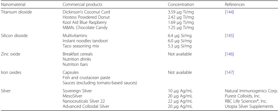
Engineered nanoparticles are used widely in a variety of products. In the USA, the Center for Food Safety main-tains a comprehensive database [36] of consumer prod-ucts in the food industry believed to contain nanoparticles (NP). The database focusses exclusively on food and food contact products, covering over 300 products and 40 dif-ferent types of nanomaterials. The database was evaluated as part of this review and the results have been summa-rized in Fig. 2. Over 70 food products contained iENM based on independent testing and/or labels. In addition, iENM were present in 14 baby and infant products and 16 cooking products [36]. A sizeable category of 45 products contained iENM intended for packaging. It is also appar-ent from Fig.2that independent and more rigorous test-ing of such products to confirm the presence of iENM, along with their concentration, chemical composition, and size distributions of primary particles vs. agglomerates is needed. The most common iENM used directly in food products are TiO2(only part of the product is in the nano-scale, as noted above), SiO2(silicon dioxide), iron oxides (Fe2O3), zinc oxide (ZnO) and silver (Ag). Table1 summa-rizes the types of foods containing each type of iENM, with product examples and reported mass concentrations for each product. These materials are or contain at least one product that fulfills the strict definition of an engi-neered nanomaterial with regards to primary particle size and synthesis/processing method. TiO2E171 for example has only ~ 1/3 of particles in the nanoscale. However, our own analysis of other TiO2used in foods (and certified as food-grade E171 equivalent) revealed that some of them may contain > 50% nanoscale particles and would there-fore meet the strict definition of an iENM. Notable in such analysis is the lack of information on primary particle size distribution and agglomerates, which is useful for in vitro iENM testing relevant to the nano fraction. It is also inter-esting to note how each iENM is used in the different products. For example, TiO2 is used frequently as a
whitening agent, SiO2 (silica made by wet processes or flame pyrolysis and not to be confused with crystalline sil-ica) is used as filler, and both ZnO and Fe2O3are used as food supplements. Silver has been used as a direct food additive in various colloidal silver drinks, whereas other Ag uses are in food contact applications. This analysis also re-veals the prominence of nano ingredients such as TiO2and SiO2in several foods frequently consumed by children.
Specifications regulating use of each iENM as a food additive also are unique. Titanium dioxide (TiO2) is allowed as a color additive (whitening agent) in food by the FDA, provided the added product conforms to the recommended specifications as described in Title 21 [37] of the e-CFR (electronic Code of Federal Regulations) and does not exceed 1% by weight of the food. The FDA has no specific guidelines for use of TiO2as a dietary supple-ment or as an antimicrobial agent in food products.
Silicon dioxide is permitted by FDA as a“food additive for direct addition to food for human consumption”provided that it is manufactured by vapor phase hydrolysis [38], does not exceed 2% by weight of the food, and conforms to the recommended specifications. Silica’s intended use as an anticaking agent is subject to the following condi-tions: (i) it is only permitted in foods in which the additive has been demonstrated to have an anticaking effect, (ii) it can be used in an amount not more than what is reason-ably required to produce its intended effect, and (iii) it can be used in an amount not to exceed 2% by weight of the food. Iron oxide and its hydrated forms are allowed by the FDA as a color additive in food, provided it conforms to the recommended specifications [39] and does not exceed 0.1% by weight of the final food product. In the USA, ZnO is also allowed as a color additive in foods, as well as in cosmetics, provided it is manufactured by the French process (described as the indirect process whereby zinc metal isolated from the zinc-containing ore is vaporized and then oxidized), conforms to the recommended speci-fications [39], and is used in accordance with good manu-facturing practices. Of note, these regulations are for their microscopic bulk materials and not specifically for the nanomaterials themselves.
Model test systems
Sixteen of the 24 in vitro studies reviewed utilized Caco-2 monocultures or a sub-clone of Caco-2 known as C2BBe1 cells, Table 2), both of which, even though originating from human epithelial colorectal adenocarcinoma cells, can be induced to differentiate into morphologically and functionally mature cells that resemble the enterocytes lining the small intestine. Although it has been suggested that C2BBe1 monolayers are more representative of the small intestinal epithelium than Caco-2 due to similar
transepithelial electrical resistance, morphological homo-geneity and BB myosin I expression levels similar to that of a human enterocyte, only 2 studies were found using them [40,41]. Additionally, the epithelial cell line used in vitro should be allowed to grow, form tight junctions and differentiate to enterocytes to form an intact barrier– rep-resentative of the GI tract epithelium, which is verified by measuring TEER (Trans-epithelial electrical resistance) values and expression of tight junction proteins before nanotoxicological assessment [42–44]. Other in vitro models include the cell lines representative of gastric epi-thelium (GES-1), mucus-secreting cells (HT29-MTX), colon epithelium (SW480, DLD-1), and mucus-secreting colon epithelium (NCM460). Of note was the use of MET-1 bacterial community to represent an in vitro model of gut microbial community [45]; and combina-tions of Caco-2/HT29-MTX or Caco-2/Raji-B cell lines in co-culture models to represent mucus-secreting epithe-lium and follicle-associated epitheepithe-lium, respectively [46].
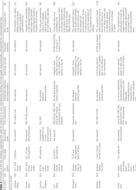
The Sprague Dawley rat model and CD-1 (ICR) mouse model were used in 13 out of 19 of the in vivo studies reviewed (Table 3). In one case, an ex vivo animal model comprising of Peyer’s patches and ileum was used. Nanoparticles were delivered by gavage as dispersions in a food matrix.
Test ENM identity: Food or industrial grade?
Table2 summarizes the in vitro studies intending to as-sess toxicity of iENM, including any information in each study regarding the nanomaterial’s grade, nanomaterial characterization, consideration of dosimetry and physio-logical relevance, as well as the primary findings of each study. In our literature survey from 2007 to 2017, only 19% of the studies (8 out of 42) used food-grade nano-materials to assess their toxicity on intestinal/gastric
Table 1Most frequent ingested engineered materials and nanomaterials (iENM) used in foods together with products and their
concentrations, as reported in the literature
Nanomaterial Commercial products Concentration References
Titanium dioxide Dickinson’s Coconut Curd
Hostess Powdered Donut Kool Aid Blue Raspberry M&Ms. Chocolate Candy
3.59μg Ti/mg
2.42μg Ti/mg
1.69μg Ti/mg
1.25μg Ti/mg
[144]
Silicon dioxide Multivitamins
Instant noodles tandoori Taco seasoning mix
6.4μg Si/mg
6.0μg Si/mg
5.3μg Si/mg
[145]
Zinc oxide Breakfast cereals
Nutrition drinks Nutrition bars
Not available [146]
Iron oxides Capsules
Fish and crustacean paste
Sauces (excluding tomato-based sauces)
Not available [147]
Silver Sovereign Silver
MesoSilver
Nanoceuticals Silver 22 Advanced Colloidal Silver
10μg Ag/mL
20μg Ag/mL
22μg Ag/mL
20μg Ag/mL
Table 3 Overview of the key findings regarding the state of science in in vivo nanotoxicity testing of food-grade nanomaterials, categorized by nanomateria l type First author Year Test system Dose range Nanomaterial grade PCM characterization Standardized dispersion and characterization Dose range rationale Dissolution biokinetics Main conclusions from study Ref Titanium dioxide Jiangxue Wang 2007 CD-1 (ICR) mouse model 5 g/kg bw Not reported XRF analysis only Not reported Not reported Not reported Ti O2 ret ained in liver, spleen , ki d n e ys an d lun g ti ss ue s, suggesting u pt ake by gastrointest in al tr ac t [ 21 ] Yanmei Duan 2010 CD-1 (ICR)
female mouse model
62.5, 125 and 250 mg/kg bw Not reported (self-synthesized) XRD, ICP-MS analysis Not reported Not reported Not reported In tr ag as tr ic Ti O2 admin ist ra tion in mice dama ges h o meo stas is b loo d sys te m and g ene rat es imm u n e re spo n se re sultin g in dis rupti on of liv er fu n cti on [ 85 ]
Carolina M. Nogueira
2012
Bl
57/6
male mouse model
100 mg/kg bw Commercially available for use in food, pharmaceuticals and cosmetics DLS, XRD No standard dispersion protocol specified Not reported Not reported Ti O2 micro an d nano p ar ticles indu ce a Th1-mediate d inflammatory response in th e small intestine, especially ileum [ 48 ] Yun Wang 2013
Sprague Dawley male
rat model 10, 50 and 200 mg/kg bw Not reported TEM, ICP-AES, XRD, FTIR, SSA by BET method, hydrodynamic size, zeta potential No standard dispersion protocol specified Intragastric doses selected based on the intake of dietary TiO 2 particles in the UK ICP-MS and ICP-OES to measure Ti content in tissues Young rats seem more sus cept ible to TiO 2 nanopar ticle exp osur e, which ca n p rovoke redu ctive stress in the p lasma of b oth young and old ra ts but through di fferent mechanisms [ 96 ] Zhangjian Chen 2014
Sprague Dawley male
rat model 10, 50 and 200 mg/kg bw/ day for 30 days Not reported Previously characterized [ 96 ] No standard dispersion protocol specified Intragastric doses selected based on the intake of dietary TiO 2 particles in the UK Not reported TiO 2 nanoparticles induce DNA double strand breaks in rat bone marrow cells after repeated oral administration for 30 days. It might be practical to control the application of TiO 2 nanoparticles as food additives [ 62 ] Roberta Tassinari 2014
Sprague Dawley rat
model 1 and 2 mg/kg bw/day for 5 days Not reported TEM, SEM, ICP-MS No standard dispersion protocol specified Dose levels selected based on the available data on the effects of TiO 2 nanomaterials ICP-MS to measure Ti content in tissues TiO 2 nanoparticles target endocrine-active tissues at dose levels relevant to human dietary intake; with no observable general toxicity and limited tissue deposition and damage in spleen [ 126 ] Emilie Brun 2014 Peyer ’ s patches and regular ileum (ex vivo),
mice model (in
Table 3 Overview of the key findings regarding the state of science in in vivo nanotoxicity testing of food-grade nanomaterials, categorized by nanomateria l type (Continued) First author Year Test system Dose range Nanomaterial grade PCM characterization Standardized dispersion and characterization Dose range rationale Dissolution biokinetics Main conclusions from study Ref Not reported for in vivo experiments Zhangjian Chen 2015
Sprague Dawley rat
model 2, 10 and 50 mg/kg bw/day for 30 or 90 days Not reported TEM, ICP-AES, XRD, FTIR spectroscopy, SSA by BET, hydrodynamic diameter (DLS), zeta potential No standard dispersion protocol specified Intragastric doses for rats selected based on the daily oral intake of TiO 2 nanoparticles for children under the age of 10 years in the US Not reported TiO 2 nanoparticles alone or in combination with glucose induce liver, kidney and heart injuries as well as changes in white blood cells and red blood cells in young rats. Interactions between TiO 2 nanoparticles and glucose was different in different body systems, leading to synergistic or antagonistic effects accordingly [ 123 ] Fashui Hong 2015 ICR male mice model 2.5, 5 and 10 mg/kg bw/day for 60 days Not reported (self-synthesized) TEM, XRD, SSA by BET, hydrodynamic diameter (DLS), zeta potential No standard dispersion protocol specified Dose levels were selected based on a report of the World Health Organization from 1969 Not reported TiO 2 nanoparticles cause testicular toxicity, reduced sperm production, and induced sperm lesions in a dose dependent manner. These effects are in close relation to reductions in daily food and water intake, biochemical dysfunctions and oxidative stress [ 65 ] Zhangjian Chen 2015
Sprague Dawley rat
model 2, 10 and 50 mg/kg bw/day for 30 or 90 days Not reported TEM, ICP-AES, XRD, FTIR spectroscopy, SSA by BET, hydrodynamic diameter (DLS), zeta potential No standard dispersion protocol specified Intragastric doses for rats selected based on the daily oral intake of TiO 2 nanoparticles for children under the age of 10 years in the US Not reported Long-term (90 days) daily ingestion of TiO 2 nanoparticles can exert mild and temporary cardiovascular toxicity by reduction in heart rate and systolic blood pressure, and increase in diastolic blood pressure [ 149 ] Ismael M. Urrutia- Ortega 2016 BALB/c male mice model 5 mg/kg bw for 10 weeks Food grade TiO 2 (E171) SEM, TEM, Raman spectroscopy, hydrodynamic diameter (NTA), zeta potential No standard dispersion protocol specified Intragastric doses justified based on collective exposure to TiO 2 from nominal consumption estimates and other sources Not reported TiO 2 E171 nanoparticles enhance tumor formation in the distal colon of chemical induced colitis-associated cancer (CAC) model of male BALB/c adult mice, marked by increase in CAC tumor progression markers. [ 49 ] Hanqing Chen 2017 CD-1 (ICR)
male mouse model
Table 3 Overview of the key findings regarding the state of science in in vivo nanotoxicity testing of food-grade nanomaterials, categorized by nanomateria l type (Continued) First author Year Test system Dose range Nanomaterial grade PCM characterization Standardized dispersion and characterization Dose range rationale Dissolution biokinetics Main conclusions from study Ref sp ecie s d iv er sity. Ti O2 n ano par tic le s di d no t in duce ob vi ous ch ang es in GI T hi stolo g y o r g ut m icr obi o ta co m p os itio n Maria G. Ammendolia 2017 Sprague Dawley rat model 1 and 2 mg/kg bw/day for 5 days Not reported TE M, SE M, hydrody n amic diameter (DL S) ,P d I. SSA and p urity (p ro vided
by man
u fa ct ur e r) No standard dispersion protocol specified Dose levels
selected based on
the available data on the effects of TiO 2 nanomaterials ICP-MS to measure Ti in gut tissue Higher dose of TiO 2 nanoparticles in male rats causes increase in height and width of villus, and dose-related increase in density of goblet cells. No such ef-fects are seen on female rats. TiO 2 nanoparticles penetrate intestinal mu-cosa (suggested by ICP-MS data) [ 125 ] Fashui Hong 2017 ICR male mice model 1.25, 2.5 and 5 mg/kg bw/day for 9 months Not reported (self-synthesized) TEM, XRD, SSA by BET, hydrodynamic diameter (DLS) [ 151 ] No standard dispersion protocol specified Dose levels were selected based on a report of the National Inst it u te for Oc cu pat ional Sa fe ty & H e al th (N IO SH ) from 201 1 [ 152 ] ICP-MS to measure Ti in gastric tissues Long term exposure to nano TiO 2 results in dysfunction of gastric secretion, inflammation, atrophy, and other lesions of gastric mucosa, which is closely associated with alterations of inflammation responding signal pathways in the stomach. [ 100 ] Sarah Bettini 2017 Adult male Wistar rat model 10 mg/kg bw/day for 7 days Food grade TiO 2 (E171) TEM, TEM-EDX, XANES, hydrodynamic diameter, PdI, zeta potential TiO 2 products prepared following the generic Nanogenotox dispersion protocol [ 153 ] Not reported TEM-EDX analysis in liver and intestine, and NanoSIMS analysis in Peyer ’ s Patches Intragastric food-grade TiO 2 administration for one week impairs intestinal immune homeostasis through Th17-driven autoimmune complications. Chronic exposures correlating with the development of an
inflammatory microenvironment, may
Table 3 Overview of the key findings regarding the state of science in in vivo nanotoxicity testing of food-grade nanomaterials, categorized by nanomateria l type (Continued) First author Year Test system Dose range Nanomaterial grade PCM characterization Standardized dispersion and characterization Dose range rationale Dissolution biokinetics Main conclusions from study Ref Hanqing Chen CD-1 (ICR)
male mouse model
2.5 mg/kg bw/day for 7 days Not reported to be food-grade TEM, hydrodynamic diameter (DLS), zeta potential No standard dispersion protocol specified Oral gavage doses justified based on estimated daily intake of TiO 2 and SiO 2 , and recommendation of OECD for Ag [ 150 ] Ag nanoparticles cause colitis-like symptoms in intestinal tract, and changes in gut microbiome. SiO 2 nanoparticles cause significant increase in proinflammatory cytokines and microbial species diversity. TiO 2 nanoparticles did not induce obvious changes in GIT histology or gut microbiota composition Zinc oxide Vyom Sharma 2012
Swiss albino male mouse model
50 and 300 mg/kg bw Not reported DLS and TEM No standard dispersion protocol specified Followed OECD guidelines [ 154 ] Not reported Sub-acute oral exposure to ZnO nanoparticles leads to their accumulation in liver causing oxidative stress-mediated DNA damage and apoptosis [ 81 ] Surekha Pasupuleti 2012
Sprague Dawley rat
model 5, 50, 300, 1000 and 2000 mg/kg bw Not reported DLS, zeta potential, SEM Not reported Followed OECD guidelines [ 155 ] Not reported Nano-sized ZnO exhibit toxic effects (increased AST and ALT serum levels, microscopic lesions in various organs) at lower doses in comparison to micron-sized ZnO [ 79 ] Miri Baek 2012
Sprague Dawley rat
epithelial cells in an ingested nanoparticle exposure sce-nario (Fig. 3). Ingestion of food containing ENM is the primary exposure route of the GI tract to exogenous ENM. A lifecycle study of food grade SiO2found that 10 out of 14 foods contained SiO2 of the same morphology and size as the pristine food grade SiO2[14]. For 4 out of 14 foods; however, they may also have contained non-food grade SiO2. In this context, using food-grade ENM for toxicity testing is critical. Importantly, a small fraction of inhaled nanoparticles can be transferred to the GI tract through mucociliary escalator clearance mechanisms and swallowing [12]. This was demonstrated after an intratra-cheal instillation of a single dose of radiolabeled TiO2NPs in Wistar-Kyoto female rats, where up to 5% of the in-stilled dose reached the GI tract after 24 h, which subse-quently increased to 20% after 28 days [47]. Because workers and consumers could be exposed to a variety of nanoparticle types via inhalation, which are categorically far more diverse than food grade ENM, this may lead to GI tract exposure of a larger variety of nanoparticles. However, the dose and dose rate employed must be realis-tic and reflect the peculiarities of such ingestion through inhalation exposure pathway. Among in vivo studies, we found that 14 out of 16 studies did not report the grade of the test nanomaterial (Table3). The study by Nogueira et al. [48], which used TiO2commercially available for use in food, pharmaceuticals and cosmetics, reported that these ENM induced a Th-1-mediated inflammatory response in the small intestine, with more pronounced cytokine pro-duction in the ileum. Urrutia-Ortega et al. [49], which used food-grade TiO2 (E171), showed that TiO2 nanoparticles used as food additive can enhance tumor formation in the distal colon in a colitis-associated cancer (CAC) model of male BALB/c adult mice, accompanied by a marked by in-crease in CAC tumor progression markers (COX2, Ki67 and β-catenin). Bettini et al. further showed that chronic exposure to TiO2E171 promoted ACF formation in normal
mucosa, demonstrating the ability of food-grade TiO2 to promote the development of preneoplastic lesions in rats without pre-existing epithelial barrier injuries [50]. Such studies provide more direct evidence on safety concerns of the ENM added to food products, and further supports the relevance of testing these nanomaterials.
There is an abundance of nanotoxicology literature doc-umenting the impact of variations in physico-chemical and morphological (PCM) properties of an ENM on toxi-cological outcomes. Differences in the primary particle size, surface area and chemistry, along with metal impur-ities, surface functionalization, or particle morphology, impact their behavior in and interaction with biological systems in vitro and in vivo [51]. The most compelling ex-amples come from the‘safe-by-design’cases, where delib-erate modifications in the surface chemistry, such as encapsulation of surfaces of ENM with amorphous silica [52, 53], or doping of ZnO with small amounts of Fe to suppress dissolution [54, 55], have a striking impact on the toxicity outcome. Food grade ENM are no exception. Toxicological findings on GI toxicity from industrial grade TiO2 (different size range, crystalline phase, and impur-ities) do not extend to food grade TiO2 and vice-versa. The same can be said for other iENM types.
Thus, when conducting toxicity assessment of ENM in the GI tract, it is important to use materials intended for or used in food, regardless of the test system. Several ingested nanoparticle toxicity studies used commercially available nanomaterials with no specification of the nanomaterials’ grade (Table2and Table3) and its commercial use or ap-plication [56–64]. In other studies, tested nanomaterials were synthesized in the lab [46,65–67]. Numerous studies examining the toxicity of TiO2nanoparticles on intestinal cells reported use of photocatalytic TiO2 [68–70] rather than food grade/pigment TiO2(Table2), even though food grade TiO2is the primary source of TiO2in food products. Photocatalytic TiO2 has antimicrobial properties and is used in food contact materials such as food preparation surfaces, self-cleaning and de-polluting paints and micro-bial surfaces [71], which can act as a secondary source of TiO2introduction into food products. There are significant differences between industrial and food grade TiO2 in terms of size, size distribution, specific surface area, surface properties and their agglomeration in aqueous phases, as discussed in detail by Dudefoi et al. [72]. For example, the primary particle sizes in photocatalytic P25 TiO2 were below 100 nm, whereas only 17–35% of the primary parti-cles were under 100 nm in diameter in the food-grade TiO2 [73]. Yang et al. reported that cationic dyes adsorbed more readily to food grade TiO2than P25 TiO2[73], presumably due to the presence of phosphate groups on the surface of food grade TiO2but not P25 TiO2[72]. These differences in surface chemistry implies different potential for inter-action with organics, proteins, and other micronutrients in
the food matrix. In another scenario, where TiO2used in sunscreens ends up being ingested (via swimming pool water, spoiled clothing in the workplace, or a child ingesting sunscreen by accident), surface treated TiO2nanoparticles used commercially in sunscreens (such as T-Lite™) must be used for in vitro and/or in vivo studies [74]. Chen et al. [16] used nano TiO2extracted from several chewing gums and found that its cytotoxicity was higher than that of commer-cially available P25 TiO2. At low concentrations of 350 ng/ mL (100 ng/cm2), food grade/pigment TiO2can also cause subtle changes in cell morphology, such as disruption of the brush border epithelium, but these concentrations did not acutely damage intestinal epithelium [26].
Food/product matrix effect
Selection of test ENM that are representative of the ingested exposure scenario and contained in the product that is actually ingested is critical for the relevance of a study. Using food grade variants instead of any available commercial forms of the test ENM will not only enable exposure scenario-relevant study designs but could also contribute towards reproducible observations across labs and more relevant toxicological outcomes [16, 73]. In other scenarios, such as assessing the hazard or risk of iENM resulting from ingestion of ENM from cosmetics and sunscreens, using food grade variants of those ENM would be of little utility. Instead, using the nanomaterials present in these cosmetic products lead to more relevant tests. Another critical factor to be considered for such tests is the matrix in which these iENM reside. When used in food and cosmetic formulations, ENM are immersed in a complex matrix of organic and inorganic additives, which interact with and become absorbed onto the surface of ENM, resulting in the formation of coronas with organic biomolecules such as proteins, lipids, and sugars. Such surface modifications may influ-ence cellular uptake of ENM, their biomolecular recog-nition, dissolution behavior, and eventually their toxicity. The food matrix effect has been largely ignored in in vitro and many in vivo studies until very recently. Argu-ably, the food matrix is a bigger challenge to address properly in in vitro studies, but it is an important con-sideration [75]. In such cases, simulating exposure sce-narios that closely resemble the process by which iENM are incorporated into or used in a food matrix, and vari-ations in the food matrix itself, are essential. Further-more, one should consider the complex journey that iENM undergo as they pass through the GI tract and the many PCM changes that they experience as a result of their transition through various compartments of the GI tract (e.g. exposure to different acidity, pH, digestive en-zymes, food components, etc.). Exposing intestinal cells to pristine iENM makes unrealistically bold assumptions about representativeness of this test system. This aspect
of the test methodology would benefit from more guid-ance, consensus documents, standardized protocols, and reference materials.
Comprehensive PCM characterization in dry state
The physicochemical and morphological (PCM) proper-ties play a pivotal role in determining the kinetics of nanoparticles, their dissolution and interaction with cells in cell culture medium and its impact on biological re-sponses [76, 77]. It is widely accepted in the scientific community that PCM characterization of ENM are of paramount importance in order to correlate PCM prop-erties with biological/toxicological responses [78]. As per the European Food Safety Authority (EFSA) guidelines, adequate characterization of ENM used in food products should include chemical composition, particle size/size distribution, physical form and morphology, particle and mass concentration, specific surface area, surface chem-istry, surface charge, redox potential, and chemical re-activity/catalytic activity [7]. [see Additional file1: Table S1] highlights some of the most important
physicochem-ical properties of nanoparticles and common
characterization methods. In our survey from 2007 to 2016, we identified several studies (Fig.4), especially per-taining to iENM, which had minimal to no PCM characterization [21, 48, 56, 58, 69, 79–81]. Inadequate PCM characterization of iENM, in the dry state and in food matrices persists to this day.
Our literature review identified 53% of studies (23 out of 43) lacking adequate characterization of nanomater-ials in the dry state (Fig. 4). We also observed that in vivo studies (10 out of 19) lacked more in comprehen-sive PCM characterization than in vitro studies (see Ta-bles 2 and 3). To relate the potential toxicity of iENM determined from in vitro or in vivo studies to the spe-cific features of iENM [82, 83], various metrics of their
PCM properties should be evaluated in powder form and in biological matrices [84]. Such parameters may in-clude among others size, morphology, mass, surface area, aspect ratio, charge, solubility and surface chemis-try. There appears to be an increase in the number of studies with more adequate PCM characterization in more recent years. Before 2015, 45% (13 out of 29) of the studies performed adequate PCM characterization, which increased to 71% (10 out of 14) in 2015–2017; yet, 29% of those studies still had insufficient characterization. A good example of the importance of an adequate PCM characterization approach is that of Gerloff et al. [68], which enabled identification of dis-tinctive toxicity of TiO2 anatase/rutile mixed phase on Caco-2 cells over pure anatase TiO2. However, in 20 out of 43 studies, little or no characterization was performed [66, 67, 85]. PCM characterization is indispensable in a mechanistic study investigating the biological effects of TiO2, SiO2and ZnO nanoparticles on different cell lines intended to represent the intestinal epithelium [64] (al-though no single cell line can accomplish that), as the PCM properties of nanoparticles alone are capable of in-stigating unique biological responses [77, 86, 87]. We encourage future studies, especially in vivo, to imple-ment comprehensive PCM characterization of iENM that exclusively use relevant food grade nanomaterials.
Standardized nanoparticle dispersions and their characterization
ENM properties are typically measured in dry powder state (e.g. mass or surface area per volume) to compare biological responses to ENM exposure in terms of ad-ministered dose. These comparisons do not take into ac-count particle-particle and particle to physiological fluid interactions in the liquid suspension [82, 88–90]. These interactions largely depend on the dispersion protocol, PCM characteristics of nanoparticles [91], and the prop-erties of the suspending media (pH, protein content, ionic strength, etc.) [92]. For in vitro testing, ENM, which are normally agglomerated in nanopowder form, are dispersed in certain liquid medium, typically DI water. The dispersed ENM are then transferred into a physiologically relevant dispersion media, which are ei-ther cell culture media for in vitro studies or body fluids for in vivo studies [32]. The methods used to disperse ENM in physiologically relevant media for in vitro stud-ies can have a substantial impact on the size, size distribution and the overall dispersion stability (re-agglo-meration state). Additionally, the effective density of the agglomerates formed when ENM are dispersed in physiologically relevant media differs from the density of the raw material, at times by several fold, primarily due to the protein corona formation and intra-particle trap-ping of the media [34]. The effective density and
agglomeration potential of the ENM affect their fate and transport in physiologically relevant media and impact the dissolution rate as well as available surface area for bio-interactions. The fate and transport of ENM in a media determines their settling rate, as well as other dose metrics such as delivered mass, surface number and particle number, each of which are discussed under dosimetry consideration. Consequently, in several in vitro studies, it has been shown that the agglomerates of nanoparticles exert different biological responses in comparison to well-dispersed nanoparticles [93–95]. Striking, but perhaps not surprising, is the finding that preparation of dispersions for nanotoxicity tests con-tinues to be highly variable and non-reproducible across different labs, a practice that continues today.
The dispersion protocols used in the published literature of iENM to date are highly variable. In an in vivo study [96], TiO2nanoparticles were dispersed in ultrapure water and ultrasonicated for 15 min before administering an intragastric dose. Another in vitro study by Zhao et al. [58] followed a protocol where TiO2NP in methanol were bath sonicated for 30 min followed by diluting the stock solution to 10 μg/mL in complete cell culture medium, and then further bath sonicating the suspension for 10 min before cell culture treatment. Another set of in vitro studies examining toxicity of several nanoparticles in Caco-2 cells suspended all materials in serum-free media and bath sonicated for 7–10 min before adding the testing concentration to the cells [61, 68, 80]. McCracken et al. [59] and Tassinari et al. [97] both procured similar TiO2 (< 25 nm particle size, 35–65 m2
protocols will enable comparisons and evaluations of in vitro as well as in vivo studies across labs. Furthermore, studies attempting to understand the effects of surface-coated or surface-treated nanoparticles [74] should avoid extensive sonication as it can cause removal of surface coating through large, but very localized, forces produced by cavitation [102, 103], which in turn can po-tentially alter biological responses to each type of treated nanoparticles.
Highly reproducible and standardized methodologies for nanoparticle dispersion, along with fate and transport modeling (discussed in greater detail in the next sec-tion), have been developed over the past few years, with the first papers on this topic appearing in 2012. Taurozzi and Hackley [104] published a detailed study on prepar-ing standardized nanoparticle dispersions and reportprepar-ing on the precise conditions so that the dispersion protocol is reproducible among other labs. Fully validated and transferable dispersion and transport modeling protocols are now available for common cell lines and ENM in the context of inhalation nanotoxicology, and they can be adopted for iENM [31–33, 105]. Yet, the practice has not changed significantly across the broader scientific community, and as a result, progress has been slow. The impact of such practices has not been properly docu-mented among in vitro nanotoxicology studies of iENM, but based on our literature review, we hypothesize that it could be significant. Some important factors related to sonication conditions, for which there is strong evidence in the published literature, include variation in size dis-tribution, dispersion stability, ion release/concentration, generation of free radicals and non-radical oxidants such as hydrogen peroxide during the sonication process that get transferred to the cell culture medium, and other modifications to nanoparticle surface properties. Spuri-ous oxidant production may negatively affect assay per-formance and/or confound in vitro results.
Dose range, rationale and dosimetry considerations
Dose range and rationale
The importance of dose rate and dose range in nanotox-icology testing has been documented in at least two re-cent nanotoxicology studies [106, 107]. The same logic holds true for testing iENM. Establishing a dose range that is realistic and physiologically relevant should take into consideration real-world ingestion (and inhalation) exposures, their frequency and other important features, such as administration of iENM in complex and diverse food matrixes. The challenges of selecting a defensible dose range is greatest in in vitro nanotoxicology of ingested ENM, for the simple reason that cell monocul-tures or co-culmonocul-tures in a plate represent a very different environment compared to that of cells within an organ. Assessment of a relevant dose or dose range requires
extrapolation of estimated human daily intake or expos-ure data for a specific ENM to equivalent in vitro doses for a relevant cell culture model [108]. As an example, if an individual ingests 1 mg of TiO2 through chewing gum, the amount of dose and time for which the buccal cavity cells are exposed will be different from the intes-tinal epithelial cells. Even if either cell type ends up be-ing exposed to the same amount, the surface area of each tissue will change the amount the cells are exposed to per unit surface area. Although there have been several toxicity studies where in vitro doses (μg/mL) are con-verted to equivalent in vivo (mg/kg) doses for studies in mice, any attempt to extrapolate published human expos-ure values to equivalent in vitro ingested doses in nano-toxicology has not been reported so far.
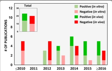
In our survey of the literature from 2007 to 2017, we found that 40% (17 out of 42) of the iENM toxicity stud-ies provided a dose range rationale, of which 14 were in vivo studies (Fig. 5). In in vitro studies (Table 2), the dose range, dose metric and time points varied across the study. For example, in studies assessing cytotoxicity of TiO2 nanoparticles on Caco-2 cells, groups have re-ported the following: 100 and 1000 μg/mL as acute and chronic exposure doses, respectively [56]; 0.35 to 35μg/ mL dose range [26]; 20 and 80 μg/cm2 for 4 and 24 h exposures [68, 80], respectively; 1 to 20 μg/cm2 for 6 and 24 h exposures [60]; 0 to 200μg/mL for 24 h expo-sures [16, 69]; 0 to 500 μg/mL for 48 h exposures [46]; 50 and 200 μg/mL for 24 h exposures [99]; and 1 mg/L for 0 h, 2 h, 4 h, 6 h, 8 h and 24 h exposures [109]. None of these reports provided a rationale for the indicated dose amounts. It is also not clear how any of these doses compared to the tissue doses (small intestines in this case) in vivo in animal studies and/or in humans. Al-though in most studies, Caco-2 cells were treated with nanoparticles only after verifying the formation of an in-tact epithelium by following a standard procedure of
allowing the cells to grow for 19–21 days, measuring TEER (Trans-epithelial electrical resistance) values and expression of tight junction proteins [42–44]. In other studies, cells were treated within 12 h (overnight) to 4 days of cell growth without confirmation of intact in-testinal epithelium [60, 69,80,109]. In an in vitro study examining the effects of surface-treated TiO2 nanoparti-cles widely used in sunscreens on Caco-2 cells, Fisichella et al. [74] deliberately chose the dose range to be higher than predicted environmental concentrations (10 to 100 μg/mL versus an expected 0.0007 to 0.016 μg/mL) under the assumption of a possible increase in the local environment, such as a child ingesting sunscreen by accident.
The dose ranges used in existing in vitro iENM tox-icity studies differ widely between groups and are not validated based on in vivo or in-human daily intake or exposure data (Table 2). More importantly, even though the estimated human daily intake differs for each ENM, the same dose range is used for different nanomaterials in multi-nanomaterial in vitro studies [61,64, 80]. How-ever, the use of high doses may be desirable for com-parative assessment and hazard ranking but is of limited utility if the study relies on a single unrealistically high dose or when it lacks a dose range and dose-response analysis. These dose issues continue to be prevalent in in vitro nanotoxicology of iENM. On the other hand, we found that dose ranges used in most in vivo iENM tox-icity studies (14 out of 19) were well justified based on
OECD guidelines (Organization for Economic
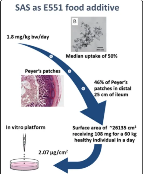
Co-operation and Development) or estimated daily diet-ary intake of iENM (Table 3). For in vitro iENM toxicity studies, we recommend using a mathematical approach for calculation of nominal doses based on published esti-mated daily dietary intake values to equivalent in vitro doses, from which a range of doses can then be selected. Such an extrapolation, although not reported so far, would require consideration of several factors such as estimated daily intake (mg/kg bw/day), exposure vari-ability (also known as median exposure dose), exposure/ dose at the tissue site, estimated surface area of the ex-posure site (cm2
), and other biokinetic considerations. Figure6 illustrates conceptually the process of calculat-ing a nominal equivalent in vitro dose for Caco-2 cells.
Dosimetry consideration
An in vitro cell culture system, which is usually a sim-plistic representation of a complex biological system, is a valuable tool to study cell biological, physiological and pathological processes under stress. However, as a plat-form, in vitro mono- or bi/tri-cultures have their own problems and limitations. One such challenge relates to delivery of nanomaterials in dispersions. The dynamics of the in vitro system can have a profound effect on the
outcome and/or the interpretation of the results. Most in vitro studies report administered doses in terms of ei-ther an initial mass concentration or of a total adminis-tered mass, but it is of great importance to consider the actual dose delivered to cells over time. The in vitro dos-imetry concept, and its importance and applicability, has been discussed in much detail in several recent and import-ant papers [88, 110,111]. The delivered dose (in the form of mass or ions) is the fraction of the administered dose that ends up depositing on the cell monolayer in an in vitro system in a given time, which eventually interacts with the cells to trigger a biological response. Dose delivered is largely dependent on the intrinsic PCM properties of the suspended nanomaterial, the extrinsic properties of culture media in which the nanomaterial is suspended, and the time course of exposure. Presence of larger agglomerates and effective density also impact the dose deposition kinet-ics [33, 34, 112], which emphasizes the significance of standard dispersion protocols and their characterization. Furthermore, presence of a sticky mucus layer, such as in co-cultures of epithelial (Caco-2/C2BBe1) and goblet cell lines (HT29-MTX), could drastically change the dose de-position and uptake kinetics in comparison to a monocul-ture of Caco-2/C2BBe1, which is devoid of mucus. Thus, any applied dose should be representative of a relevant dose
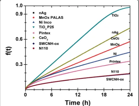
experienced by the specific cell culture system under a real-istic exposure scenario. In other words, delivered dose (or the dose range) is mainly dependent on the cell type used in the study, and the administered dose should take into consideration the in vitro dosimetry. As illustrated in Table 2, our evaluation affirms that, to our knowledge, since 2007, no in vitro study in the realm of iENM toxicity con-sidered dosimetry and its implications, which can poten-tially have a profound impact on the outcome and/or the interpretation of results. In an assessment of the impact of dosimetry, Pal et al. [32] found that after taking dosimetry into consideration, the slopes of administered/delivered dose-response relationships changed 1:4.94 times and were ENM-dependent, which significantly changed the toxico-logical ranking of engineered nanomaterials. Moreover, the resultant overall relative ranking of ENM intrinsic toxicity matched the in vivo inflammation data much better (Fig.7). With this in mind, an in vitro cell culture model is of great utility if it closely resembles or validates the in vivo condi-tions [113]. Future in vitro iENM toxicity studies should consider better modeling of exposures and equivalency that are relevant between exposure scenarios and in vitro dosimetry.
Dissolution kinetics
The majority of nanoparticle toxicity studies require a dosing protocol in which the test material is required to be in a liquid phase (culture media), where the term “dispersion” instead of“solution” is used. Dissolution, in the case of nanomaterials, denotes release of ions or molecules from the surface of a nanomaterial and their distribution throughout the available liquid volume as a result of entropy [114]. Although, there is sizeable litera-ture on dissolution and biodurability of natural and
synthetic micro-sized particles and fibers, studies of iENM toxicity lack dissolution evaluation of these nano-materials (Fig.8) [59,115]. The dissolution of nanopar-ticles in a culture media is largely driven by the concentration gradient that exists between the surface of NPs and the culture media. This, in turn, depends on the intrinsic PCM properties of nanoparticles, which include particle size, composition, shape, crys-tallinity, surface area and modification, and dispersion state of the nanoparticles. It also depends on the ex-trinsic properties of the culture media in which the NPs are dispersed, which includes parameters such as pH, ionic strength, constituent solvated molecules, temperature, ion concentration and availability of con-stituents to form complexes with released ions. This results in different dissolution rates for the same nanoparticles in different culture media with different order kinetics [116]. Such differences in dissolution rates necessitates its consideration when reporting the biological effects of nanoparticles. In addition, the dis-solution state of nanoparticles (particulate form or dissolved state) in a dispersion medium is a key com-ponent of the dynamic process that determines their uptake pathway, mechanism of toxicity, and the bio-logical compartment in which the NP will have high-est potential impact [116]. It has been shown through in vivo studies that even when no nanopar-ticles could be seen in TEM images, accumulation of nanoparticles was evident on ICP-MS analysis, im-plying an ionized fate in the cells or tissues [96, 117]. Ionization is important in driving another phenomenon, tissue redistribution, translocation and formation of new nanoparticle species with different chemical composition (e.g., as phosphates, oxalates or carbonates) at a distal site [118]. This phenomenon has been documented well for cerium oxide nanoparticles (Ce3+
in Ce2O3 or Ce4+ in
Fig. 7VCM-ISDD model-based calculations for nm delivered dose for different ENM formulation in RPMI + 10% FBS medium. Reproduced in parts with permission from Pal et al. [32]
CeO2), where surface Ce3+ sites, the main driver of toxicity, become complexed with phosphate to form cerium phosphate (CePO4) completely reverts their toxicity and stimulates growth [119–121].
Dissolution of iENM in relevant GI tract media con-tinues to be lacking in the literature. Only one out of 24 in vitro studies in the realm of iENM toxicity research addressed dissolution kinetics and its biological rele-vance (Table 2 and Fig. 8). Angelis et al. [60] in an in vitro study have shown significantly different dissolution kinetics of ZnO nanoparticles in serum or serum-free culture media, which drastically influences the cytotox-icity of these nanoparticles in Caco-2 cells. They showed that in serum-free media, toxicity of ZnO NP at lower concentrations was predominantly due to their dissol-ution into Zn2+ions, whereas at higher concentrations it was caused by both ZnO nanoparticles and Zn2+ions. It is, therefore crucial to consider the dissolution kinetics of nanoparticles in relevant cell culture media and its impact to in vitro studies.
Among in vivo studies, only 21% (4 out of 19) of the studies from 2007 to 2017 addressed dissolution kinetics (Fig.8). Most of those in vivo studies measured the ionic forms of respective nanomaterials in various tissues [63, 96,97,117] (Table3). However, it is also essential to de-termine the dissolution kinetics, biodurability and bio-persistance of nanomaterials in digestive fluids where nanoparticles interact with several complex fluids of varying pH, ionic strength and enzymatic activity during their course traveling through the gastrointestinal tract.
Summary of toxicological endpoints and outcomes
Among in vitro studies, the effects of iENM on cell prolif-eration, cellular energetics (WST-1, WST-8, live/dead kit, CellTiter-Glo, XTT, MTT and NRU assay), membrane damage (LDH and Trypan blue assay), apoptosis initiation (Annexin V-FITC and monodansylcadaverine staining), necrosis (Sytox red staining), DNA damage (Fpg-modified comet assay), morphology (electron microscopy), barrier permeability (Dextran-FITC transport), reactive oxygen species generation (electron paramagnetic resonance, total glutathione content, DCFH-DA assay), proinflammatory and inflammatory cytokine release (ELISA), and gene ex-pression (qRT-PCR) have been explored. Ion release from test nanomaterial and their subcellular location of accu-mulation has also been investigated using fluorescent la-belling of Zn2+ions [63]. The effects of iENM (e.g. TiO2) on in vitro models of gut microbiome has also been ex-plored by monitoring gas production (gas chromatog-raphy), analysing fatty acid production (fatty acid methyl ester analysis), and microbiome diversity (16S rRNA 454 pyrosequencing).
Upon exposure to iENM, in vivo studies have deter-mined the coefficients of liver, kidneys, stomach and
spleen, biochemical analysis of the blood for biomarkers of liver, kidney, cardiac, thyroid and reproductive func-tion, histopathological and ICP-MS/ICP-AES analysis of the tissues, hematological parameters, cytokine release and inflammatory cells quantification in GI tract seg-ments, semen evaluation (sperm count, motility and % abnormal sperms, biochemical assay of enzyme activities and oxidative stress) for testicular toxicity, heart rate and blood pressure, tumor progression biomarkers (COX2, β-catenin and Ki67) and IL-1β, IL-2, IL-6, TNF-α, IFN-γ, IL-8, IL-10, IL-17 and GM-CSF levels in colon tissue, gut microbiome composition (16S rRNA by 454 pyrosequencing), Ti concentration in tissues (con-focal microscopy, micro X-ray fluorescence imaging and NanoSIMS imaging), intestinal permeability (51Cr-EDTA radioactivity), and formation of aberrant crypts (preneo-plastic lesions) using Bird’s procedure [122]. Table4 pro-vides an extensive summary of various toxicological endpoints and respective assays used in in vitro and in vivo iENM toxicity literature as well as the studies of the gut microbiome.
Titanium dioxide
encoding for proteins involved in epithelial structure maintenance [46].
Persistence of TiO2 nanoparticles in specialized gut cells could possibly induce chronic damage. Indeed, this has been demonstrated by several long-term in vivo
ingested exposure studies. An early single high dose study by Wang et al. demonstrated uptake of TiO2 nanoparticles through the GI tract and their retention in liver, spleen, kidneys and lung tissues in CD-1 (ICR) mouse [21]. TiO2 nanoparticles induced lung, kidney
Table 4Toxicological endpoints used and/or recommended in in vitro and in vivo iENM toxicity investigations, and studies of the
gut microbiome
Endpoint Used and/or recommended assays or procedures References
In vitro
Nanoparticle ion release and accumulation location Fluorescent labeling of ions [156], ICP-MS [63,129]
Cell proliferation Cell count using hemocytometer [128]
Cellular energetics WST-1, WST-8, live/dead kit, CellTiter-Glo, XTT, MTS, MTT, NRU,
Prestoblueassay
[46,69,127]
ROS generation Electron paramagnetic resonance, total glutathione content,
DCFH-DA assay
[63,64,68,127]
Cell membrane damage LDH and trypan blue assay [59]
Apoptosis initiation Annexin V-FITC, monodansylcadaverine staining [59,64]
Necrosis Sytox red and propidium iodide (PI) staining [59,63]
Pro-inflammatory and inflammatory cytokine release ELISA, Wester blotting [129]
DNA damage Fpg-modified comet assay [62,68]
Brush border morphology Immunocytochemistry, electron microscopy (TEM and SEM) [66]
Barrier integrity Trans-epithelial electrical resistance measurement [46,128]
Barrier permeability Dextran-FITC and Lucifer yellow transport [46,128]
Gene expression qRT-PCR [46,64,66,68]
In vivo
Coefficients of organs Ratio of tissue (wet weight) to body weight [21,85]
Changes in tissues Histopathological evaluation [48,123,149]
Testicular toxicity Sperm count, motility and % abnormal sperms [65]
Tissue accumulation ICMP-MS or ICP-AES [100]
Reductive stress GSH/GSSG ratio in plasma [96]
Tissue function Blood biochemical and hematological analysis [123,149]
Inflammatory cells quantification in blood and the GI segment of interest
Flow cytometry,imaging flow cytometrya [48,50]
Apoptosis in the GI segment of interest TUNEL assay [81,125]
Cytokine release in blood and the GI segment of interest ELISA (IL-1β, IL-2, IL-6, TNF-α, IFN-γ, IL-8, IL-10, IL-17 and GM-CSF),
Western blotting
[48,49]
Tumor progression biomarkers in colon tissue Immunohistochemistry (COX2,β-catenin and Ki67),ELISAa,
Western blottinga
[49]
Intestinal permeability 51Cr-EDTA radioactivity [50]
Aberrant crypts formation in the GI tract Bird’s procedure [122] [50]
Local tissue concentration Micro X-ray fluorescence, NanoSIMS imaging [46,50]
Gut microbiome composition 16S rRNA pyrosequencing,Shotgun metagenomic sequencinga,
Microbial transcriptomicsa [124]
Gut microbiome models
Gas production Gas chromatography [45]
Fatty acid production Fatty acid methyl ester analysis [45]
Microbiome diversity 16S rRNA 454 pyrosequencing,Shotgun metagenomic sequencinga,
Microbial transcriptomicsa
[45,124]
a
and heart injuries as well as changes in red and white blood cell count in Sprague Dawley rats in a dose, time and gender-dependent manner after a 90 day exposure to 0–50 mg/kg bw/day [123]. Reduction in sperm pro-duction and sperm lesions were induced in ICR male mouse in a dose-dependent manner upon exposure to 0–10 mg/kg bw/day for 60 days [65]. A 10 week expos-ure to TiO2 E171 for 5 mg/kg bw for 5 days/week en-hanced tumor formation in the distal colon of chemical induced colitis-associated cancer (CAC) model of male BALB/c adult mice, marked by increase in CAC tumor progression markers in BALB/c male mice model [49]. Such colitis-like symptoms were not observed in CD-1 (ICR) male mouse after a 7-day exposure to 2.5 mg/kg bw/day of TiO2 nanoparticles [124] but Bettini et al. demonstrated that exposure to 10 mg/kg bw/day of food-grade TiO2for the same time period impaired in-testinal immune homeostasis through Th17-driven auto-immune complications in adult male Wistar rats [50], which was also observed in a similar study by Nogueira et al. after a single dose exposure TiO2micro and nano-particles commercially used in food products [48]. Bet-tini et al. further demonstrated that a 100 day exposure to food-grade TiO2 correlated with development of an inflammatory microenvironment, which promoted and could potentially initiate preneoplastic lesions in the colon [50]. A 9-month long exposure to nano TiO2 at 0–5 mg/kg bw/day also resulted in dysfunction of gastric secretion, inflammation, atrophy, and other lesions of gastric mucosa in ICR male mouse [100]. Interestingly, we noted that acute studies, in contrast to chronic ingested exposure studies, were much more likely to conclude with no observable toxic effects of TiO2 nano-particles [46,124–126].
Silicon dioxide, zinc oxide and iron oxide
In the past decade, a total of 7, 9 and 2 studies were published in regard to ingested exposure to SiO2, ZnO and Fe2O3 nanoparticles, respectively. Pertaining to the safety assessment of ingested SiO2 nanoparticles, all studies, except one, were conducted in vitro and there was limited agreement between them. Gerloff et al. in two studies [80, 127] and a study by Gehrke et al. [57] have shown that 24 h exposure to 80μg/cm2SiO2 nano-particles induced cytotoxic effects, DNA damage and glutathione depletion in Caco-2 cells, and 24 h exposure to ~ 150 μg/cm2 SiO2 nanoparticles stimulated HT29 cells proliferation, interfered with glutathione biosyn-thesis and the toxicity was found to be dependent on concentration, size and FCS (fetal calf serum) content of the culture medium, respectively. On the other hand, SiO2 nanoparticles have been reported to be relatively safe and exhibited no/minimal toxic effects after 24 h exposure to C2BBe1 cells at 10 μg/cm2 [59], 24 h
exposure to GES-1 and Caco-2 cells up to 100μg/ml of food additive silica [128] and 12 h exposure to three in-testinal cell lines (DLD-1, SW480 and NCM 460) at 1000 μM [64]. No overall toxicity beyond production of pro-inflammatory cytokines (IL-1β, IL-6 and TNFα) was observed when male CD-1 (ICR) mouse were adminis-tered 2.5 mg/kg bw/day SiO2 nanoparticles for 7 days but interestingly, microbiome analysis demonstrated creased microbial species diversity with an obvious in-crease in the genusLactobacillus[124].
With regards to ingested ZnO nanoparticles, 5 studies were conducted in vitro, 3 in vivo and one study had both in vitro and in vivo aspects. All in vitro studies re-ported mild to significant toxic effects on different intes-tinal cell lines (Caco-2, C2BBe1, GES-1, DLD-1, SW480, NCM 460) when exposed to ZnO nanoparticles alone [59,64,80,127, 129] or in combination with Vitamin C [63]. In vivo studies reported possible accumulation of ZnO nanoparticles (more likely in their ionic form) in the liver, lung and kidney with the smaller particles clearing from the body, primarily via feces, more rapidly than the larger ones [130]. A single dose of 5 to 2000 mg/kg bw to Sprague Dawley rats resulted in in-creased serum levels of ALT (alanine aminotransferase) and APT (aspartate aminotransferase), and microscopic lesions in liver, pancreas, heart and stomach at lower doses after 14 days [79]. Moreover, a 14-day consecutive exposure at 300 mg/kg bw/day to male Swiss albino mice also elevated serum ALT and alkaline phosphatase (ALP) levels, induced oxidative stress-mediated DNA damage and apoptosis – leading to pathological lesions in the liver [81].
In the past decade, only 2 in vitro studies investigated the effects of Fe2O3on intestinal epithelial cells. The stud-ies demonstrated size-independent adsorption of hematite nanoparticles on Caco-2 cells, which triggered dynamic reorganization of the brush border epithelium, disruption of tight junctions, drop in TEER, and differential expres-sion of tight junctions-maintaining genes [66,67].
To summarize, lack of in vitro and in vivo studies per-taining to ingestion of iron oxide nanoparticles, incon-sistencies among similarly designed in vitro iENM toxicity studies of TiO2 and SiO2, and lack of in vivo studies in case of SiO2 are sizeable knowledge gaps in the safety assessment of iENM. Future studies, especially in vitro, should be mindful to not propagate similar methodological issues discussed earlier and inconsistent findings.
Other factors