M E T H O D O L O G Y
Open Access
Active centromere and chromosome
identification in fixed cell lines
Thian T. Beh
1,2, Ruth N. MacKinnon
3,4and Paul Kalitsis
1,2*Abstract
Background:The centromere plays a crucial role in ensuring the fidelity of chromosome segregation during cell divisions. However, in cancer and constitutional disorders, the presence of more than one active centromere on a chromosome may be a contributing factor to chromosome instability and could also have predictive value in disease progression, making the detection of properly functioning centromeres important. Thus far, antibodies that are widely used for functional centromere detection mainly work on freshly harvested cells whereas most cytogenetic samples are stored long-term in methanol-acetic acid fixative. Hence, we aimed to identify antibodies that would recognise active centromere antigens on methanol-acetic acid fixed cells.
Results:A panel of active centromere protein antibodies was tested and we found that a rabbit monoclonal antibody against human CENP-C recognises the active centromeres of cells fixed in methanol-acetic acid. We then tested and compared combinations of established methods namely centromere fluorescencein situhybridisation (cenFISH), centromere protein immunofluorescence (CENP-IF) and multicolour FISH (mFISH), and showed the usefulness of CENP-IF together with cenFISH followed by mFISH (CENP-IF-cenFISH-mFISH) with the aforementioned anti-CENP-C antibody. We further demonstrated the utility of our method in two cancer cell lines with high proportion of centromere defects namely neocentromere and functional dicentric.
Conclusions:We propose the incorporation of the CENP-IF-cenFISH-mFISH method using a commercially available rabbit monoclonal anti-CENP-C into established methods such as dicentric chromosome assay (DCA), prenatal karyotype screening in addition to constitutional and cancer karyotyping. This method will provide a more accurate assessment of centromere abnormality status in chromosome instability disorders.
Keywords:Centromere, CENP-A, CENP-C, Immunofluorescence, Fluorescencein situhybridisation (FISH), Multicolour FISH (mFISH), Neocentromere, Dicentric, Dicentric chromosome assay (DCA), Human erythroleukaemia (HEL) cell line
Background
One of the hallmarks of cancer is genome instability, often characterised by the presence of aneuploidy and genetic heterogeneity resulting from chromosome missegregation or defective DNA repair followed by the failure to enter cellular arrest or death [1, 2]. Such genetic heterogeneity ranges from the extent seen in leukaemias, generally presented with simple chromosomal rearrangements, to carcinomas that are often complex [3, 4]. It is only with cumulative method improvements and technological
advancements made over the past 60 years that we are able to better understand disease mechanisms, and then apply the knowledge to cancer diagnosis, classifi-cation, prognosis, treatment selection and monitoring after treatment using the combination of molecular pathology, molecular cytogenetics and genomics in cancer research [5, 6].
One such technical advancement is the combination of RNA or DNA fluorescence in situ hybridisation (FISH) with immunofluorescence, commonly used for detection of RNA or DNA together with the protein of interest in or on the same cell. Co-detection of both genetic and the protein (epigenetic) components is espe-cially crucial in determining the activity status of a centromere — whether it is functional (active) or non-functional (inactive). The human centromere is a DNA-* Correspondence:[email protected]
1
Murdoch Childrens Research Institute, Royal Children’s Hospital, Parkville, Melbourne, VIC 3052, Australia
2Department of Paediatrics, University of Melbourne, Royal Children’s Hospital, Parkville, Melbourne, VIC 3052, Australia
Full list of author information is available at the end of the article
protein structure consisting of the repetitive α-satellite DNA wrapping around nucleosomes containing CENP-A that specify the inner kinetochore onto which other kinetochore protein complexes assemble [7]. A properly functioning centromere is essential for correct chromo-some segregation during cell divisions.
For cytogenetic investigations in the research setting, combinations of FISH and multicolour FISH (mFISH) performed on fixed cells as well as immunofluorescence followed by FISH (Imm-FISH) performed on freshly harvested cells are routinely used. However, for the stud-ies of the centromere regions, most antibodstud-ies raised against human centromere proteins do not recognise the epitopes of their targets after fixation in methanol-acetic acid despite using the method proposed by Earnshaw et al. [8]. This includes immersing cell preparations into a low ionic strength buffer to unravel the conformation of the compact chromosome to improve accessibility of target antigen and then returning the cells to buffer at physiological ionic strength to restore the chromosome morphology [8]. As for the current Imm-FISH carried out on freshly harvested cells, the morphology of the chromosomes is often distorted due to the involvement of the cytocentrifugation step [9, 10].
In this paper, we report on and discuss (1) the screen-ing outcome of several kinetochore antibodies for fixed cells, (2) the difference between the proposed method involving immunocytochemistry, FISH and mFISH, and the combination of other methods, and (3) the potential utility of the proposed method with the positive anti-body, rabbit monoclonal anti-CENP-C, in identifying chromosomes with structural centromere defects in clinical samples of patients with congenital diseases or cancer as exemplified using T-47D, a breast cancer cell line, and SN12C, a renal cancer cell line, from the NCI-60 cancer panel.
For the comparison of methods aforementioned, the human erythroleukaemia (HEL) cell line was used be-cause MacKinnon et al. [11] have shown by FISH that HEL has two large, rearranged chromosomes positive for multiple nucleolar organiser regions and three rear-ranged chromosomes that contain centromere DNA se-quences from two different chromosomes. HEL has been widely used for cell biology and differentiation studies in addition to the extensive data generated from a variety of techniques namely whole chromosome painting, single nucleotide polymorphism (SNP) array, OncoMap sequencing, mFISH, multicolour chromosome banding (M-BAND) and targeted FISH [11].
Autoimmune antibodies in the sera of scleroderma patients were known to react to intranuclear antigens of tissue sections but a subset were pinpointed to stain the centromere region after substantiation with mitotic cells by Moroi et al. [12]. In 1985, the anti-centromere
antibodies (ACAs) from the sera of CREST (calcinosis, Raynaud’s phenomenon, esophageal dismotility, sclero-dactyly, telangiectasia) variant of scleroderma patients led to the discovery of the first three centromere pro-teins namely CENP-A (17 kDa), CENP-B (80 kDa) and CENP-C (140 kDa), named from the lowest molecular weight to the highest [13].
Some ACAs had been shown to work on methanol-acetic acid fixed cells. However, they usually recognise multiple centromere proteins depending on the individ-ual serum and are limited in supply since they are re-stricted to individual autoimmune patients. Hence, ACAs are limited in their use for functional centromere identification where CENP-A and CENP-C are found ex-clusively on active centromeres but CENP-B is localised to the 17-bp CENP-B box of the centromeric repetitive DNA sequence regardless of its activity status [10, 14].
CENP-A and CENP-C are both part of the constitutive centromere-associated network (CCAN) that forms the inner kinetochore plate onto which other protein complexes assemble. CENP-A is a histone-H3 variant constituting the nucleosome core in a portion of the centromeric chromatin [15]. For CENP-A related stud-ies, a mouse monoclonal antibody against human CENP-A (Clone: 3–19) generated by Ando et al. [16] has been widely used as it is known to give punctate signals that mark the inner kinetochore of the centromere re-gion. Nonetheless, the binding of this antibody to CENP-A is obliterated if cells are fixed in methanol-acetic acid solution [16]. On the other hand, a rabbit polyclonal serum generated against full-length human CENP-C by the Earnshaw laboratory was shown to work on methanol-acetic acid fixed cells as reported by Warburton et al. [9]. In addition, CENP-C perfectly co-localises with CENP-A and both are constitutive markers of active centromeres [17] but the supply of the rabbit serum against CENP-C is limited.
Results and discussion Centromere antibody screening
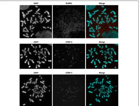
With the awareness that most samples in the cytogenetic laboratories are stored long-term in methanol-acetic acid fixative and with the expectation that centromere status screening will provide useful information for these la-boratories, we decided to screen several commercially available antibodies that target components of an active centromere using fixed HCT-15, a near diploid and lowly rearranged human colon cancer cell line [4].
protein of the BUB1B gene that plays a role in sensing proper chromosome-microtubule attachments during prometaphase to metaphase when it localises to the kinetochore (Fig. 1) [18]. The mouse monoclonal anti-body against HEC1 (NDC80) [9G3] (Abcam), a protein of the NDC80 complex that stabilises microtubule-kinetochore binding, was also negative for centromere signals on fixed cells. Rabbit monoclonal against CENP-E [CENP-EPR4542(2)] (Abcam), a kinesin-like motor protein that accumulates at the kinetochore throughout meta-phase [19], gave positive signals at the centromere re-gions but its staining was not homogeneous across all human chromosomes (Fig. 1), making it not ideal for the utilisation we were aiming for. Rabbit monoclonal against CENP-C [EPR15939] (Abcam) probed on fixed HCT-15 showed positive punctate signals (Fig. 1) similar to the signals seen in immunocytochemistry on non-fixed, cytocentrifuged cells even when it was used at a high dilution factor of 1 in 3,000 (data not shown).
Combining cenFISH, CENP-IF and mFISH methods
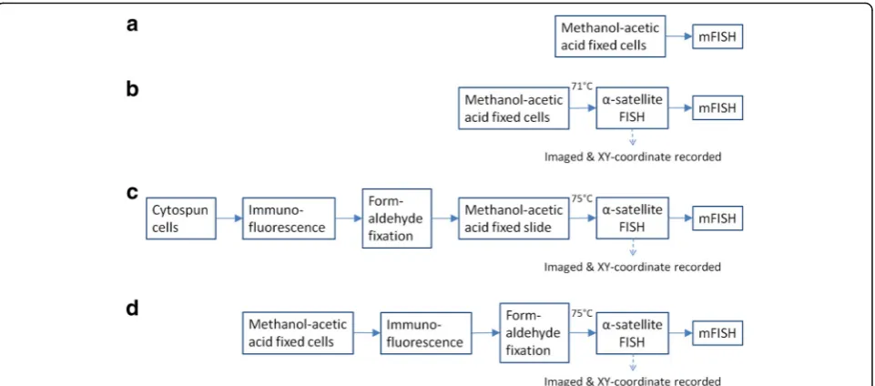
After validating that the rabbit monoclonal against human CENP-C worked on fixed cells, we then cross-linked the primary and secondary antibodies before performing α -satellite FISH (cenFISH). This combined method of centro-mere protein immunofluorescence (CENP-IF) followed by cenFISH will be referred to as CENP-IF-cenFISH here on-wards. In addition to the epigenetics and DNA-based infor-mation of the centromere activity, we believe that knowing the identity of the chromosomes would further add value to cytogenetic analysis. Hence, we attempted to carry out mFISH on the same cell spread on which CENP-IF-cenFISH had been performed prior and to ensure that mFISH would work sufficiently well post-CENP-IF-cenFISH, we compared the combination of methods as out-lined in Fig. 2 using the HEL cell line (Fig. 3).
In general, the chromosome morphology of cytocentri-fuged cells was not well-maintained compared to the meta-phase spread of cells stored in methanol-acetic acid fixative (unpublished observation). However, we were still able to distinguish the chromosome identities with the mFISH pro-cedure followed by image analysis (Fig. 3c) as seen in the comparable karyotypes generated from all methods (Fig. 3).
In our experiments, the cross-linking step with formalde-hyde for antibodies against CENP-A and CENP-C prior to cenFISH in methods C and D respectively caused the red fluorescent signals at the active centromere regions to carry over into the mFISH analysis (Fig. 4b, lower panel). The green fluorescent signals from cenFISH were not detectable. Thus, by taking the carrying over of fluorescent antibody signal into account, we suggest the preparation of an add-itional sample but using a differently coloured secondary antibody against anti-CENP-A or anti-CENP-C for more accurate chromosome identification by mFISH post- CENP-IF-cenFISH if necessary.
Comparison between CENP-IF-cenFISH-mFISH with CENP-A and CENP-C
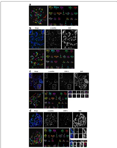
Referring to Fig. 2, method A is the standard mFISH per-formed according to the protocol recommended by the manufacturer (MetaSystems) and it is a useful method to identify interchromosomal translocations but is unable to identify rearrangements involving α-satellite DNA espe-cially the ones without observable constrictions. However, with method B, cenFISH followed by mFISH, multiple α -satellite signals were observed on two large chromosomes designated (i) and (ii) (Fig. 3b). Even in combination with DAPI staining of the DNA where chromosome constric-tions may be identified, identification of the centromere was proven to be not trivial in HEL cell line especially on the chromosome denoted (i). However, through methods Table 1List of antibodies targeting components of active centromere. Antibodies were first tested on non-fixative treated cells to deter-mine optimal concentration or dilution for immunocytochemistry and then tested on cells stored in methanol-acetic acid fixative
Antibody Species Concentration or
dilution
Cells stored in methanol-acetic acid
1 BubR1 [EPR12259(2)] ab183496 (Abcam) Rabbit IgG monoclonal
0.527μg/ml No
2 CENP-A (Clone: 3–19) Mouse IgG
monoclonal
1 : 500 No
3 CENP-A phospho Ser18 (Active Motif) Rabbit IgG polyclonal 1 : 400 No
4 CENP-C (Serum from rabbit 554; gift from William Earnshaw)
Rabbit polyclonal 1 : 1000 Yes
5 CENP-C [EPR15939] ab193666 (Abcam) Rabbit IgG monoclonal
0.677μg/ml Yes
6 CENP-E [EPR4542(2)] ab133583 (Abcam) Rabbit IgG monoclonal
0.275μg/ml Not homogeneous across all chromosomes
7 HEC1 [9G3] ab3613 (Abcam) Mouse IgG
monoclonal
C and D (both are CENP-IF-cenFISH-mFISH), the pres-ence of CENP-A and CENP-C signals indicates which of the two α-satellite positive constrictions was the active centromere [Fig. 3c (i) and 3D (i)].
As reported in MacKinnon et al. [11], three derivative chromosomes, namely der(4;20)t(4;11;20) and another two that resulted from whole arm translocations between two chromosomes, der(5;17) and der(10;19), contain centromere sequences originated from two different chro-mosomes. We were able to detect the presence of two active centromeres based on CENP-C signals on at least one der(10;19) chromosome in 2 out of 6 metaphase spreads with method C [Fig. 3c (iii) and (iv)] and in 4 out of 7 metaphase spreads using method D [Fig. 3d (iii)]. Der(5;17) and der(4;20) chromosomes were always show-ing only a pair of punctate CENP-A and CENP-C signals. Besides, the der(20)t(11;15;20) [Fig. 3d (iv)] in the meta-phase spread of Fig. 3d was the only der(20) that showed 2 pairs of signals for CENP-C out of 7 metaphases
analysed. This raised the possibility of the centromere of chromosome 15 being present on der(20) which could not be tested by MacKinnon et al. [11].
Detection of neocentric and functional dicentric chromosomes
To demonstrate the method’s utility, method D which used anti-CENP-C was carried out on two cell lines, T-47D and SN12C. T-47D is a breast cancer cell line known to us for having a stable neocentric chromosome while SN12C is a renal cancer cell line that shows a high proportion of dicentric chromosomes in the metaphase spreads that we analysed for another study (in preparation). In both in-stances, our method was able to first detect centromere ab-normalities within the metaphase spreads via CENP-IF-cenFISH (Fig. 4a & b, upper panels), followed by identifica-tion of the chromosomes that were involved in the final rearranged and centromere defect-bearing chromosomes (Fig. 4a & b, lower panels). The neocentromere in T-47D
was found to be on a segment of chromosome 3 and the neocentric chromosome was a product of re-arrangement between chromosome 3 and 5 (Fig. 4a, lower panel) whereas the dicentric chromosome in SN12C was formed from the rearrangement between chromosome 10 and 21 (Fig. 4b, lower panel).
In our study, we have demonstrated that our method, CENP-IF-cenFISH-mFISH, was able to (1) identify rearrangements implicating the centromeric DNA, (2) identify active centromere based on the presence of the constitutive kinetochore protein, CENP-C, and (3) reveal the identity of the chromosomes involved in the rear-rangements for methanol-acetic acid fixed cells.
We hereby propose that this method will be particularly helpful in studying clinicopathologically complex groups of tumours, for example liposarcomas, potentially as an additional criterion in subcategorising them. Both atypical lipoma/well-differentiated liposarcoma (ALP-WDLPS) and dedifferentiated liposarcoma (DDLPS) are identified by the presence of similar marker chromosomes, namely the supernumerary ring or giant rod chromosome con-taining the amplified 12q14-15 region with amplification of theMDM2andCDK4genes. However, ALP-WDLPS is classified as having intermediate aggressiveness compared to DDLPS which is malignant [20]. In addition, a high proportion of ALP-WDLPS has a marker chromosome with a neocentromere in contrast to DDLPS that also has the marker chromosome containing amplified 12q14-15 but with alphoid-centromere [21, 22].
Functionally dicentric chromosomes with two active centromeres have been thought to be involved in the breakage-fusion-bridge cycle and have recently been suggested to also contribute to chromothripsis based on the modelling of a subset of pa ediatric acute lympho-blastic leukaemia (ALL) with intrachromosomal amplifi-cation of chromosome 21 [23]. Furthermore, in refining the treatment for patients of pa ediatric ALL, the pres-ence or abspres-ence of a dicentric chromosome was consid-ered alongside other cytogenetic and genomic criteria [24], and in a recent acute myeloid leukaemia (AML) study, the difference in median survival between patients with one dicentric (5.8 months) and those with three dicentric chromosomes (1.8 months) was shown to be significant [25]. Taken together, our method which has the capability to detect functional dicentric chromo-somes in methanol-acetic acid fixed cytogenetic prepara-tions could assist in understanding the involvement of dicentric chromosomes in disease mechanism and also in risk stratification of patients for the treatment of diseases other than childhood ALL and AML.
Furthermore, this method could also be applied to pre-natal and congenital cases with chromosomal rearrange-ments containing centromere abnormalities that are not detected with current genomic technologies such as SNP microarrays and massively parallel sequencing [26]. An example of such chromosomal rearrangements is the isodicentric Y [idic(Y)], commonly found in children with disorders of sex development. Patients presented
a
b
c
d
with idic(Y) are often mosaic with 45,X cells and a high proportion of females presented with 45,X Turner syn-drome have the X chromosome of maternal origin, which together suggest that idic(Y) is mitotically un-stable [27, 28]. In addition, idic(Y) is the most common structural anomaly of chromosome Y in infertile men exhibiting an abnormal Y chromosome [28]. An azoo-spermic prospective father with idic(Y) may seek assisted reproductive technologies (ART) to achieve parenthood but he risks transmitting the idic(Y) to his offspring. Hence, the detection of idic(Y) is important for pre- and
postnatal genetic counselling as well as for genetic screening and recommendation of ART for infertile men. However, for routine screening, anti-CENP-C CENP-IF used together with chromosome specific FISH probe would be a more economical approach.
Another important application of the method is in the refinement of dicentric chromosome assay (DCA), the gold standard for biodosimetry assessment of individuals after exposure to radiation [29]. The International Atomic Energy Agency recommends an analysis of more than 1000 cells for better ascertainment of dicentric
a
b
Fig. 4CENP-IF-cenFISH-mFISH performed on cancer cell lines T-47D and SN12C with neocentric and dicentric chromosome respectively.aUpper panel: immunofluorescence images for T-47D showingα-satellite (green), CENP-C (red) and DAPI (blue); boxed in red is the neocentric chromosome and insets are enlarged images of the neocentric chromosome. Lower panel: mFISH colour profile of the neocentric chromosome indicating it is a rearranged chromosome containing segments from chromosomes 3 and 5.bUpper panel: immunofluorescence images for SN12C showing α-satellite (green), CENP-C (red) and DAPI (blue); boxed in red is the dicentric chromosome and insets are enlarged images of the dicentric chromosome. Lower panel: mFISH colour profile of the dicentric chromosome indicating it is a rearranged chromosome from chromosomes 21 and 10
(See figure on previous page.)
Fig. 3Representative images of HEL metaphase for each method, with marked (i) der(9) and (ii) psu dic(22;9).aStandard mFISH. Metaphase spread (left) and karyotype (right) in false colour.bcenFISH-mFISH. Upper panel: immunofluorescence images showingα-satellite (green) and DAPI (blue); insets are enlarged images of (i) der(9) and (ii) psu dic(22;9). Lower panel: metaphase spread and karyotype in false colour.c CENP-IF-cenFISH-mFISH with CENP-A. Upper panel: immunofluorescence images of cytospun cells showingα-satellite (green), CENP-A (red) and DAPI (blue); insets are enlarged images of (i) der(9) and (ii) psu dic(22;9). Lower panel: metaphase spread and karyotype in false colour display. (iii) and (iv) are der(10;19) with two pairs of CENP-A signals.dCENP-IF-cenFISH-mFISH with CENP-C. Upper panel: immunofluorescence images showingα-satellite
(green), CENP-C (red) and DAPI (blue). Lower panel: metaphase spread and karyotype in false colour display; (i) and (ii) are der(9) and psu dic(22;9)
chromosome formation for low radiation dose of less than 100 mSv, equivalent to a computed tomography (CT) scan [30, 31]. A recent study by Abe et al. [31] re-ported that the analysis of 1000 metaphase spreads probed with cenFISH was sufficient to yield comparable precision to 2000 conventional Giemsa stained meta-phases. However, these DCA methods only assess for the occurrence of a dicentric chromosome based on the presence of primary constrictions and centromere DNA, therefore, not providing any information on whether the affected chromosome contains one or two active centro-meres which may have impacts on downstream genome instability. This gap of information could be addressed with the truncated version of our method D (excluding the mFISH step) as a DCA. With an additional 2 h and 40 min approximately for immunocytochemistry on top of cenFISH, it will provide the information on both the centromere DNA and its associated active kinetochore protein CENP-C, further improving the precision of the current DCA methods.
Conclusions
The CENP-IF-cenFISH method using pan-centromeric FISH probe and commercially available rabbit monoclo-nal anti-CENP-C to detect active centromere performed on methanol-acetic acid fixed cells is an improvement on the existing DCA methods and prenatal and congeni-tal testings for chromosome structure anomalies such as isodicentric Y. Furthermore, the CENP-IF-cenFISH-mFISH method which additionally reveals chromosome identity could potentially add diagnostic value and in-crease our understanding of disease mechanism if it was to be incorporated into established procedures including constitutional and cancer cytogenetic tests.
Methods Cell culture
Cell lines namely HEL, HCT-15, T-47D and SN12C were cultured in Roswell Park Memorial Institute (RPMI) 1640 with 10 % fetal bovine serum, 2 mM L-glutamine, penicillin and streptomycin with 5 % CO2at 37 °C [32].
Cell preparation & immunocytochemistry
For immunocytochemistry performed on freshly har-vested cells, colcemid (KaryoMAX, Thermo Fisher Scientific) was first added to cell medium to a final con-centration of 0.1 μg/ml for 1.5 h. Cells were harvested via mitotic shake-off and subjected to hypotonic treat-ment (0.075 M KCl) for 15 min. 1500 cells were used for each slide preparation. Cells cytospun onto the glass slides were washed thrice with cold KCM (120 mM KCl, 20 mM NaCl, 10 mM Tris–HCl pH 7.5, 0.5 mM EDTA, 0.1 % Triton X-100) for 5 min each time and first incu-bated with mouse anti-human CENP-A (1:500) then
followed by Alexa594 conjugated secondary antibody against mouse (1:1000) (Thermo Fisher Scientific), both diluted in KCM buffer with 1 % BSA, at 37 °C for 1 h and 40 min respectively, with three 5 min KCM buffer washes after each incubation. Cells were then cross-linked in KCM containing 4 % v/v formaldehyde (Merck Millipore) for 10 min at room temperature (RT), washed twice with distilled water, briefly once and 5 min for an-other, and air-dried before being fixed in ice-cold methanol-acetic acid (methanol:acetic acid, 3:1 volume ratio) at 4 °C for 30 min, air-dried and left to age for at least 48 h at RT. Antibody screening was performed on HCT-15 cells with the primary antibodies diluted ac-cording to Table 1 followed by Alexa594 conjugated secondary antibodies (1:1000) (Thermo Fisher Scientific) against the respective species. The cells were visualised without being fixed with methanol-acetic acid.
For immunocytochemistry on methanol-acetic acid fixed cells, cells were dropped onto glass slides and
dipped immediately into TEEN buffer (1 mM
triethanolamine-HCl pH 8.5, 0.2 mM Na EDTA, 25 mM NaCl) once the fixative had dried. TEEN buffer was changed twice, after 3 min. Cells were blocked with 0.1 % Triton X-100 (Sigma-Aldrich) and 0.1 % BSA (Sigma-Aldrich) in TEEN at 37 °C for 15 min followed by incubation in rabbit anti-human CENP-C (1:3000) (Abcam) in TEEN at 37 °C for 1 h. Slides were washed with KB buffer (10 mM Tris–HCl pH 7.7, 150 mM NaCl, 0.1 % BSA) thrice, 4 min each time before incu-bating with Alexa594 conjugated secondary antibody against rabbit (1:1000) (Thermo Fisher Scientific) diluted in KB buffer at 37 °C for 40 min. Slides were then washed twice with KB for 4 min each time before being fixed in KB containing 4 % formaldehyde for 10 min at RT, washed twice with water, briefly once and 5 min for another, and then air-dried. For antibody screening, after the washing step post-secondary antibody incubation, slides were mounted with Vectashield antifade mounting medium (Vector Laboratories) added with DAPI before visualisation.
Pan-centromeric probes
0.1 volumes NaOAc and 2.5 volumes of 100 % ethanol. Precipitated DNA was spun down, washed with 70 % ethanol and subsequently air-dried. The DNA pellet was then resuspended in 40 μl hybridisation buffer (30 % formamide, 2 X SSC, 10 % dextran sulfate) before being denatured at 95 °C for 5 min and then placed on ice. This was a modification based on Roche’s Nick Transla-tion Kit protocol.
FISH
For cells that were to be probed directly, colcemid was added to the cell medium to a final concentration of 0.1 μg/ml for 1.5 h. Cells were trypsinised, spun down for 4 min and washed once with phosphate-buffered sa-line (PBS) before being subjected to hypotonic treatment in 0.075 M KCl at 37 °C for 15 min. Cells were then fixed by adding ice-cold methanol-acetic acid, spun down, resuspended with the fixative after discarding the supernatant and this process was repeated once and the pellet was resuspended in a final 200–800μl of fixative to yield an optimal cell density for metaphase spread preparation. Fixed cells were dropped onto glass slides and aged for at least 48 h at RT before performing FISH. Biotinylated probes against α-satellites were co-denatured with DNA on the slides at 71 °C for 5 min and incubated in a humidified chamber at RT for 16– 18 h. Slides were then (i) washed using 2 X saline-sodium citrate (SSC) buffer twice followed by 1 X SSC buffer thrice, each time at RT for 5 min, (ii) blocked with Tris-NaCl-Blocking (TNB) [0.1 M Tris-HCl pH 7.5, 150 mM NaCl, 0.5 % w/v Blocking Reagent (Roche)] buffer at 37 °C for 30 min, (iii) incubated with avidin conjugated with Alexa-488 (dilution 1:500) (Thermo Fisher Scientific), (iv) washed thrice with 4 X SSC with 0.05 % v/v Tween-20 at 37 °C for 5 min each time and (v) mounted with Vectashield antifade mounting medium containing DAPI.
For cells that had undergone immunocytochemistry with CENP-A, aged slides were co-denatured with α -satellites probes at 75 °C instead of 71 °C for 5 min and incubated in a humidified chamber at RT for 16–18 h. Subsequent steps were the same as aforementioned.
mFISH
mFISH was carried out with 24 XCyte (MetaSystems) according to the manufacturer’s instructions and with omission of a few early steps for slides that had under-gone FISH and Imm-FISH. Slides that had underunder-gone FISH only and immunocytochemistry followed by FISH were washed in 1 X PBS at RT for 3 min and 2 X SSC at 70 °C for 30 min, allowed to cool to RT for about 20 min, washed in 0.1 x SSC at RT for 1 min, denatured in 0.07 M NaOH at RT for 1 min, washed in 0.1 X SSC followed by 2 X SSC at 4 °C for 1 min each wash and
then sequentially dehydrated in 30, 50, 70 and 100 % ethanol at RT for 1 min each before being air dried. Denatured 24 XCyte mFISH probes were then put onto the slides and incubated in a humidified chamber at 37 °C for 2 days. Post-hybridisation slides were washed in 0.4 X SSC at 72 °C for 2 min, 2 X SSCT (2 X SSC containing 0.05 % v/v Tween-20) at RT for 0.5 min and rinsed briefly in water before being air dried and mounted with Vecta-shield antifade mounting medium containing DAPI for visualisation.
Microscopy and analysis
All images were taken using a Zeiss Axio Imager.M1 microscope/AxioCam Mrm camera. All Imm-FISH im-ages were captured and analyzed with Axio Vs40 vs4.6.1.0 software (Carl Zeiss) while mFISH images were taken and analyzed with Isis colour fluorescence and FISH imaging system (MetaSystems).
Abbreviations
cenFISH:centromere fluorescencein situhybridisation; CENP-IF: centromere protein immunofluorescence; Imm-FISH: immunofluorescence followed by FISH; mFISH: multicolour FISH; RT: room temperature.
Competing interests
The authors declare that they have no competing interests.
Authors’contributions
All experiments were carried out and the manuscript was drafted by TTB. The manuscript was reviewed and edited by PK and RNM. All authors read and approved the final manuscript.
Acknowledgements
We would like to thank Professor William Earnshaw at the Wellcome Trust Centre for Cell Biology, University of Edinburgh for the rabbit serum against CENP-C and Professor Hamish Scott at SA Pathology, Adelaide for the HEL cell line. This work was supported by the National Health and Medical Research Council (NHMRC), Australia Project Grant 1031089 and by the Victorian Government’s Operational Infrastructure Support Program.
Author details
1Murdoch Childrens Research Institute, Royal Children’s Hospital, Parkville, Melbourne, VIC 3052, Australia.2Department of Paediatrics, University of Melbourne, Royal Children’s Hospital, Parkville, Melbourne, VIC 3052, Australia. 3Victorian Cancer Cytogenetics Service, St Vincent’s Hospital, Fitzroy, Melbourne, VIC 3065, Australia.4Department of Medicine, St Vincent’s Hospital, University of Melbourne, Fitzroy, Melbourne, VIC 3065, Australia.
Received: 6 February 2016 Accepted: 17 March 2016
References
1. Hanahan D, Weinberg RA. Hallmarks of cancer: the next generation. Cell. 2011;144:646–74.
2. Gordon DJ, Resio B, Pellman D. Causes and consequences of aneuploidy in cancer. Nat Rev Genet. 2012;13:189–203.
3. Hoglund M, Gisselsson D, Hansen GB, Mitelman F. Statistical dissection of cytogenetic patterns in lung cancer reveals multiple modes of karyotypic evolution independent of histological classification. Cancer Genet Cytogenet. 2004;154:99–109.
4. Roschke AV, Tonon G, Gehlhaus KS, McTyre N, Bussey KJ, Lababidi S, et al. Karyotypic complexity of the NCI-60 drug-screening panel. Cancer Res. 2003;63:8634–47.
6. Bernheim A. Cytogenomics of cancers: from chromosome to sequence. Mol Oncol. 2010;4:309–22.
7. Kalitsis P, Choo KH. The evolutionary life cycle of the resilient centromere. Chromosoma. 2012;121:327–40.
8. Earnshaw WC, Ratrie 3rd H, Stetten G. Visualization of centromere proteins CENP-B and CENP-C on a stable dicentric chromosome in cytological spreads. Chromosoma. 1989;98:1–12.
9. Warburton PE, Dolled M, Mahmood R, Alonso A, Li S, Naritomi K, et al. Molecular cytogenetic analysis of eight inversion duplications of human chromosome 13q that each contain a neocentromere. Am J Hum Genet. 2000;66:1794–806.
10. Page SL, Earnshaw WC, Choo KH, Shaffer LG. Further evidence that CENP-C is a necessary component of active centromeres: studies of a dic(X; 15) with simultaneous immunofluorescence and FISH. Hum Mol Genet.
1995;4:289–94.
11. Mackinnon RN, Wall M, Zordan A, Nutalapati S, Mercer B, Peverall J, Campbell LJ. Genome organization and the role of centromeres in evolution of the erythroleukaemia cell line HEL. Evol Med Public Health. 2013;2013:225–40.
12. Moroi Y, Peebles C, Fritzler MJ, Steigerwald J, Tan EM. Autoantibody to centromere (kinetochore) in scleroderma sera. Proc Natl Acad Sci U S A. 1980;77:1627–31.
13. Earnshaw WC, Rothfield N. Identification of a family of human centromere proteins using autoimmune sera from patients with scleroderma. Chromosoma. 1985;91:313–21.
14. du Sart D, Cancilla MR, Earle E, Mao JI, Saffery R, Tainton KM, et al. A functional neo-centromere formed through activation of a latent human centromere and consisting of non-alpha-satellite DNA. Nat Genet. 1997;16:144–53.
15. Hori T, Fukagawa T. Establishment of the vertebrate kinetochores. Chromosome Res. 2012;20:547–61.
16. Ando S, Yang H, Nozaki N, Okazaki T, Yoda K. CENP-A, -B, and -C chromatin complex that contains the I-type alpha-satellite array constitutes the prekinetochore in HeLa cells. Mol Cell Biol. 2002;22:2229–41.
17. Warburton PE, Cooke CA, Bourassa S, Vafa O, Sullivan BA, Stetten G, et al. Immunolocalization of CENP-A suggests a distinct nucleosome structure at the inner kinetochore plate of active centromeres. Curr Biol. 1997;7:901–4. 18. Baker DJ, Dawlaty MM, Wijshake T, Jeganathan KB, Malureanu L, van Ree JH,
et al. Increased expression of BubR1 protects against aneuploidy and cancer and extends healthy lifespan. Nat Cell Biol. 2013;15:96–102.
19. Yen TJ, Li G, Schaar BT, Szilak I, Cleveland DW. CENP-E is a putative kinetochore motor that accumulates just before mitosis. Nature. 1992;359:536–9.
20. Bridge JA. The role of cytogenetics and molecular diagnostics in the diagnosis of soft-tissue tumors. Mod Pathol. 2014;27 Suppl 1:S80–97. 21. Garsed DW, Marshall OJ, Corbin VD, Hsu A, Di Stefano L, Schroder J, et al.
The architecture and evolution of cancer neochromosomes. Cancer Cell. 2014;26:653–67.
22. Marshall OJ, Chueh AC, Wong LH, Choo KH. Neocentromeres: new insights into centromere structure, disease development, and karyotype evolution. Am J Hum Genet. 2008;82:261–82.
23. Li Y, Schwab C, Ryan SL, Papaemmanuil E, Robinson HM, Jacobs P, et al. Constitutional and somatic rearrangement of chromosome 21 in acute lymphoblastic leukaemia. Nature. 2014;508:98–102.
24. Moorman AV, Enshaei A, Schwab C, Wade R, Chilton L, Elliott A, et al. A novel integrated cytogenetic and genomic classification refines risk stratification in pediatric acute lymphoblastic leukemia. Blood. 2014;124:1434–44.
25. Sarova I, Brezinova J, Zemanova Z, Ransdorfova S, Izakova S, Svobodova K, et al. Molecular cytogenetic analysis of dicentric chromosomes in acute myeloid leukemia. Leuk Res. 2016;43:51–7.
26. Prakash S, Guo D, Maslen CL, Silberbach M, Milewicz D, Bondy CA, Gen TACI. Single-nucleotide polymorphism array genotyping is equivalent to metaphase cytogenetics for diagnosis of Turner syndrome. Genet Med. 2014;16:53–9.
27. Lange J, Skaletsky H, van Daalen SK, Embry SL, Korver CM, Brown LG, et al. Isodicentric Y chromosomes and sex disorders as byproducts of homologous recombination that maintains palindromes. Cell. 2009;138:855–69. 28. Kim JW, Park SY, Ryu HM, Lee DE, Lee BY, Kim SY, et al. Molecular and
clinical characteristics of 26 cases with structural Y chromosome aberrations. Cytogenet Genome Res. 2012;136:270–7.
29. Crespo RH, Domene MM, Rodriguez MJ. Biodosimetry and assessment of radiation dose. Rep Pract Oncol Radiother. 2011;16:131–7.
30. IAEA. Cytogenetic dosimetry: applications in preparedness for and response to radiation emergencies, EPR-biodosimetry. Vienna: International Atomic Energy Agency; 2011.
31. Abe Y, Miura T, Yoshida MA, Ujiie R, Kurosu Y, Kato N, et al. Increase in dicentric chromosome formation after a single CT scan in adults. Sci Rep. 2015;5:13882.
32. Shoemaker RH. The NCI60 human tumour cell line anticancer drug screen. Nat Rev Cancer. 2006;6:813–23.
33. Choo KH, Vissel B, Earle E. Evolution of alpha-satellite DNA on human acrocentric chromosomes. Genomics. 1989;5:332–44.
• We accept pre-submission inquiries
• Our selector tool helps you to find the most relevant journal
• We provide round the clock customer support
• Convenient online submission
• Thorough peer review
• Inclusion in PubMed and all major indexing services
• Maximum visibility for your research
Submit your manuscript at www.biomedcentral.com/submit